Original Article
3 7 3 Arq Bras Oftalmol. 2014;77(6):373-6 http://dx.doi.org/10.5935/0004-2749.20140093
INTRODUCTION
Many studies have contributed to the development of foldable IOLs of different materials and designs, and have been performed to help cataract surgeons to select an IOL that could provide the best quality of vision for pseudophakic patients(1). Some of those studies have shown that the human cornea has a positive value of spherical aberration (SA), which is stable throughout the entire life(2).
Implantation of a spherical IOL could theoretically increase the positive SA of the eye and subsequently degrade the quality of vi-sion(3-5). The aspheric IOLs were developed as an option to generate less higher-order aberration (HOAs) after cataract surgery, providing patients a better contrast sensitivity (CS) and, therefore, enhancing patients’ quality of life(6-10).
Frequency-doubling technology (FDT) testing for the detection of glaucomatous visual field (VF) loss consists of determining CS
mea sures for detecting the frequency-doubled stimulus(11). Maddess et al.(12) proposed that a CS test based on the frequency-doubling illusion should be effective in the detection of early glaucomatous VF loss. However, previous studies have yielded divergent views regarding the diagnostic performance of FDT perimetry to standard automatic white-on-white perimetry (SAP) for the detection of glaucoma(13-15).
Because previous studies have demonstrated an improvement in CS in eyes implanted with aspheric IOLs(6-10), is important to inves-tigate its influence on the results of FDT. As the number of aspheric IOLs implanted all over the world increases and the FDT is progressi-vely incorporated in clinical practice, understanding the effect of aspheric IOLs on this mode of perimetry is of considerable clinical importance. A possible improvement in the perimetry values with such lenses may mask the diagnosis of glaucoma or lead to other dia g nostic confusions.
Influence of aspheric intraocular lens on frequency doubling technology and
contrast sensitivity: a fellow eye study
Inluência da lente intraocular asférica na perimetria de frequência dupla e sensibilidade ao contraste:
estudo contralateral
RodRigo FRançade espíndola1, MaRcony R. santhiago1, MáRio luiz RibeiRo MonteiRo1, newton KaRa-JunioR1
Submitted for publication: July 31, 2014 Accepted for publication: October 12, 2014
Study conducted at Division of Ophthalmology, University of São Paulo, São Paulo, SP, Brazil.
1 Division of Ophthalmology, University of São Paulo, São Paulo, SP, Brazil.
Funding: No specific financial support was used for this study.
Disclosure of potential conflicts of interest: None of the authors have any potential conflicts of interest to disclose.
Corresponding author: Rodrigo F. Espindola. Praça das Hortências, 70 - Itu - SP - 13301-690 - Brazil - E-mail: [email protected]
ABSTRACT
Purpose: To evaluate whether implantation of an aspheric intraocular lens (IOL) results in reduced ocular aberrations and improved contrast sensitivity after cataract surgery and, therefore, changes on frequency-doubling technology (FDT ) testing. Methods: The present prospective clinical study enrolled 25 patients with bila-teral cataract (50 eyes), who randomly received either an aspheric (Akreos AO) or a spherical (Akreos Fit) IOL in one eye and the other IOL in the second eye. Assessment 12 months postoperatively included photopic and mesopic contrast sensitivity testing. Higher-order aberrations (HOAs) were computed. FDT testing was divided into four areas to evaluate the variation of the values at different points. The median values of the local pattern thresholds (median area contrast sensitivity [MACS]) obtained with that division were calculated.
Results: The Akreos AO group obtained statistically significantly lower values of HOAs and spherical aberration compared with the Akreos Fit group. There was a statistically significant between-group difference in contrast sensitivity under mesopic conditions at all spatial frequencies. No statistically significant differences were observed in mean deviation and pattern standard deviation. The aspheric IOL exhibited higher MACS in all areas, although a statistically significant difference was reached only in the 20-degree field area (P=0.043).
Conclusions: Aspheric IOLs significantly reduced spherical aberration and HOAs, improving mesopic contrast sensitivity. Although there was a trend toward slightly improved FDT in the aspheric IOL group, it was not statistically significant.
Keywords: Cataract; Phacoemulsification; Contrast sensitivity; Lenses, intraocular
RESUMO
Objetivo: Avaliar se o implante de uma lente intraocular (LIO) asférica reduz a aberra ção ocular (alta ordem e esférica), melhora a sensibilidade ao contraste e consequen-temente, provoca mudanças nos valores da perimetria de frequência dupla (FDT ). Métodos: Trata-se de um estudo prospectivo envolvendo 25 pacientes com catarata bilateral (50 olhos) que foram randomizados para receberem uma LIO asférica (Akreos AO) em um olho e uma LIO esfércia (Akreos Fit) no olho contralateral. Com 12 meses de pós-operatório a sensibilidade ao contraste (condições fotópicas e mesópicas) e a aberração de alta ordem foram computados. A FDT foi dividida em 4 áreas para análise da variação dos valores em diferentes localizações. Os valores médios dos limiares locais (área média de sensibilidade ao contraste [MACS]) obtidos com essa divisão foram calculados.
Resultados: O grupo da Akreos AO apresentou menores valores de aberração de alta ordem e aberração esférica. Houve diferença estatisticamente significativa na sensibilidade ao contraste em condições mesópicas em todas frequências. Não houve diferença estatística nos valores do mean deviation e pattern standard deviationda FDT. A LIO asférica apresentou maiores MACS em todas as áreas, embora diferença estatística foi encontrada apenas na área de 20 graus de campo visual (P=0,043). Conclusão: A lente asférica reduziu significativamente a quantidade de aberração de alta ordem e esférica, melhorando a sensibilidade ao contraste em condições mesópicas. Embora houve uma melhora nos valores da FDT no grupo da LIO asférica, essa diferença não foi estatisticamente significativa.
Influence of aspheric intraocular lens on frequency doubling technology and contrast sensitivity: a fellow eye study
374 Arq Bras Oftalmol. 2014;77(6):373-6
The aim of the present prospective randomized clinical study was to investigate whether implantation of an aspheric IOL results in re-duced ocular aberrations and improved CS (measured using the VCTS 6000) after cataract surgery and, therefore, changes in FDT values.
METHODS
The present prospective, randomized double-blinded study included 25 patients with bilateral cataracts. Patients randomly recei-ved an aspheric IOL Akreos AO in one eye (25 eyes; Bausch & Lomb, Inc), and a spherical IOL Akreos Fit (25 eyes; Bausch & Lomb, Inc.) in the other eye. The present study was conducted in adherence with the tenets of the Declaration of Helsinki, and approval of the study was obtained from the institutional review board of the Clinical Hos-pital, São Paulo, Brazil. All patients received a detailed explanation of the study, including the necessity for an examination, after which they provided written informed consent.
Patients without any kind of glaucoma observed during the ophthalmologic examination were eligible for inclusion in the study. The optic discs were evaluated and the retinal nerve fiber layers were examined using Cirrus optical coherence tomography (OCT; Carl Zeiss Meditec) and fundus photography. Exclusion criteria were any other ocular disease, such as corneal opacities or irregularity, am blyopia, retinal abnormalities, surgical complications, posterior capsule opacification or incomplete follow-up. Ocular evaluations (vi-sual acuity, intraocular pressure measurements, gonioscopy, central corneal thickness and fundus examination) were performed in each study visit as a basic visual care parameter to avoid bias of low visual acuity on FDT perimetry and to ensure that glaucomatous patients were not included.
Patients were examined before surgery and at one, seven and 30 days, and three, six and 12 months after surgery. At 12 months, best-corrected visual acuity (BCVA), distance uncorrected visual acuity (UCVA), CS, wavefront analysis and FDT values were measured.
Visual acuity was measured using the Early Treatment of Diabetic Retinopathy Study charts under photopic conditions (target lumi-nance of 85 cd/m2). The values were converted to the logarithm of the minimal angle resolution scale (logMAR).
CS was measured using the VCTS 6000 (Vistech Consultants Inc, Dayton, Ohio, USA) with best-spectacle correction under photopic (85 cd/m2) and mesopic (3 cd/m2) conditions. Light conditions were controlled using a luxometer (Gossen-Starlite, Nürnberg, Germany). The log base 10 CS values were used to construct a graphic for each spatial frequency tested and then presented in the original test scale. Wavefront analysis was performed using the OPD-Scan aberro-meter (Nidek Co, Gamagori, Japan), which uses dynamic retinoscopy technology to obtain wavefront data. Measurements were repeated at least three times to obtain a well-focused and aligned image of the eye, and analyzed for 5 mm and 6 mm pupils. All aberrations were measured up to the sixth Zernike order.
Pupils were dilated using two drops of cyclopentolate 1% gi-ven 15 min apart, and the size measurements (in millimeters) were obtained 45 min after the last cyclopentolate drop was administered (under scotopic [1.5 cd/m2], mesopic [3 cd/m2], and photopic [85 cd/m2] conditions). Pupil diameter was measured using the Colvard pupillo-meter (Oasis Medical, Glendora, California, USA).
FDT perimetry (Carl Zeiss Meditec, Dublin, CA, USA) was per-for med using best-spectacle correction in a darkened room (target lu minance of 3 cd/m2). Patients were assessed using the N-30 full-threshold program. The strategy tested four locations in each qua-drant (10º × 10º squares), one central location (10º of the arc of a central circle) totaling 17 locations tested in the central 20º and two in the lateral 30o VF. In each area, the FDT device presents frequency-doubling stimuli with low spatial frequency, co-sinusoidal grating (0.25 cycles/ degree) and high temporal frequency (25 Hz counter-phase flicker) on a square background.
The N30 fullthreshold protocol determines the minimum con -trast necessary to detect the stimulus for each of the 19 target lo-cations in the stimulus display. This is accomplished by means of a modified binary search (MOBS) type of staircase. If the stimulus is detected, the contrast is decreased in the following presentation; if the stimulus is not detected, the contrast is increased. Thus, the test is continued until the stimulus with the lowest contrast is detected(16).
Parameters on FDT included in the analysis were mean deviation (MD) and pattern standard deviation (PSD). Only reliable fields were accepted in the study (<33% false-positive or false-negative respon-ses and <20% fixation losrespon-ses).
A screening test (C-20-5) was first performed to minimize the learning curve. An interval of at least 5 min was allowed between the screening test and the full-threshold test in order to improve reliability, as suggested by previous studies(17). These tests were not included in the analysis.
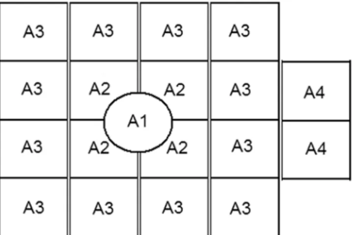
In addition, the VF was also divided into four areas (Figure 1) to evaluate the variation of the values in different areas: the central area (A1); the 10o field area (A2); the 20o field area (A3) and the 30o lateral field area (A4). The median thresholds for each of the four areas were calculated; each of these medians are referred to as the median area CS (MACS).
Statistical analysis was performed using SPSS for Windows (ver sion 15; SPSS, Inc, Chicago, Illinois, USA). For primary outcome measures, P<0.05 was considered to be statistically significant. The normality of the quantitative variables was verified using the Kol-mogorov-Smirnov test. The nonparametric Wilcoxon paired test was used to compare data between the two IOL groups. The differences between MD, PSD and MACS were compared using an unpairedt test.
RESULTS
A total of 25 patients (50 eyes; 12 men [48.0%] and 13 women [52.0%]) were enrolled in the present study. The mean age of the pa-tients was 57.80 ± 6.48 years. No significant difference was observed between the IOL groups with regard to mean IOL power (P=0.736) and mean axial length (P=0.431) (Table 1).
No significant difference was observed between the aspheric and spherical groups for distance-corrected vision (0.00 ± 0.11 and 0.01± 0.07 logMAR, respectively) (P=0.432).
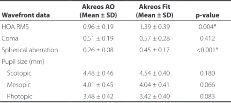
The mean pupil diameter was similar between Akreos AO and Akreos Fit under photopic (P=0.083), mesopic (P=0.066), and scotopic conditions (P=0.180) (Table 2).
Figure 1. Representation of the frequency-doubling technology division in four areas:
Espíndola RF, et al.
3 7 5 Arq Bras Oftalmol. 2014;77(6):373-6 At 12 months, the spherical equivalent was 0.03 ± 0.29 in the
Akreos AO group and 0.06 ± 0.43 in other group (P=0.296). There was no significant difference between the IOL groups with regard to distance UCVA (P=0.379) and distance BCVA (P=0.331). The UCVA was 0.08 ± 0.05 in the Akreos AO group and 0.09 ± 0.06 in the Akreos Fit group. The BCVA was 0.01 ± 0.10 in the Akreos AO group and 0.02 ± 0.09 in the Akreos Fit group.
Figures 2 and 3 present the CS results. Under photopic conditions, the Akreos AO IOL was associated with significantly better CS than the Akreos Fit IOL only at the spatial frequency of 12 cycles per de-gree (cpd) (P=0.028). Under mesopic conditions, the Akreos AO IOL was associated with significantly better CS than the Akreos Fit IOL at all spatial frequencies (1.5, 3, 6, 12, and 18 cpd; P=0.004, P=0.042,
P=0.017, P=0.0017, and P=0.001, respectively).
Table 2 shows wavefront data after cataract surgery. The Akreos AO group had statistically significantly lower values of mean HOA and SA compared with the Akreos Fit group (P=0.004andP<0.001, respectively). There were no statistically significant differences in coma values between the two IOL groups (P=0.412).
The average MDs of eyes implanted with aspheric and spherical IOLs were -1.01 ± 1.68 dB and -3.15 ± 3.78 dB, respectively, on FDT testing. There was no significant difference in either MD (P=0.051) or PSD (P=0.233) between the groups (Table 3).
Considering the VF division, the MACS of the aspheric IOL had higher values than the spherical IOL in all areas (A1 to A4), although the difference was statistically significant only in the peripherical area (A3) (P=0.043).
DISCUSSION
Functional and structural changes associated with glaucoma require special consideration in the patient who is undergoing ca-taract/IOL surgery. The decreased CS observed in glaucoma may be improved by the use of aspheric IOLs(18).
Contrast is one of the most important parameters activating cor-tical cells involved in vision processing(19). The perception of median arcuate ligament frequency doubling may be mediated by a group of retinal ganglion cells called My cells or by cortical mechanisms(20-23).
Given the similarities between the frequency-doubling illusion and CS, one might deduce that cataract extraction results in recovery of sensitivity in FDT(29). Previous studies showed that the MD of FDT and SITA-fast perimetry were improved after cataract extraction but PSD remained unchanged(24,25).
Similarly, we suspected that changes after aspheric IOL implanta-tion may influence CS and its thresholds. Therefore, the present con-tralateral eye study was conducted to analyze the theoretical benefits of aspheric IOL in improving CS and its consequences in FDT parame-ters. To our knowledge, no previous study comparing the effect of an aspheric IOL on FDT perimetry has been published to date.
Table 1. Demographic characteristics of patients enrolled in the study
Characteristics Akreos AO Akreos Fit p-value
No. of eyes 25 25
Age (years) 57.80 ± 6.48 57.80 ± 6.48
Sex (M/F) 12/13 12/13
Eye (OD/OS) 11/14 14/11
IOL power (D) 21.48 ± 2.23 21.51 ± 2.20 0.878
Axial length (mm) 22.35 ± 2.20 22.20 ± 2.43 0.630 M= male; F= female; OD= right eye; OS= left eye; lOL= intraocular lens; D= diopters.
Table 2. Wavefront data and pupil size of eyes implanted with Akreos AO and Akreos Fit
Wavefront data
Akreos AO (Mean ± SD)
Akreos Fit
(Mean ± SD) p-value
HOA RMS 0.96 ± 0.19 1.39 ± 0.39 0.004*
Coma 0.51 ± 0.19 0.57 ± 0.28 0.412
Spherical aberration 0.26 ± 0.08 0.45 ± 0.17 <0.001* Pupil size (mm)
Scotopic 4.48 ± 0.46 4.54 ± 0.40 0.180
Mesopic 4.01 ± 0.45 4.04 ± 0.41 0.066
Photopic 3.48 ± 0.42 3.42 ± 0.40 0.083
HOA= higher-order aberration; RMS= root mean square; SD= standard deviation; mm= millimeters; *= statistically significant.
Figure 2. Graph showing contrast sensitivity under photopic conditions in the aspheric
and spherical groups.
Figure 3. Graph showing contrast sensitivity under mesopic conditions in the aspheric
and spherical groups.
Table 3. Frequency doubling technology (FDT) data of eyes implanted with Akreos AO and Akreos Fit at 12 months postoperatively
FDT data
Akreos AO (Mean ± SD)
Akreos Fit
(Mean ± SD) p-value
MD (dB) -1.01 ± 1.68 -3.15 ± 3.78 0.051
PSD (dB) -5.05 ± 1.21 -6.30 ± 3.39 0.233
Influence of aspheric intraocular lens on frequency doubling technology and contrast sensitivity: a fellow eye study
376 Arq Bras Oftalmol. 2014;77(6):373-6
In the present study, under mesopic conditions, postoperative VCTS CS testing revealed significant differences between the two groups at all spatial frequencies, indicating that the Akreos AO IOL group performed better than the Akreos Fit group in larger pupil sizes. However, under photopic conditions, the Akreos AO IOL per-formed better than Akreos Fit only at 12 cpd (P=0.028). In addition, we found that the amount of HOAs was significantly lower in the aspheric group.
Although previous studies comparing aspheric and spherical IOLs have also reported the reduction of SA in patients implanted with aspheric IOLs, its influence in improved CS results is still contro-versial. Some studies show improvement in CS with aspheric IOLs(6-10), while others have showed no improvement(26,27).
The increase in optical quality obtained with aspheric IOLs in our study does not play a significant role in changing FDT parameters. Our results showed that both MD (P=0.051) and PSD (P=0.233) did not differ significantly between groups.
A previous study by Ueda et al.(25) reported findings that were similar to those in the present study. They analyzed the effect of clear (VA60BB, HOYA) and yellow-tinted lenses (YA60BB, HOYA) on FDT values, and concluded that both MD and PSD did not differ sig-nificantly. It is important to notice that this study was not conducted with aspherical IOLs.
Others studies have analyzed the influence of IOLs on short-wa-velength automated perimetry (SWAP)(28,29). Jang et al analyzed clear (AcrySof SA60AT; Alcon Laboratories) and yellow-tinted lenses (AcrySof SN60AT or SN60WF; Alcon Laboratories) on SWAP(28). The SN60WF IOL is designed with negative SA to compensate for the positive aberration of the average cornea. The authors demonstrated that yellow-tinted IOLs may affect the SWAP results, although they did not separately analyze the effect of the aspheric IOL used (SN60WF) on perimetry.
Kara-Junior et al.(29) reported different results than Jang et al.(28).They suggested that yellow-tinted IOLs (AcrySof SN60AT) did not interfere with the results of SWAP. It should be noted that both IOLs studied generated a considerable amount of SA.
With respect to mean sensitivities of the different zones as defi-ned in the present study, FDT testing resulted in improved CS after aspheric IOL implantation. We found higher mean values in the eye with Akreos AO in all areas (A1 to A4), but a statistical difference (P=0.043) was found only in the 20o field area (A3). Although the chan ges were not statistically significant, it is noteworthy that an improvement in the perimetry values with such lenses could mask the diagnosis of glaucoma or lead to other diagnostic confusions, especially considering that the functional abnormalities detected by FDT perimetry were shown to be predictive of the future onset and location of VF loss among suspected glaucoma patients(13-15).
The results of the present study indicate that MD values were higher in the Akreos AO group, and the PSD was higher in the Akreos Fit group. Note that a more negative MD indicates a worse visual per-formance than expected from the normative database, and a higher PSD indicates greater changes in sensibility. Previous studies in pa-tients with yellow-tinted IOLs and papa-tients with clear IOLs have also showed higher MD values in patients with yellow-tinted IOLs(25,28,29). However, both indices in our study were not statistically significant.
The present study had several limitations, including the absence of preoperative FDT perimetry data, the small number of cases and the absence of concurrent standard automated perimetry. A larger number of cases (approximately 100 cases in each group) could make a difference in the findings between each group, or confirm the result observed in the present study.
In addition, the present study specifically investigated these effects in patients without glaucoma. It stands to reason that in a disease process that decreases CS, an attempt to increase CS would be beneficial to the patient.
In summary, aspheric IOLs significantly reduced SA and HOAs, improving mesopic CS. Although there was a trend toward slightly
improved FDT in the aspheric IOL group, it was not statistically signi-ficant. Further studies with large sample sizes should be performed to confirm or reject this hypothesis.
REFERENCES
1. Montés-Micó R, Ferrer-Blasco T, Cerviño A. Analysis of the possible benefits of aspheric intraocular lenses: review of the literature. J Cataract Refract Surg. 2009;35(1):172-81. 2. Wang L, Dai E, Koch DD, Nathoo A. Optical aberrations of the human anterior cornea.
J Cataract Refract Surg. 2003;29(8):1514-21.
3. Guirao A, Redondo M, Geraghty E. Corneal optical aberrations and retinal image quality in patients in whom monofocal intraocular lenses were implanted. Arch Ophthalmol. 2002;120(9):1143-51.
4. Kasper T, Bühren J, Kohnen T. Intraindividual comparison of higher-order aberrations after implantation of aspherical and spherical intraocular lenses as a function of pupil diameter. J Cataract Refract Surg. 2006;32(1):78-84.
5. Packer M, Fine IH, Hoffman RS, Piers PA. Prospective randomized trial of an anterior surface modified prolate intraocular lens. J Cataract Refract Surg. 2002;18(6):692-6. 6. Mester U, Dillinger P, Anterist N. Impact of a modified optic design on visual function:
clinical comparative study. J Cataract Refract Surg. 2003;29(4):652-60.
7. Kershner RM. Retinal image contrast and functional visual performance with aspheric, silicone, and acrylic intraocular lenses; prospective evaluation. J Cataract Refract Surg. 2003;29(9):1684-94.
8. Packer M, Fine IH, Hoffman RS, Piers PA. Improved functional vision with a modified prolate intraocular lens. J Cataract Refract Surg. 2004;30(5):986-92.
9. Bellucci R, Scialdone A, Buratto L, Morselli S, Chierego C, Criscuoli A, et al. Visual acuity and contrast sensitivity comparison between Tecnis and AcrySof SA60AT intraocular lenses: a multicenter randomized study. J Cataract Refract Surg. 2005;31(4):712-7. 10. Santhiago MR, Netto MV, Barreto Jr B, Gomes BA, Mukai A, Guermandi AP, et al.
Wa-vefront analysis, contrast sensitivity, and depth of focus after cataract surgery with aspherical intraocular lens implantation. Am J Ophtalmol. 2010;149(3):383-8. 11. Johnson CA. Recent developments in automated perimetry in glaucoma diagnosis
and managements. Curr Opin Ophthalmol. 2002;13(2):77-84.
12. Maddess T, Henry GH. Performance of nonlinear visual units in ocular hypertension and glaucoma. Clin Vision Sci. 1992;7(5):371-83.
13. Sample PA, Medeiros FA, Racette L, Pascual JP, Boden C, Zangwill LM, et al. Identifying glaucomatous vision loss with visual-function-specific perimetry in the diagnostic innovations in glaucoma study. Invest Ophthalmol Vis Sci. 2006;47(8):3381-9. 14. Racette L, Medeiros FA, Zangwill LM, Ng D, Weinreb RN, Sample PA. Diagnostic
accuracy of the Matrix 24-2 and original N-30 frequency-doubling technology tests compared with standard automated perimetry. Invest Ophthalmol Vis Sci. 2008;49(3): 954-60.
15. Burgansky-Eliash Z, Wollstein G, Patel A, Bilonick RA, Ishikawa H, Kagemann L, et al. Glaucoma detection with Matrix and standard achromatic perimetry. Br J Ophthalmol. 2007;91(7):933-8.
16. Wall M, Neahring RK, Woodward KR. Sensitivity and specificity of frequency doubling perimetry in neuro-ophthalmic disorders: a comparison with conventional automa-ted perimetry. Invest Ophthalmol Vis Sci. 2002;43(4):1277-83.
17. Monteiro ML, Portes ALF, Moura FC, Regensteiner DB. Using frequency-doubling perime-try to detect optic neuropathy in patients with Graves’ Orbitopathy. Jpn J Ophthalmol. 2008;52(6):475-82.
18. Teichman JC, Ahmed IIK. Intraocular lens choices for patients with glaucoma. Curr Opin Ophthalmol. 2010;21(2):135-43.
19. Caporossi A, Casprini F, Tosi GM, Baiocchi S. Preliminary results of cataract extraction with implantation of a single-piece AcrySof intraocular lens. J Cataract Refract Surg. 2002;28(4):652-5.
20. Kim YY, Kim JS, Shin DH, Kim C, Jung HR. Effect of cataract extraction on blue-on-yellow visual field. Am J Ophthalmol. 2001;132(2):217-20.
21. Albrecht DG, Hamilton DB. Striate cortex of monkey and cat: contrast response function. J Neurophysiol. 1982;48(1):217-37.
22. Tolhurst DJ, Movshon JA, Dean AF. The statistical reliability of signals in single neurons in cat and monkey visual cortex. Vision Res. 1983;23(8):775-85.
23. Shadlen MN, Newsome WT. Noise, neural codes and cortical organization. Curr Opin Neurobiol. 1994;4(4):569-79.
24. Kook MS, Yang SJ, Kim S, Chung J, Kim ST, Tchah H. Effect of cataract extraction on frequency doubling technology perimetry. Am J Ophthalmol. 2004;138(1):85-90. 25. Ueda T, Ota T, Yukawa E, Hara Y. Frequency doubling technology perimetry after clear
and yellow intraocular lens implantation. Am J Ophthalmol. 2006;142(5):856-8. 26. Su PY, Hu FR. Intraindividual comparison of functional vision and higher order
aber-rations after implantation of aspheric and spherical intraocular lenses. J Refract Surg 2009;25(3):265-72.
27. Yu AY, Wang QM, Sun J, Xue AQ, Zhu SQ, Wang SL, et al. Spherical aberration after implantation of an aspheric versus a spherical intraocular lens in high myopia. Clin Experiment Ophthalmol. 2009;37(6):558-65.
28. Jang SY, Ohn YH, Kim SW. Effect of Yellow-Tinted Intraocular Lenses on Short-Wave-length Automated Perimetry. Am J Ophthalmol. 2010;150(2):243-7.