Rev. odonto ciênc. 2010;25(4):427-429 427
Case Report
Received: May 10, 2010 Accepted: September 29, 2010
Conflict of Interest Statement: The authors state that there are no financial and personal conflicts of interest that could have inappropriately influenced their work.
Copyright: © 2010 Rajashekhara et al.; licensee EDIPUCRS. This is an Open Access article distributed under the terms of the Creative Commons Attribution-Noncommercial-No Derivative Works 3.0 Unported License.
Bilateral fusion of primary mandibular lateral incisors
and canines: A report of a rare case
Fusão bilateral de dentes decíduos incisivo e canino inferior:
relato de um caso raro
BS Rajashekhara a
Bhavna Dave a
BS Manjunatha b
KS Poonacha a
Sunanda G Sujan a
a Department of Pedodontics and Preventive Dentistry, K M Shah Dental College & Hospital, Pipariya, Waghodia (T), Vadodara (D), Gujarat State, India
b Department of Oral and Maxillofacial Pathology, K M Shah Dental College & Hospital, Pipariya, Waghodia (T), Vadodara (D), Gujarat State, India
Correspondence:
B.S. Manjunatha
K M Shah Dental College & Hospital, Pipariya, Waghodia (T), Vadodara (D) Gujarat State – India India 391760
E- mail: [email protected]
Abstract
Purpose: Synodontia or fusion is a developmental anomaly of shape of tooth formed by union of two independently developing primary or secondary teeth. Prevalence of tooth fusion is estimated at 0.5-2.5% in the primary dentition and less in permanent dentition. The bilateral type of fusion in the primary dentition is rare and is about 0.02%. This paper describes a rare case of bilateral fusion of primary mandibular lateral and canine teeth.
Case description: An 8 year old girl had a complaint of unusually large sized teeth in her mandible. After physical examination and use of periapical radiographs and study models bilateral fused teeth in the mandibular lateral incisor and canine region was diagnosed. Conclusion: The bilateral fusion of primary mandibular lateral and canine teeth is a rare condition and should be carefully evaluated to diagnose any associated pathology. Key words: Fusion; synodontia; primary teeth; developmental anomaly; double tooth
Resumo
Objetivo: Sinodontia ou fusão é uma anomalia de desenvolvimento da forma do dente formado pela união de dois dentes decíduos ou permanentes em desenvolvimento de forma independente. A prevalência de fusão dental é estimada em 0,5 a 2,5% na dentadura decídua e menor na permanente. O tipo de fusão bilateral na dentadura decídua é rara e aproximadamente de 0,02%. Este artigo descreve um caso raro de fusão bilateral de dentes decíduos incisivos laterais e caninos inferiores.
Descrição do caso: Uma menina de 8 anos de idade tinha uma queixa clínica de dentes de tamanho grande anormal em sua mandíbula. Após exame físico e uso de radiografias periapicais e modelo de estudo, a fusão bilateral de dentes decíduos incisivos laterais e caninos inferiores foi diagnosticada.
Conclusão: A fusão bilateral de dentes decíduos incisivos laterais e caninos na mandíbula é uma condição rara e deve ser cuidadosamente avaliada para diagnosticar quaisquer patologias associadas.
428 Rev. odonto ciênc. 2010;25(4):427-429
Synodontia of primary teeth
Introduction
Fusion is commonly identiied as the union of two distinct dental sprouts, which may occur in any stage of the dental organ. Teeth are joined by the dentin region; pulp chambers and canals may be linked or separated depending on the developmental stage when the union occurs. This process involves epithelial and mesenchymal germ layers resulting in irregular tooth morphology (1). Moreover, the number of teeth in the dental arch is reduced. The literature shows controversial concepts to correctly differentiate between teeth fusion and gemination. For a differential diagnosis between these anomalies, the dentist must carry out a highly judicious radiographic and physical examination.
Prevalence of fusion of tooth is about 0.5-2.5% in the primary dentition with a lower prevalence in permanent dentition (2).
The aetiology of fusion is still unknown, but the inluence of pressure or physical forces producing close contact between two developing teeth has been reported as one possible cause (3). Genetic predisposition and racial differences have also been reported as contributing factors.
This anatomic irregularity occurs more often in the deciduous (0.5%) than in the permanent dentition (0.1%) (4,5). In the anterior region this anomaly also causes an unpleasant aesthetic tooth shape due to the irregular morphology. These teeth also tend to be greatly predisposed to caries and periodontal disease and, in some cases, endodontic treatment is very complicated (6,7).
The bilateral type of fusion in the primary dentition occurs less frequently than unilateral type and is about 0.02% (4,5). Only 14 cases have been previously reported in the English literature. Hence this article aimed at reporting a case of this rare condition and evaluating the presence of any associated pathology.
Description of the case
An 8 year old girl was referred to the outpatient section of the Department of Pedodontics and Preventive Dentistry, KM Shah Dental College and Hospital, Vadodara, India, with a chief complaint of large teeth in her mandible. Her medical history was taken but showed no relevant association with her chief complaint. Intraoral examination revealed bilateral presence of unusually large teeth in the mandibular incisor canine region. The patient was in early mixed dentition with the presence of the following teeth:
6 E D C B 1 1 B C D E 6 6 E D <CB> 1 1 <BC> D E 6
Both left and right mandibular primary lateral incisor and canines were fused to form a large tooth (Fig. 1). These extra large teeth had buccal and lingual vertical grooves and were not affected either by dental caries or periodontal problems. The primary mandibular canines were missing, conirming that it was a case of fusion and not gemination. Dental caries was present in primary mandibular second molars bilaterally.
The rest of the dentition was normal without any variations or anomalies.
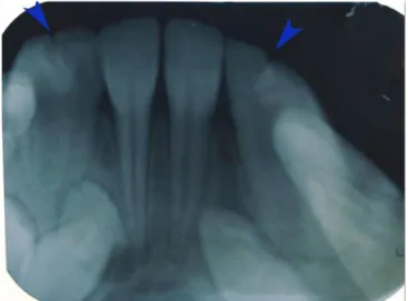
Intraoral periapical radiographs conirmed the bilateral fusion of mandibular primary lateral incisor and canines, and underlying permanent successor teeth were also evident (Fig. 2). Impressions and diagnostic casts were made (Fig. 3). The patient was diagnosed to be a case of bilaterally fused mandibular primary lateral incisors and canines.
Fig. 3. Diagnostic cast of the mandibular dental arch showing the bilateral fused teeth (shown with black arrows).
Fig. 1. Clinical aspect of the bilateral fusion of mandibular primary lateral incisor and canines forming a single large tooth on both right and left sides (shown with black arrows).
Rev. odonto ciênc. 2010;25(4):427-429 429 Rajashekhara et al.
Discussion
A rare case of fusion between the mandibular primary late-ral incisor and canine occurring bilatelate-rally is presented here.
The prevalence of tooth fusion is estimated as 0.5-2.5% in the primary dentition with a lower prevalence in
permanent dentition (2). The anomaly of conjoined teeth has
been described undera variety of names. DeJonge (1955)
proposed the term ‘Synodontia’ for those formed by the
inability of adjacent toothgerms to retain their individuality.
Although the term “doubleteeth” as suggested by Miles in
1954 is widelyaccepted and may be more appropriate (8).
Tooth fusion is deined as the union between the dentin
and/orenamel of two or more separate developing teeth (1).
Thefusion may be partial or total depending upon the stage
oftooth development at the time of union, a distinguishing
feature between fusio-totalis, partialis-coronaries and
partialis-radicularis (2,6).
The etiology of fusion is still not known. Shafer et al.(3)
speculated that the pressure produced by some physical force prolongs the contact of the developing teeth causing fusion. Lowell and Soloman (9) believe that fused teeth result from some physical action that causes the young tooth germs to come into contact, thus producing necrosis of the intervening tissue and allowing the enamel organ and dental papilla to fuse together. Others have also suggested the hereditary involvement as an autosomal dominant trait with reduced penetrance (10)
Duncan (5) reviewed and analyzed 38 published papers in the dental literature and reported the prevalence of unilateral double primary teeth at 0·5% and that of bilateral at 0·02%. Case history and clinical and radiographic examinations can provide the adequate information required for the diagnosis of such abnormalities. Fusion of primary teeth occurs less frequently, and the bilateral type is very rare and less commonly found than the unilateral type. A survey of the literature has revealed prevalence estimates for bilateral fused teeth ranging from 0·01 to 0·04% in the primary
dentition and 0·05% in the permanent dentition (11). Only 14 cases of bilateral fusion of primary mandibular lateral incisor and canines have been reported in the literature since 1940 by Tinn (12). Teeth with this abnormality are unaesthetic due to their irregular morphology. They also present a high predisposition to caries and periodontal disease, and spacing problems. The main periodontal complication in fusion cases occurs due to the presence of issures or grooves in the union between the teeth involved.
Several clinical problems in the permanent dentition follow fused primary teeth, such as physiological root resorption of fused deciduous teeth being retarded, leading to delayed or ectopic eruption of the permanent successors. When fused primary teeth are found in the clinic, the application of issure sealants on the grooves between the two components is recommended to prevent dental caries (13). Radiographs should also be taken to check the development of the permanent teeth. Careful check-ups and surgical intervention at the appropriate time are necessary to prevent delayed exfoliation and eruption of the successors. The greater root surface area of fused primary teeth may delay in exfoliation by root resorption (14). Several different approaches for the treatment of these abnormalities are available, but the morphology of
fused teethvaries so greatly that one can only decide on
individual basis.Various methods include selective grinding,
surgicalseparation or extraction followed by prosthesis (8).
Fusion of primary teeth usually is asymptomatic, but the
squeal of such teeth may result in various disturbances
in eruption of permanent teeth. Dificult and rare cases
pose a wide spectrum of problems, and the best way to manage such cases depends on variety of factors mainly the knowledge and technical skills of the practitioner. Hence the proper diagnosis by clinical and radiographic methods and
intervention at appropriate time isof paramount importance.
In conclusion, a multidisciplinary approach with different practitioners with expertise in several areas of dentistry is important to achieve functional and esthetic success to treat these rare cases.
References
Tannenbaum KA, Alling EE. Anomalous tooth development: case 1.
reports of gemination and twinning. Oral Surg Oral Med Oral Pathol 1963;16:883-7.
Hülsmann M, Bahr R, Grohmann U. Hemisection and vital treatment 2.
of a fused tooth – literature review and case report. Endod Dent Traumatol 1997;13:253-8.
Shafer WG, Hine MK, Levy BM. Developmental disturbances in shape 3.
of teeth. In: A Textbook of Oral Pathology, 4th ed. Philadelphia: WB
Saunders Company; 1983. p. 38-9.
Neville BW, Damn DD, Allen CM, Bouquot JE. Oral and Maxillofacial 4.
Pathology. 2nd ed. Elsevier publishing: Philadelphia: Pennsylvania; 2002.
Duncun WK, Helpin ML. Bilateral fusion and gemination: a literature 5.
analysis and case report. Oral Surg Oral Med Oral Pathol 1987;64:82-7. Peyrano A, Zmener O. Endodontic management of mandibular 6.
lateral incisor fused with supernumerary tooth. Endod Dent Traumatol 1995;11:196-8.
Pereira AJ, Fidel RA, Fidel SR. Maxillary lateral incisor with two 7.
root canals: fusion, gemination or dens invaginatus? Braz Dent J 2000;11:141-6.
Gupta S, Singla S, Marwah N, Dutta S, Goel M. Synodontia 8.
between Permanent Maxillary Lateral Incisor and A Supernumerary Tooth: Surgical Treatment Perspective. J Oral Health Comm Dent 2007;1:52-5.
Lowell RJ, Soloman AL. Fused teeth. J Am Dent Assoc 1964; 68: 9.
762.
Stewart R, Prescott GH. Genetic aspects of anomalous tooth 10.
development. Oral Facial Genetics. St. Louis: Mosby Co;1976. Neves AA, Neves ML, Farinhas JA. Bilateral connation of 11.
permanent mandibular incisors:a case report. Int J Paediatr Dent 2002;12:61-5.
Tinn CA. Excess, deficiency and gemination in the deciduous and 12.
permanent dentition of school children. Br Dent J 1940;68:236-8. Surmont PA, Martens LC, Craene LG. A complete fusion in the 13.
primary human dentition: a histological approach. ASDC J Dent Child 1988;55:362-7.
Brook AH, Winter GB. Double teeth. A retrospective study of 14.