Received: November 04, 2011 Accepted: January 31, 2012
Conflict of Interests: The authors state that there are no financial and personal conflicts of interest that could have inappropriately influenced their work.
Copyright: © 2011 Freitas et al.; licensee EDIPUCRS. This is an Open Access article distributed under the terms of the Creative Commons Attribution-Noncommercial-No Derivative Works 3.0 Unported License.
The diagnostic challenge of vertical root fracture
in endodontically treated teeth: A case report
O desafio diagnóstico de fratura radicular vertical em dentes
endodonticamente tratados: relato de caso
Pollyanna Queiroz Freitas a
Paulo Maria Santos Rabêlo-Júnior b
Claúdia Maria Coelho Alves c
Soraia de Fatima Carvalho Souza d
a School of Dentistry, Federal University of
Maranhão, São Luis, MA, Brazil
b Department of Oral and Maxillofacial Surgery,
School of Dentistry, Federal University of Maranhão, São Luis, MA, Brazil
c Department of Periodontology, School of Dentistry,
Federal University of Maranhão, São Luis, MA, Brazil
d Department of Endodontics, School of Dentistry,
Federal University of Maranhão, São Luis, MA, Brazil
Correspondence:
Soraia de Fatima Carvalho Souza Universidade Federal do Maranhão Faculdade de Odontologia
Av. dos Portugueses s/n – Campus do Bacanga São Luis, MA – Brasil
65085-580
E-mail: [email protected]
Abstract
Purpose: To present the diagnostic challenge of a clinical case of vertical root fracture (VRF) in an endodontically treated mandibular left lateral incisor and discuss the diagnostic methods employed to achieve the conclusive diagnosis.
Case Description: At 16 months after endodontic treatment, a 60-year-old female patient reported pain during mastication. Clinically, she presented with an active distolingual fistula and a probing depth of 9 mm on the distal aspect of tooth 32. A radiographic examination indicated pear-shaped distal bone loss. The fistula was mapped, which confirmed that the lesion had a periodontal origin. A diagnostic hypothesis of a VRF was established. Exploratory surgery revealed the VRF on the distolingual aspect of the root without separation of the root fragments.
Conclusion: Knowledge of the diagnostic aspects and the correct interpretation of radiographic images was enough to establish the diagnostic hypothesis of a VRF. However, the conclusive diagnosis was only confirmed during exploratory surgery.
Key words: Diagnosis; Endodontics; vertical root fracture
Resumo
Objetivo: Apresentar o desafio de diagnosticar Fratura Radicular Vertical (FRV) em um incisivo lateral inferior esquerdo tratado endodonticamente e discutir os métodos diagnósticos empregados para obtenção do diagnóstico conclusivo.
Descrição do Caso: Após 16 meses do término do tratamento, a paciente relatou dor à mastigação. Clinicamente apresentava uma fístula disto-lingual ativa e profundidade de sondagem de 9-mm na face distal do dente 32. Radiograficamente apresentava perda óssea distal em forma de “pêra”. Foi realizado o mapeamento da fístula, confirmando tratar-se de uma lesão de origem periodontal. Estabeleceu-se a hipótese diagnóstica de FRV. Para a confirmação do diagnóstico realizou-se cirurgia exploratória. Foi constatada a FRV na face disto-lingual da raiz sem o afastamento dos fragmentos radiculares.
Conclusão: Concluiu-se que o conhecimento dos achados semiotécnicos e a correta interpretação das imagens radiográficas foram suficientes para se estabelecer a hipótese diagnóstica de FRV. Entretanto, o diagnóstico conclusivo só foi confirmado durante a cirurgia exploratória.
Introduction
Vertical root fractures (VRFs) may affect any human tooth, either vital or non-vital. Teeth that morpho-
logically present roots with mesiodistal lattening and
undergo endodontic therapy are considered the most susceptible to a VRF occurrence. The prevalence of VRFs is 52% in maxillary and mandibular premolars, followed by the mesial roots of mandibular molars (24%), maxillary and mandibular central and lateral incisors (14%) and the mesiobuccal and palatal roots of the maxillary molars (10%) (1).
A VRF in endodontically treated teeth may be deined as
a line in the longitudinal direction, initiating at the internal root canal wall and extending to the external root surface toward the periodontal tissues. It occurs in any third of the root canal and tends to split the root in the buccolingual direction (2,3). The characteristic signs and symptoms may manifest in days or years after the fracture (1,4): sensitivity to vertical percussion (VP) and digital palpation (DP), the
presence of a istula and a deep and isolated periodontal
pocket. Radiographically, periapical and lateral radiolucency is present with a “halo” shape in the supposedly affected tooth (5).
VRFs are considered severe complications among those affecting endodontically treated teeth due to the following factors: (1) an inability of the dentist, endodontist or periodontist to determine the conclusive diagnosis of this clinical condition because the signs and symptoms are similar to those of an unsuccessful endodontic treatment or periodontal disease (5); and (2) patient anxiety about solving the problem (6).
This paper presents the case of an endodontically treated tooth affected by a VRF and discusses the diagnostic methods employed to achieve the conclusive diagnosis.
Description of the case
A 60-year-old female, presenting with good general health, was referred for endodontic treatment due to a pain caused by temperature changes in the mandibular left lateral incisor. The intraoral visual examination indicated that the patient was using a mandibular removable partial denture (RPD) and that tooth 32 was in supra-occlusion. A clinical
examination identiied a Class III composite resin on the
mesiolingual aspect. The periodontal tissues appeared normal and were unresponsive to the VP or DP tests. However, the cold test (Endo-Ice, Maquira, Maringá, PR, Brazil) caused exacerbation of the pain, with a slow decrease; the pain was sustained for several minutes. A radiographic examination revealed the periapical region was normal. Irreversible pulp
inlammation was diagnosed, and a root canal procedure was
immediately indicated.
After anesthesia and rubber dam placement, endodontic access was performed following the conventional guidelines. The pulp canal chamber was then irrigated using 1% sodium
hypochlorite (NaOCl). The working length was established
1 mm from the radiographic apex, and the root canal was prepared using #1 and #2 Gates-Glidden burs (Dentsply Maillefer, Ballaigues, Switzerland) in the coronal and
middle thirds. The apical third was enlarged to a K-ile #35, followed by the step-back technique using K-type hand iles (Dentsply Maillefer, Ballaigues, Switzerland). A inal rinse
with 17% ethylenediaminetetraacetic acid (EDTA) was also performed to remove the smear layer. The canal was dried with absorbent paper points, and a corticosteroid and antibiotics (Otosporin otosolução®, FQM, Rio de Janeiro,
RJ, Brazil) were placed into the root canal. Coronal sealing
was performed (Coltosol®, Dentalville, Joinville, SC,
Brazil), and the intracanal medication was kept in place for 72 h.
The patient did not report painful symptomatology to VP and DP. A root canal obturation was performed by the lateral condensation technique combined with cold vertical condensation using gutta-percha and a resin-based root canal sealer (Sealer-26®, Dentsply, Petrópolis, RJ,
Brazil). Cervical sealing was immediately performed with
a restorative glass ionomer cement (Vitro Fill LC®, DFL,
Rio de Janeiro, RJ, Brazil), followed by restoration with a composite resin (Opallis®, FGM, Joinville, SC, Brazil) and
a dentin adhesive system (Prime & Bond 2.1, Dentsply, Petrópolis, RJ, Brazil).
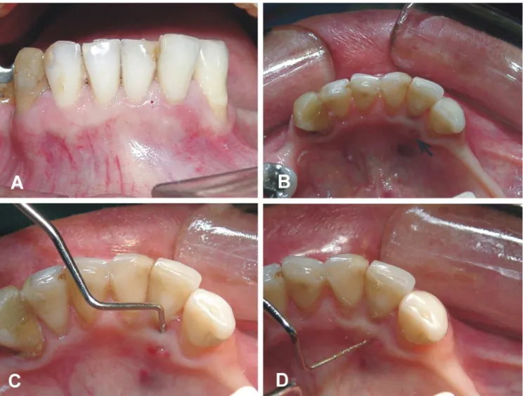
At 16 months after treatment, the patient returned
with complaint of pain during mastication. Clinically, the
mandibular left lateral incisor was still in supra-occlusion
(Fig. 1A) and exhibited an active distolingual istula
(Fig. 1B). Periodontal probing revealed a probing depth
of 9 mm on the distal aspect (Fig. 1C) and a horizontal istula depth of 5 mm (Fig. 1D). Considering the quality
of the root canal obturation (Fig. 2A), the depth of the isolated periodontal pocket and the pear-shaped distal bone loss only in this tooth (Fig. 2B), a suspected
diagnosis of VRF was reached. The istula was mapped (Fig. 2C), which conirmed that the lesion had a periodontal origin. Exploratory surgery was performed to conirm
the diagnosis (Fig. 3A), and it revealed a VRF on the distolingual aspect of the root without separation of the root fragments; the tooth was extracted (Fig. 3B). The granulation tissue removed from the socket was sent for
a histopathological examination (Fig. 3C). The extent of
the fracture was observed after cleaning the specimen; the VRF extended from the cervical region to the apical region of tooth 32 (Fig. 3D). The histopathological diagnosis
revealed a non-speciic chronic inlammatory process, with a mononuclear inlammatory iniltrate and collagen
Fig. 1. Clinical examination: (A) mandibular left lateral incisor in supra-occlusion; (B) distolingual fistula (arrow); (C) periodontal pocket probing; (D) horizontal fistula probing.
Discussion
VRFs are considered to be a severe complication, with a hardly conclusive and often confusing diagnosis, which unavoidably leads to tooth extraction (7-9). The differential diagnosis between a VRF and failed endodontic treatment or periodontal disease requires an analysis of complementary examinations in addition to the clinical data and the history of the affected tooth. Among these, the most used is the periapical radiographic examination. Notwithstanding, VRFs are rarely observed in this examination because the central X-ray beam does not fall on the fracture plane. A VRF is only observed on a periapical radiograph if the root fragments are separated. However, some characteristics of the radiographic image may suggest a VRF, such as radiopaque
signs due to sealer overlow at the external root surface,
isolated horizontal bone loss, unexplained bone loss at the furcation region, diffuse V-shaped bone loss, enlargement of the periodontal ligament space and a radiolucent “halo” around the entire root surface (10).
Other diagnostic dificulties of this type of fracture are
related to endodontically treated teeth because the line of fracture may be shadowed by the obturation material (4),as in the present case. The diagnostic radiographs suggested the presence of a VRF in the radiographic image, exhibiting a longitudinal radiolucent “halo” that nearly encompassed
the entire distal aspect of the root, conirming the horizontal
and isolated bone loss on the mandibular left lateral incisor. In addition, an occlusal adjustment was not performed after the endodontic procedure, and the tooth was still in supra-occlusion, which could be a causal factor of the VRF. Initially, this radiographic aspect was hypothetically
interpreted as a sequel of chronic inlammation induced by the VRF because this inding is non-speciic and might be
confused with manifestations of periodontal disease or failed endodontic treatment (8).
Advanced imaging examinations may be requested, including conventional computed tomography (9), cone-beam or digital volume computed tomography (11,12) and coherence optical tomography (13). To date, coherence
optical tomography is not available for dental use; it is a diagnostic unit for atherosclerotic plaques found in heart catheter laboratories and could be a valuable tool for the diagnosis of VRFs (13). Unfortunately, conventional computed tomography and cone-beam or digital volume computed tomography are not available to most of the population because of their high cost. The conventional periapical radiograph is still the most employed method, despite its low sensitivity, and this is likely due to the accessibility of low cost of X-ray machines. Furthermore, the diagnosis of a VRF is only conclusive when the affected tooth is surgically explored to visualize the fracture line (1,14,15), which was confirmed in the present case.
Advanced imaging techniques are good alternate methods for enhancing the diagnosis of VRFs (11,12) because
periapical radiographs are unable to detect the fracture line. Tomographs present the advantages of high-resolution and three-dimensional images, in addition to the low patient exposure to radiation. However, tomography requires sophisticated machines, which precludes its accessibility, especially in Brazil, where the Public Health System (SHS) does not provide these machines for dental use.
Currently, no scientiic evidence indicates the availability
of an accurate, safe and accessible method for the diagnosis of VRFs. Despite the technological advances of imaging examinations, the diagnosis of a VRF remains a clinical challenge. Nonetheless, the diagnostic hypothesis in this case was established through a careful clinical examination, accurate interpretation of the periapical radiograph and
conirmation of the VRF by surgical exposure of the
suspected tooth.
Tamse A, Fuss Z, Lustig J, Kaplavi J. An evaluation of endodontically treated vertically 1.
fractured teeth. J Endod 1999;25:506-8.
Howe CA, Mckendry DJ. Effect of endodontic acces preparation on resistance to crow-root 2.
fracture. J Am Dent Assoc 1990;121:712-5.
Lertchirakarn V, Palamara JE, Messer HH. Load and strain during lateral condensation and 3.
vertical root fracture. J Endod 1999;25:99-104.
Fachin EV. Vertical root fracture: a case report. Quint Int 1993;24:479-500. 4.
Tamse A, Zilburg I, Halpern J. Vertical root fractures in adjacent maxillary premolars: na 5.
endodontic-prosthetic perplexity. Int Endod J 1998;31:127-32.
Pack AR. A report on two patientes with vertical root fracture: a dilema for the periodontist, 6.
endodontist, and patient. N Z Dent J 1994;90:103-6.
Pitts DL, Natkin E. Diagnosis and treatment of vertical root fractures. J Endod 1983;9: 7.
338-46.
Youssefzadeh S, Gahleitner A, Dorffner R, Bernhart T, Kainberger FM. Dental vertical root 8.
fractures: value of CT in detection. Radiology 1999;210:545-9.
Nair MK, Nair UP, Grondahl HG, Wallace JA. Detection of artificially induced vertical 9.
radicular fractures using tuned aperture computed tomography. Eur J Oral Sci 2001;109:375-9.
Moule A, Kahler B. Diagnosis and management of teeth with vertical root fractures. Aust 10.
Dent J 1999;44:75-87.
Kamburoglo K, Murat S, Yüksel SP, Cebeci AR, Horasan S. Detection of vertical root fracture 11.
using cone-beam computerized tomography: an in vitro assessment. Oral Surg Oral Med Oral Pathol Oral Radiol Endon 2010;109:74-81.
Hassan B, Metska ME, Ozok AR, van der Stelt P, Wesselink PR. Detection of vertical root 12.
fractures in endodontically treated by a cone beam computed tomography scan. J Endod 2009;35:719-22.
Shemesh H, van Soest G, Wu MK, Wesselink PR. Diagnosis of vertical root fractures with 13.
optical coherence tomography. J Endod 2008;34:739-42.
Lustig JP, Tamse A, Fuss Z. Patterno bone resorption in vertically fractured, endodontically 14.
treated teeth. Oral Surg Oral Med Oral Pathol Oral Radiol Endod 2000;90:224-7. Tamse A. Vertical root fractures in endodontically treated teeth: diagnostic signs and clinical 15.
management. Endod Topics 2006;13:84-94.