©Revista Brasileira de Fisioterapia
O
RIGINALA
RTICLEEffects of low-level laser therapy after nerve
reconstruction in rat denervated soleus
muscle adaptation
Efeitos do laser de baixa potência após reconstrução nervosa na adaptação do
músculo sóleo de rato
Marcela A. Silva-Couto, Davilene Gigo-Benato, Carla R. Tim, Nivaldo A. Parizotto, Tania F. Salvini, Thiago L. Russo
Abstract
Background: Peripheral nerve injury (PNI) rehabilitation remains a challenge for physical therapists because PNI effects are very disabling. Low-level laser therapy (LLLT) has been described as a physical resource that is able to influence enzymes called metallopeptidases (MMPs) associated with extracellular matrix (ECM) turnover, thus accelerating neuromuscular recovery after nerve crush injuries. However, the effects of LLLT in the treatment of severe nerve injuries and denervated slow-twitch muscles are still inconclusive.
Objectives: The aim of this study was to evaluate the effects of different wavelengths and energy densities of LLLT irradiation, applied to a severe nerve injury after reconstruction, on denervated slow-twitch skeletal muscle adaptation. Method: Rats were submitted to a neurotmesis of the sciatic nerve followed by end-to-end neurorrhaphy. They received transcutaneous LLLT irradiation at the lesion site. The LLLT parameters were: wavelengths - 660 or 780 nm; energy densities - 10, 60 or 120 J/cm2; power - 40 mW; spot - 4 mm2. Sciatic
functional index (SFI), histological, morphometric, and zymographic analyses were performed. One-way ANOVA followed by Tukey’s test was used (p≤0.05). Results: An atrophic pattern of muscle fibers was observed in all injured groups. The MMP activity in the soleus muscle reached normal levels. On the other hand, SFI remained below normality after PNI, indicating incapacity. No difference was found among PNI groups submitted or not to LLLT in any variable. Conclusions: LLLT applied to the nerve post-reconstruction was ineffective in delaying degenerative changes to the slow-twitch denervated muscles and in functional recovery in rats. New studies on recovery of denervated slow-twitch muscle are necessary to support clinical practice.
Keywords: neurologicalrehabilitation; nerve injury; laser therapy; skeletal muscle; physical therapy.
Resumo
Contextualização: A reabilitaçao das lesões nervosas periféricas (LNP) ainda é um desafio para a fisioterapia. A terapia com o laser de baixa potência (LBP) é descrita como um recurso físico capaz de interagir com enzimas relacionadas à alteração da matrix extracelular. Denominadas metalopeptidases (MMPs), essas enzimas atuam durante a recuperação neuromuscular após LNP. No entanto, os efeitos da LBP no tratamento de músculos desnervados de contração lenta após LNP graves ainda são inconclusivos. Objetivo: Avaliar os efeitos de diferentes comprimentos de onda e densidades de energia de irradiação de LBP, aplicado sobre o local do nervo após LNP grave e reconstrução. Método: Ratos foram submetidos a neurotmese do nervo isquiático e neurorrafia término-terminal. Os parâmetros do laser são: comprimento de onda: 660 ou 780 nm; densidades de energia: 10, 60 ou 120 J/cm2; potência: 40 mw; spot: 4 mm2. O
índice funcional isquiático (IFC) e análises histológicas, morfométricas e zimografia foram realizados. ANOVA one-way e teste de Tukey (p≤0,05) foram utilizados. Resultados: Um padrão atrófico das fibras musculares foi observado em todos os grupos com LNP. A atividade das MMPs no músculo sóleo alcançaram níveis normais. Entretanto, o IFC permaneceu inferior à normalidade após a LNP, indicando incapacidade. Não houve diferença entre os grupos de LNP submetidos ou não à LBP em qualquer variável. Conclusão:
O LBP é incapaz de retardar alterações degenerativas em músculos sóleos desnervados e é ineficaz na recuperação funcional de ratos. Novos estudos sobre a recuperação do músculo de contração lenta desnervados são necessários para apoiar a prática clínica.
Palavras-chave: reabilitação neurológica; desnervação; laserterapia; músculo esquelético; fisioterapia.
Received: 01/30/2012 – Revised: 03/02/2012 – Accepted: 03/20/2012
Physical Therapy Department, Universidade Federal de São Carlos (UFSCar), São Carlos, SP, Brazil
Introduction
Peripheral nerve injury (PNI) rehabilitation remains a challenge for physical therapists. his type of injury causes pa-ralysis and causes profound degenerative alterations to skeletal muscle, leading to atrophy1 and force deicits2, thus impairing funcionality3. According to a Brazilian study, out of 456 cases analyzed, 41% of PNIs are neurotmesis4. In this type of PNI, there is nerve discontinuity as well as perineural disruption and in many cases loss of nerve tissue5. he patient is impaired both economically and socially in the occurrence of PNI, there-fore post-operative treatment should aim for maximal restora-tion of patient funcrestora-tionality by stimulating neuronal growth and maintaining muscle trophism until reinnervation occurs.
he denervated skeletal muscle is a rich scenario of modii-cations that still has not been fully clariied. PNIs usually gener-ate not only muscle iber atrophy, but also incite alterations to the extracellular matrix (ECM) surrounding these ibers6. For example, denervated muscles have extensive endomysium and perimysium proliferation4. Often such proliferation can be as-sociated with lexibility reduction, ibrosis, and deicits in the conduction of tension forces7. In this sense, ECM reorganisa-tion is an important element to understanding the mecha-nisms of muscle adaptation in denervation.
MMPs are a zinc-dependent proteolytic enzyme family in-volved in the ECM remodeling process. hey can be synthesized and secreted in the skeletal muscle by Schwann cells, satellite cells, and ibroblasts, speciically in the intramuscular nerves and the neuromuscular junction (NMJ)8,9. Among these en-zymes, the MMP-2 (gelatinase A) and the MMP-9 (gelatinase B) are key to the ECM remodeling process in the skeletal muscle during changes in the intensity of physical activity or in cases of changes to task demands and to the process of injury repair8.
hese enzymes are known for acting on a non-ibrillar form of type IV collagen degradation and interstitial collagen hy-drolysis10. he investigation concerning MMP activity is clini-cally relevant because MMPs act directly on collagen turnover and, therefore, on ibrosis formation, lexibility reduction, and mechanical force alterations in denervated muscles7. Further-more, previous studies reported that MMPs can be involved in the reinnervation process of denervated muscle ibers6,10 and probably allow axonal growth cones to advance into the muscle ECM.
In this context, the regulation of MMPs in denervated muscles has great importance to clinical practice. Understand-ing how the resources normally used by the rehabilitation team can afect MMP activation can provide a scientiic basis for its use in humans. Among the possible candidates that promote neuromuscular recovery in PNI is low-level laser therapy (LLLT).
Recently, it was demonstrated that LLLT accelerates muscle iber cross-section area (CSA) recovery in denervated fast-twitch muscles of rats when LLLT is applied to crushed nerves3. hese authors concluded that LLLT irradiation accelerated neuromus-cular recovery by increasing MMP-2 activation in the injured nerve and inhibiting the activation of MMP-9 and -2 in injured nerves and denervated muscles, respectively. hese changes in MMP activation were also associated with walking recovery3. his study has brought subsidies for future indications of LLLT use in humans. However, studies are needed on slow-twitch muscle adaptation during severe nerve injuries.
he objective of this study was to evaluate the response of a denervated slow-twitch skeletal muscle (soleus) to LLLT irradiation applied to an injured nerve. A severe nerve injury model (neurotmesis), followed by end-to-end neurorrhaphy reconstruction, was used in the present study in an attempt to mimic clinical situations. Furthermore, special attention was given to the selection of irradiation parameters, muscle function and trophism, and ECM adaptation in denervated muscles. his work is relevant to neurological rehabilitation because it considers a common situation in physical therapy practice. Moreover, the use of the animal model in this study is justiied due to ethical reasons surrounding the biopsy of denervated muscles in humans. Finally, the hypothesis of this study was that LLLT irradiation in injured-reconstructed nerves is able to accelerate nerve recovery and muscle reinner-vation, improving function and reestablishing soleus muscle trophism via the regulation of MMP activity.
Method
Animal care and experimental groups – Sixty-four male 3-month-old Wistar rats (275 g) were used. he animals were housed in plastic cages in a room with controlled environmental conditions and free access to water and standard food. he Eth-ics Committee of Universidade Federal de São Carlos (UFSCar), São Carlos, SP, Brazil, approved the experimental procedures (Process 001/06), and the study was conducted in accordance with the national guide for care and use of laboratory animals.
Experimental groups
neurorrhaphy irradiated with LLLT 660 nm 10 J/cm2 (TT660 10); (4) transected nerve and end-to-end neurorrhaphy irradiated with LLLT 660 nm 60 J/cm2 (TT660 60); (5) transected nerve and end-to-end neurorrhaphy irradiated with LLLT 660 nm 120 J/cm2 (TT660 120); (6) transected nerve and end-to-end neurorrhaphy irradiated with LLLT 780 nm 10 J/cm2 (TT780 10); (7) transected nerve and end-to-end neurorrhaphy irradiated with LLLT 780 nm 60 J/cm2 (TT780 60); (8) transected nerve and end-to-end neuror-rhaphy irradiated with LLLT 780 nm 120 J/cm2 (TT780 120).
Surgery procedure
he animals were anesthetised with an intraperitoneal in-jection of a premixed solution containing ketamine (95 mg/kg) and xylazine (12 mg/kg). he skin was shaved and cleaned with 10% povidone iodine. A 2-cm-long incision was made on the skin through a gluteal approach and the left sciatic nerve was exposed. he sciatic nerve was cut and sutured with a nylon monoilament 8.0 in the epineural region only. his microsur-gical procedure was performed by visualization of a surmicrosur-gical magnifying lens with 4x magniication. he nerves were kept moist with 37ºC sterile saline solution throughout the surgi-cal intervention. After surgery11,12, the animals were housed in single cages and fed rat chow and water ad libitum. For the irst four days, acetaminophen (13.5 mg/100 mL) was added to the water for pain reduction. A single dose of the antibiotic Ter-ramycin (1 mg/0.1 mL) was administered to prevent secondary complications related to possible infections.
LLLT protocol and experimental design
Biostimulation was carried out using a gallium–alumi-num–arsenide laser device (TWIN LASER, MM Optics, São Carlos, SP, Brazil) with the following parameters: continuous radiation, wavelength: 660 or 780 nm, power: 40 mW, spot area: 4 mm², energy density at the point of entry: 10, 60 or 120 J/cm2. he time of stimulation was predetermined by the device fol-lowing the abovementioned parameters. All parameters were obtained from Gigo-Benato et al.3 and are described in detail in Table 1. Calibration was performed by MM Optics (São Carlos, SP, Brazil). Briely, a calibrated powermeter was used to verify the power of the laser device. his veriication was approved only if the deviation was not higher than 20% of mean value.
Radiation was applied transcutaneously after shaving the skin over the site of the surgery (recognizable for the pres-ence of the surgical scar) at two points along the sciatic nerve, one above and one below the scar site, and two centimetres apart. Applications were made daily for 10 consecutive days
beginning on the irst day after surgery and on alternative days for another month. he animals were handled with care. Laser biostimulation did not cause any pain or distress to the ani-mals, therefore it was not necessary to use anesthesia.
Assessment of nerve function recovery
he assessment of nerve function recovery was carried out by calculating the sciatic functional index (SFI) as described by Bain, Mackinnon and Hunter13. Animals were tested in a con-ined walkway 42 cm long and 8.2 cm wide, with a dark shelter at the end. A white sheet of paper was placed on the loor of the rat walkway. he rats’ hind paws were pressed down onto a inger paint-soaked sponge, and they were then allowed to walk down the walkway leaving their hind footprints on the paper. hree measurements were taken from the footprints: (1) the print length (PL), i.e. the distance from the heel to the third toe; (2) the toe spread (TS), i.e. distance from the irst to the ifth toe; and (3) the intermediate toe spread (ITS), i.e. distance from the second to the fourth toe. All three measurements were taken from the experimental (E) and normal (N) sides. he SFI was calculated according to the following equation14:
SFI = -38.3[(EPL-NPL)/NPL]+109.5[(ETS-NTS)/ NTS]+13.3[(EITS-NITS)/NITS]-8.8
Muscle evaluation
he right soleus muscles were carefully dissected to avoid mechanical injuries. he muscles were then divided in half at the middle of the belly. he proximal fragment was used for the histological and morphometric measurements. he distal fragment was immediately frozen in liquid nitrogen and stored at -80°C (Forma Scientiic, Marietta, OH) for the zymographic analysis.
Zymography
Tissue extraction and zymographic analysis was performed according to current methodology15,16. he molecular mass of gelatinolytic activities was determined by comparison to refer-ence protein molecular mass marker PageRuler Prestained Pro-tein Ladder (Fermentas Life Sciences, Burlington, ON, Canada). Activity bands were identiied following a previous description17, according to their molecular weights (pro-MMP-2: 72 kDa; intermediate-MMP-2: 64 kDa; and active-MMP-2: 57 kDa and pro- MMP-9: 92 kDa; intermediate-MMP-9: kDa; active-MMP-9: 81 kDa). Densitometric quantitative analysis of the protein bands in the zymography was performed using the software GeneTools v3.06 (Syngene, Cambridge, UK).
Statistical analysis
he Shapiro–Wilk test and Levene’s test were applied to evalu-ate the normality and homogeneity of the results, respectively. Repeated measures ANOVA was performed for the SFI. For the muscle-iber cross-sectional area and the MMP activity variables, one-way ANOVA was used to identify possible diferences among groups. When diferences were observed, Tukey’s test was per-formed. For all tests, the signiicance level was set at 5% (p≤0.05).
Results
Sciatic functional index (SFI)
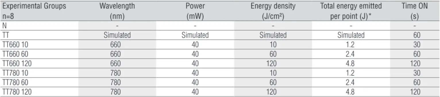
he pre-neurotmesis (pre-TT) SFI values were considered normal (-7.44). No diference was found among the experimental groups in the pre-TT moment (p>0.05; Figure 1). As expected, on the irst day after injury there was a reduction in the SFI com-pared with pre-TT in all injured groups (p>0.05; Figure 1). On the last day (84th day), a partial functional recovery was observed in all injured groups when compared to day 1 (p<0.05; Figure 1).
Nevertheless, these values remained inferior to those values ob-served in the normal group (p<0.05; Figure 1), with no diference among injured groups on day 84 post-injury (p>0.05; Figure 1). No diference was detected among the groups at any point be-tween the irst and the last day (data not shown).
Muscle morphology and cross-sectional area (CSA)
he muscle morphology analysis showed an atrophic pat-tern of muscle ibers for all injured groups (LLLT irradiated or not) when compared to the normal group (Figure 2). Connec-tive tissue proliferation was observed in the denervated soleus muscles (Figure 2B-G), specially surrounding the muscle ibers (endomysium) and iber bundles (perimysium) compared to Normal group (Figure 2A).
Moreover, angulated and degenerated ibers were observed in all denervated groups (Figure 2B-G). hese characteristics are a direct indication of skeletal muscle modiications caused by the absence of innervation. In these groups, central nuclei were also observed, conirming the myopathic phenotype (Figure 2I).
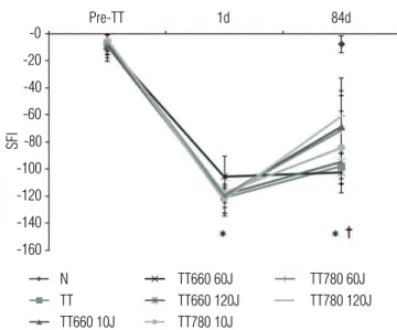
Muscle iber atrophy was conirmed by muscle iber CSA measurement. All denervated groups showed smaller muscle iber CSA than normal (p<0.05; Figure 3; TT: -69.2%, TT660 10: -61.5%; TT660 60: -46.1%; TT660 120: -64.1%; TT780 10: -51.3%; TT780 60: -53.8% and TT780 120: -52.6%). No diference was observed among denervated groups (p>0.05; Figure 3).
MMP activity in denervated soleus muscle
he MMP-9 activity was not detected in any samples of the soleus muscle. In comparison, three MMP-2 isoforms were located (pro, intermediate, and active) in all groups. A repre-sentative gel is shown in Figure 4A.
Densitometric analysis showed no diference among de-nervated and normal groups in any of the isoforms (p>0.05; Figure 4B-D).
Experimental Groups n=8
Wavelength (nm)
Power (mW)
Energy density (J/cm²)
Total energy emitted per point (J)*
Time ON (s)
N - - - -
-TT Simulated Simulated Simulated Simulated 60
TT660 10 660 40 10 1.2 30
TT660 60 660 40 60 2.4 60
TT660 120 660 40 120 4.8 120
TT780 10 780 40 10 1.2 30
TT780 60 780 40 60 2.4 60
TT780 120 780 40 120 4.8 120
Table 1. Parameters of LLLT application in different experimental groups.
Discussion
he present study demonstrated that LLLT applied to reconstructed nerve with the parameters investigated here is unable to avoid degenerative modiications to denervated slow-twitch muscles and inefective in recovering functional-ity in rats. he present results did not corroborate the indings observed in denervated fast-twitch muscles whose crushed nerves were irradiated with LLLT3.
Recently, Gigo-Benato et al.3 pointed out that LLLT irra-diation of crushed sciatic nerves caused acceleration in nerve regeneration and contributed toward CSA recovery in the tibi-alis anterior (TA) muscles of rats. hey also demonstrated that a wavelength of 660 nm, with energy densities of 10, 60, and 120 J/cm², was efective in increasing the MMP-2 activity of the injured nerves, possibly facilitating axonal growth through laminin, ibronectin, and type IV collagen degradation18. In ad-dition, LLLT decreased the MMP-9 activity of these nerves, which probably helped to attenuate the inlammatory process. Finally, they demonstrated a decrease in MMP-2 activity in the TA muscles of the crushed groups irradiated with 660 nm LLLT. he factors that hinder a direct comparison between this and the present study are the diferences between the experimental models, namely, the crushing model compared to the nerve section model and the diferent investigation times.
Pre-TT: pre-nerve injury; 1d: first day post-injury; 84d: eighty-fourth day post-injury (last day of the experiment). *p<0.05 for TT, TT660 10, TT660 60, TT660 120, TT780 10, TT780 60, and TT780 120 compared to their own values in 1d and N. Note the normal pre-injury (-7.44). On the 1st day post-injury, as expected, there is a significant drop (-110.40) compared to the pre-injury phase. At 84 days post-injury, recovery may be observed in the index, but still well below normal levels (†p<0.05).
SFI
-160
Pre-TT 1d 84d
-140 -120 -100 -80 -60 -40 -20 -0
N TT TT660 10J
TT660 60J TT660 120J TT780 10J
TT780 120J TT780 60J
Figure 1. Sciatic functional index (SFI) in different experimental groups.
Figure 2. Soleus muscle cross-section of the different experimental groups stained with toluidine blue: A) N; B) TT; C) TT660 10; D) TT660 60; E) TT660 120; F) TT780 10; G) TT780 60; H) TT780 120; and I) TT660 120. White arrows indicate muscle fibers with centralized nuclei; black arrows indicate the angled fibers; asterisks indicate proliferation of connective tissue; and arrow heads indicate the degenerating fibers.
Another factor that may have inluenced the indings re-lated to muscle trophism is iber composition. Gigo-Benato et al.3 investigated fast-twitch muscles (TA) composed mainly of type II ibers. However, the present study focused on a slow-twitch muscle (soleus) composed mainly of type I ibers19. It has been well described in the literature that slow-twitch muscles exert antigravity function20 and respond more to immobiliza-tion and disuse situaimmobiliza-tions if compared to fast-twitch muscles. Furthermore, the reduction in oxidative capacity, and the increase in glycolytic metabolism in denervated slow-twitch muscles combined with muscle iber phenotype transition ( from type I to type II) can also interfere with muscular adap-tative response19,21.
Figure 3. Cross-sectional area (CSA) of soleus muscles from the different experimental groups.
Muscle fiber cross-sectional area
(µ
m
2)
0
N TT TT660 10 TT660 60 TT660 120 TT780 10 TT780 60 TT780 120 200
400 600 800 1000 1200 1400 1600
*p<0.05 compared to N. Note the decrease in CSA in all groups compared to denervated N. There was no difference between the TT groups irradiated or not with LLLT.
Figure 4. MMP-2 activity in denervated soleus muscle.
N 0 1 2
TT 0.5
1.5
0 1 2
0.5 1.5
0 1 2
0.5 1.5
TT660 10
Arbitrar
y Units
Arbitrar
y Units
Arbitrar
y Units
TT660 60 Pro 62 KDa
74 KDa 57 KDa
Intermediary
Active
TT660 120 TT780 10 TT780 60 TT780 120
N TT TT660 10 TT660 60 TT660 120 TT780 10 TT780 60 TT780 120
N TT TT660 10 TT660 60 TT660 120 TT780 10 TT780 60 TT780 120
B)
C)
D)
A)
on cellular metabolism increasing the size and amount of mi-tochondria, and oxygen consumption27,28. hus, we believe that LLLT could stimulate the mitochondria of large motor units, facilitating growth cone advance. However, future studies must be performed to test this hypothesis.
Regarding MMP-2 activity, normal levels were found in all denervated soleus muscles. his can be attributed to timing. Demestre et al.6 reported an increase in MMP-2 activity in denervated soleus muscles 20 and 40 days after crush nerve in-jury and a return to normal levels 63 and 72 days after inin-jury, in accordance with the present study. hus when comparing two types of injury, it is possible to reach the conclusion that MMP activity is normal in later phases of post-neurotmesis recovery. In contrast, these normal levels of MMP-2 activity could be related to a state of balance that atrophic muscles can reach. Recent studies reported that other degradation pathways, i.e. autophagic pathway, can be involved in late denervated muscle atrophy. In addition, skeletal muscles have strategies to inhibit these pathways and preserve myoibrillar organization, such as the increase in Runx1 (Runt-related transcription fac-tor 1) expression29. he present study corroborates this inding because the muscle iber analysis of the denervated groups showed many signs of injury-regeneration cycles, such as cen-tral nucleus and iber fragmentation. Angled ibers were also observed in all denervated groups, showing a homogeneous pattern in these muscles.
Considering functional recovery, it should be observed that the injury model used in the present study is slower than crush models, with worse prognosis and longer recovery. De Sá et al.30 demonstrated that 80% of nerve function was recovered within 60 days of end-to-end neurorrhaphy. he authors sug-gested that in a few more weeks the repairing process could be complete. Hence, when evaluating the period of 84 days post-injury, we observed a signiicant, albeit incomplete, func-tional improvement in all injured groups (LLLT irradiated or not). New studies should investigate longer times after injury in order to determine the moment of total functional recovery.
It is also worth noting that the present study carried out a dose-response curve following recommendations of clini-cal protocols of LLLT application. For the wavelength, some studies demonstrated that either visible31-33 or near-infrared lasers34 are able to stimulate neuronal growth. Energy density is another important parameter to be considered when inves-tigating LLLT efects. Analysis of published results showed that
LLLT at diferent levels of energy density (ranging from <10 to 150 J/cm2) are efective in promoting nerve regeneration. he same is observed for time of irradiation which varies from <1 min to 90 min. But despite the evidence that light can afect nerve recovery, the literature is still unclear on the best combi-nation of parameters to improve the regenerative process. Fi-nally, all published studies that reported good results in nerve regeneration used continuous LLLT33,35,36. he present study followed all of these recommendations.
he results of the present study have signiicant clinical considerations. Based on our indings, therapists should keep in mind that denervated slow-twitch muscles do not recover as quickly as fast-twitch muscles3. Future studies should focus on interventions not only for the injured nerve, but also for the denervated muscle. Muscle stretching, electrical stimulation, and LLLT have already shown signiicant results in PNI
treat-ment37-40, thus, they should also be considered in future studies
for the recovery of denervated slow-twitch muscles.
he present study has some limitations that should be considered. A time-course curve to verify cellular and mo-lecular modiications could provide information about the MMP activity pattern over time and possible correlations with morphological indings. Furthermore, a comparison between fast- and slow-twitch muscles is necessary to investigate the diferences in the reinnervation process and MMP content/ activity according to muscle type. Although the present study focused on muscle investigation, other studies should consider the evaluation of morphological and molecular nerve aspects to provide evidence of interaction between LLLT and periph-eral nerves.
In conclusion, using a severe PNI standardized model and an LLLT protocol based on literature recommendations, we found that LLLT applied to injured nerves, regardless of the dose, was inefective in accelerating functional recovery and improving denervated slow-twitch muscle trophism in rats.
Acknowledgements
References
1. Russo TL, Peviani SM, Durigan JL, Gigo-Benato D, Delfino GB, Salvini TF. Stretching and electrical stimulation reduce the accumulation of MyoD, myostatin and atrogin-1 in denervated rat skeletal muscle. J Muscle Res Cell Motil. 2010;31(1):45-57.
2. Dow DE, Cederna PS, Hassett CA, Kostrominova TY, Faulkner JA, Dennis RG. Number of contractions to maintain mass and force of a denervated rat muscle. Muscle Nerve. 2004;30(1):77-86.
3. Gigo-Benato D, Russo TL, Tanaka EH, Assis L, Salvini TF, Parizotto NA. Effects of 660 and 780 nm low-level laser therapy on neuromuscular recovery after crush injury in rat sciatic nerve. Lasers Surg Med. 2010;42(9):673-82.
4. Kouyoumdjian JA. Peripheral nerve injuries: a retrospective survey of 456 cases. Muscle Nerve. 2006;34(6):785-8.
5. Sunderland S. Nerves and nerve injuries. 2ª ed. London: Churchill Livingston; 2002.
6. Demestre M, Orth M, Wells GM, Gearing AJ, Hughes RAC, Gregson NA. Characterization of matrix metalloproteinases in denervated muscle. Neuropathol Appl Neurobiol. 2005;31(5): 545-55.
7. Smith AST, Shah R, Hunt NP, Lewis MP. The role of connective tissue and extracellular matrix signaling in controlling muscle development, function, and response to mechanical forces. Semin Orthod. 2010;16(2):135-42.
8. Carmeli E, Moas M, Reznick AZ, Coleman R. Matrix metalloproteinases and skeletal muscle: a brief review. Muscle Nerve. 2004;29(2):191-7.
9. Kherif S, Dehaupas M, Lafuma C, Fardeau M, Alameddine HS. Matrix metalloproteinases MMP-2 and MMP-9 in denervated muscle and injured nerve. Neuropathol Appl Neurobiol. 1998;24(4):309-19.
10. Ahtikoski AM, Tuominen H, Korpelainen JT, Takala TE, Oikarinen A. Collagen synthesis and degradation in polyneuropathy and myopathies. Muscle Nerve. 2004;30(5):602-8.
11. Dvali L, Mackinnon S. Nerve repair, grafting, and nerve transfers. Clin Plast Surg. 2003;30(2):203-21.
12. Shamir MH, Rochkind S, Sandbank J, Alon M. Double-blind randomized study evaluating regeneration of the rat transected sciatic nerve suturing and postoperative low-power laser treatment. J Reconstr Microsurg. 2001;17(2):133-7.
13. Bain JR, Mackinnon SE, Hunter DA. Functional evaluation of complete sciatic, peroneal, and posterior tibial nerve lesions in the rat. Plast Reconstr Surg. 1989;83(1):129-38.
14. Varejão AS, Cabrita AM, Meek MF, Bulas-Cruz J, Melo-Pinto P, Raimondo S, et al. Functional and morphological assessment standardized rat sciatic nerve crush injury with a non-serrated clamp. J Neurotrauma. 2004;21(11):1652-70.
15. Marqueti RC, Parizotto NA, Chriguer RS, Perez SE, Selistre-de-Araújo HS. Androgenic-anabolic steroids associated with mechanical loading inhibit matrix metallopeptidase activity and affect the remodeling of the Achilles tendon in rats. Am J Sports Med. 2006;34(8):1274-80.
16. Peviani SM, Russo TL, Durigan JL, Vieira BS, Pinheiro CM, Galassi MS, et al. Stretching and electrical stimulation regulate the metalloproteinase-2 in rat denervated skeletal muscle. Neurol Res. 2010;32(8):891-6.
17. Carvalho RF, Dariolli R, Justulin Junior LA, Sugizaki MM, Politi Okoshi M, Cicogna AC, et al. Heart failure alters matrix metalloproteinase gene expression and activity in rat skeletal muscle. Int J Exp Pathol. 2006;87(6):437-43.
18. La Fleur M, Underwood JL, Rappolee DA, Werb Z. Basement membrane and repair of injury to peripheral nerve: defining a potential role for macrophages, matrix metalloproteinases, and tissue inhibitor of metalloproteinases-1. J Exp Med. 1996;184(6):2311-26.
19. Lieber RL, Fridén JO, Hargens AR, Feringa ER. Long-term effects of spinal cord transection of fast and slow rat skeletal muscle. II. Morphometric properties. Exp Neurol. 1986;91(3):435-48.
20. Zimowska M, Brzoska E, Swierczynska M, Wstreminska W, Moraczewski J. Distinct patterns of MMP-9 and MMP-2 activity in slow and fast twitch skeletal muscle regeneration in vivo. Int J Dev Biol. 2008;52(2-3):307-14.
21. Zhou, Z, Cornelius CP, Eichner M, Bornemann A. Reinnervation-induced alterations in rat skeletal muscle. Neurobiol Dis. 2006;23(3):595-602.
22. Kandel ER, Schwartz JH, Jessell TM. Principles of neural science. 4th ed. New York: McGraw-Hill; 2000.
23. Lundborg G, Dahlin LB. Anatomy, function, and pathophysiology of peripheral nerves and nerve compression. Hand Clin. 1996;12(2):185-93.
24. Miyauchi A, Kanje M, Danielsen N, Dahlin LB. Role of macrophages in the stimulation and regeneration of sensory nerves by transposed granulation tissue and temporal aspects of the response. Scand J Plast Reconstr Surg Hand Surg. 1997;31(1):17-23.
25. Heumann R, Hengerer B, Brown M, Perry H. Molecular mechanisms leading to lesion-induced increases in nerve growth factor synthesis. Ann N Y Acad Sci. 1991;633:581-2.
26. Karu TI. Primary and secondary mechanisms of action of visible-to-near IR radiation on cells. J Photochem Photobiol B. 1999;49(1):1-17.
27. Rizzi CF, Mauriz JL, Freitas Corrêa DS, Moreira AJ, Zettler CG, Filippin LI, et al. Effects of low-level laser therapy (LLLT) on the nuclear factor (NF)-kappaB signaling pathway in traumatized muscle. Lasers Surg Med. 2006;38(7):704-13.
28. Schroeder P, Pohl C, Calles C, Marks C, Wild S, Krutmann J. Cellular response to infrared radiation involves retrograde mitochondrial signaling. Free Radic Biol Med. 2007;43(1): 128-35.
29. Wang X, Blagden C, Fan J, Nowak SJ, Taniuchi I, Littman DR, et al. Runx1 prevents wasting, myofibrillar disorganization, and autophagy of skeletal muscle. Genes Dev. 2005;19(14): 1715-22.
30. De Sá JMR, Mazzer N, Barbieri CH, Barreira AA. The end-to-side peripheral nerve repair. Functional and morphometric study using the peroneal nerve of rats. J Neurosci Methods. 2004;136(1):45-53.
31. Rochkind S, Rousso M, Nissan M, Villarreal M, Barr-Nea L, Rees DG. Systemic effects of low-power laser irradiation on the peripheral and central nervous system, cutaneous wounds, and burns. Lasers Surg Med. 1989;9(2):174-82.
32. Hamilton GF, Robinson TK, Ray RH. The effects of helium-neon laser upon regeneration of the crushed peroneal nerve. J Orthop Sports Phys Ther. 1992;15(5):209-14.
33. Anders JJ, Geuna S, Rochkind S. Phototherapy promotes regeneration and functional recovery of injured peripheral nerve. Neurol Res. 2004;26(2):233-9.
34. Rochkind S, El-Ani D, Nevo Z, Shahar A. Increase of neuronal sprouting and migration using 780nm laser phototherapy as procedure for cell therapy. Lasers Surg Med. 2009;41(4): 277-81.
35. Gigo-Benato D, Geuna S, de Castro Rodrigues A, Tos P, Fornaro M, Boux E, et al. Low-power laser biostimulation enhances nerve repair after end-to-side neurorrhaphy: a double-blind randomized study in the rat median nerve model. Lasers Med Sci. 2004;19(1):57-65.
36. Rochkind S, Geuna S, Shainberg A. Chapter 25: Phototherapy in peripheral nerve injury: effects on muscle preservation and nerve regeneration. Int Rev Neurobiol. 2009;87:445-64.
37. Fernandes KCBG, Polacow MLO, Guirro RRJ, Campos GER, Somazz MC, Pinto VF, et al. Análise morfométrica dos tecidos muscular e conjuntivo após desnervação e estimulação elétrica de baixa frequência. Rev Bras Fisioter. 2005;9(2):235-41.
38. Reis FA, Belchior ACG, Nicolau RA, Fonseca TS, Carvalho PTC. Efeito da terapia com laser de arsenieto de gálio e alumínio (660Nm) sobre a recuperação do nervo ciático de ratos após lesão por neurotmese seguida de anastomose epineural: análise funcional. Rev Bras Fisioter. 2008;12(3):215-21.
39. Caierão QM, Betini J, Teodori RM, Minamoto VB. O efeito do intervalo da estimulação elétrica no músculo desnervado de rato. Rev Bras Fisioter. 2008;12(2):143-8.