Evaluation of the mutagenicity and antimutagenicity of
Ziziphus joazeiro
Mart.
bark in the micronucleus assay
Marcelo Fabiano Gomes Boriollo
1,2, Marielly Reis Resende
4, Thaísla Andrielle da Silva
1,2,
Juliana Yoshida Públio
2, Luiz Silva Souza
1,2, Carlos Tadeu dos Santos Dias
4,
Nelma de Mello Silva Oliveira
1,3and João Evangelista Fiorini
1,21
Laboratório de Farmacogenômica e Biologia Molecular, Faculdade de Ciências Médicas,
Universidade José do Rosário Vellano, Alfenas, MG, Brazil.
2
Centro de Pesquisa e Pós-Graduação em Ciência Animal, Área de Patologia e Farmacologia Animal,
Universidade José do Rosário Vellano, Alfenas, MG, Brazil.
3
Laboratório de Ecotoxicologia e Microbiologia Ambiental, Faculdade de Tecnologia,
Universidade Estadual de Campinas, Limeira, SP, Brazil.
4
Departamento de Ciências Exatas, Escola de Agricultura “Luiz de Queiroz”, Universidade de São Paulo,
Piracicaba, SP, Brazil.
Abstract
The aim of this study was to evaluate the mutagenicity (clastogenicity/aneugenicity) of a glycolic extract ofZiziphus joazeiro bark (GEZJ) by the micronucleus assay in mice bone marrow. Antimutagenic activity was also assessed us-ing treatments associated with GEZJ and doxorubicin (DXR). Mice were evaluated 24-48 h after exposure to positive (N-nitroso-N-ethylurea, NEU - 50 mg.kg-1
and DXR - 5 mg.kg-1
) and negative (150 mM NaCl) controls, as well as treat-ment with GEZJ (0.5-2 g.kg-1
), GEZJ (2 g.kg-1
) + NEU and GEZJ (2 g.kg-1
) + DXR. There were no significant differ-ences in the frequencies of micronucleated polychromatic erythrocytes in mice treated with GEJZ and GEJZ + DXR compared to the negative controls, indicating that GEZJ was not mutagenic. Analysis of the polychromatic:nor-mochromatic erythrocyte ratio revealed significant differences in the responses to doses of 0.5 g.kg-1
and 1-2 g.kg-1 and the positive control (NEU). These results indicated no systemic toxicity and moderate toxicity at lower and higher doses of GEZJ. The lack of mutagenicity and systemic toxicity in the antimutagenic assays, especially for treatment with GEZJ+ DXR, suggested that phytochemical compounds in Z. joazeiro bark attenuated DXR-induced muta-genicity and the moderate systemic toxicity of a high dose ofZ. joazeiro bark (2 g.kg-1
). Further studies on the genotoxicity ofZ. joazeiro extracts are necessary to establish the possible health risk in humans and to determine the potential as a chemopreventive agent for therapeutic use.
Keywords: antimutagenicity, bone marrow, doxorubicin, micronucleus assay, mutagenicity,Zizyphus joazeiroMart. (raspa-de-Juá). Received: October 3, 2013; Accepted: March 17, 2014.
Introduction
Many species of medicinal plants, such asAmburana
cearensis, Anadenanthera colubrina, Mentha x villosa,
Myracrodruon urundeuva,Plectranthus amboinicus,Ruta graveolens,Ximenia americanaandZiziphus joazeiro, are widely used by communities in the Brazilian Caatinga to treat a large spectrum of clinical conditions ranging from diseases requiring palliative care to general aches, e.g., bronchitis, sinusitis, rhinitis, nasal congestion, headaches,
flu, fever, expectorant, colic, hypertension, thrombosis, in-digestion, intestinal dysfunction, liver and kidney prob-lems, infectious and inflammatory processes and pain in
general (Cartaxo et al., 2010). Ziziphus joazeiro Mart.
(Rhamnaceae) is a native Brazilian tree resistant to dry en-vironments (Cartaxoet al., 2010). This species is an impor-tant source of water and food for animals in arid habitats (Braga, 1960; Cruz, 1985; Nuneset al., 1987).
A phytochemical analysis of Z. joazeiro Mart. has
shown that the leaf epicuticular wax is rich inn-alkanes (78.6%), very efficient compounds for impermeabilizing the leaf surface, and triterpenoids (Oliveiraet al., 2003). A similar analysis of a dichloromethane extract ofZ. joazeiro
Mart. bark identified triterpenoids with weak antibacterial activity (e.g., betulinic, alphitolic and ursolic acids) and re-Genetics and Molecular Biology, 37, 2, 428-438 (2014)
Copyright © 2014, Sociedade Brasileira de Genética. Printed in Brazil www.sbg.org.br
Send correspondence to Marcelo Fabiano Gomes Boriollo. Labo-ratório de Farmacogenômica e Biologia Molecular, Faculdade de Ciências Médicas & Centro de Pesquisa e Pós-Graduação, Universidade José do Rosário Vellano, Rodovia MG 179, km 0, Campus Universitário, 37130-000 Alfenas, MG, Brazil. E-mail: [email protected].
markable activity against Staphylococcus epidermidis
[e.g., betulinic acid ester derivatives such as 7b
-(4-hydroxy-benzoyloxy), 7b
-(4-hydro-3-methoxy-benzoylo-xy) and 27-(4-hydroxy-3-methoxy-benzoylo-(4-hydro-3-methoxy-benzoylo-xy)] (Schuhly
et al., 1999).Ziziphus joazeiroMart. bark also contains an abundance of saponins that have been used as toothpastes, with aqueous extracts showing antimicrobial action against bacteria (planktonic cells and artificial biofilms) related to
dental caries and periodontal diseases (Alviano et al.,
2008). Other popular therapeutic applications ofZ. joazeiro
Mart. include the treatment of dandruff, rheumatism, flu, fever, chronic bronchitis, gastric ulcers, indigestion, heart-burn and headaches (Schuhlyet al., 1999; Cartaxoet al., 2010). In addition, experimental studies have identified po-tential antifungal (Cruzet al., 2007), antibacterial (Schuhly
et al., 1999; Alvianoet al., 2008; Lealet al., 2010), antioxi-dant (Alvianoet al., 2008) and antipyretic (Nuneset al., 1987) activities, as well as low toxicity (Alviano et al., 2008).
Biologically active compounds have been recognized for their pharmacological properties, but many of them are of limited therapeutic use because of their toxicological, carcinogenic and mutagenic properties (Ames, 1983; Konstantoupoulouet al., 1992; Tavares, 1996). The analy-sis of genotoxicity is a major aspect of drug development since most pharmaceutical companies evaluate the poten-tial of a new therapeutic agent based on its genotoxicityin vitroandin vivo(Purveset al., 1995). In this context, the screening of popularly used plants and their isolated com-ponents for mutagenic activity is necessary and important for establishing adequate control measures. This screening can also provide insights into the mechanisms involved in the biological effects of plants used as therapeutic agents (Varanda, 2006).
As far as genotoxicity studies are concerned, thein
vivomicronucleus (MN) assay in rodent bone marrow is a
crucial part of the battery of tests used to identify hazardous mutagens (Mateucaet al., 2006). This assay is especially suited for assessing mutagenic hazards because it contem-plates various factors, such asin vivo metabolism, phar-macokinetics and DNA repair mechanisms, even though these processes vary among species and tissues and have different genetic endpoints (OECD, 1997a,b; Ribeiroet al., 2003). Since bone marrow erythroblasts develop into poly-chromatic erythrocytes (PCEs),i.e., cells generated by ex-trusion of the main nucleus, micronuclei may remain in an otherwise anucleated cytoplasm. Consequently, the fre-quency of micronucleated polychromatic erythrocytes (MNPCEs) has been the principal endpoint for MN assays. The measurement of MNPCEs in peripheral blood is possi-ble in any species in which the spleen does not remove micronucleated erythrocytes, or that is sufficiently sensi-tive to agents that cause structural or numerical chromo-somal aberrations. An increase in the frequency of MNPCEs in treated animals,i.e., a positive result, indicates
that a substance can cause the formation of micronuclei through chromosomal damage or damage to the mitotic ap-paratus of erythroblasts. On the other hand, a negative re-sult implies that the test substance does not cause micro-nucleus formation in immature erythrocytes. The number of normochromatic erythrocytes (NCEs) in peripheral blood that contain micronuclei for a given number of ma-ture erythrocytes can also be used as the endpoint of this as-say (OECD, 1997c; Ribeiroet al., 2003). Several studies
have used the mammalianin vivoMN assay to understand
the mutagenic effects induced by phytotherapeutics and foods (Indartet al., 2007; Venkateshet al., 2007; Chan-drasekaran et al., 2011; Silva et al., 2011; Alves et al., 2012).
Although several studies have examined the potential therapeutic effectiveness of Z. joazeiro Mart., there has been no systematic investigation of the genotoxic and mutagenic effects of this plant. In this work, we examined the mutagenic effects of a glycolic extract ofZ. joazeiro
Mart. bark as part of a wider study on the genotoxic poten-tial of herbal extracts. The effect of the maximum permissi-ble concentration ofZ. joazeiroMart. on the mutagenicity
of doxorubicin (DXR) in mouse bone marrow, i.e., its
antimutagenic activity, was also examined.
Material and Methods
Raw material and sample preparation
A glycolic extract ofZ. joazeiro bark (GEZJ) was
purchased commercially and stored according to the manu-facturer’s recommendations (AKSY Comercial Ltda., São Bernardo do Campo, SP, Brazil). Aliquots (1.5 L) of this extract were submitted to solvent removal proceedings by rotary evaporation (40 rpm) (Rotavapor model R-215) cou-pled to a bath heating system maintained at 50-60 °C (Bath Heating model B-491), a vacuum pump (vacuum of 500 mm Hg; Vacuum Pump V-700 with Automatic Vac-uum Controller V-855), a water recirculator (Recirculator Chiller F-100) and an evaporation bottle (Büchi Labor-technik AG, Switzerland). The final product was
trans-ferred to a 1 L reaction bottle (SCHOTT®DURAN®) and
kept at -20 °C for 24 h in order to evaluate the freezing of the final product and the efficacy of solvent evaporation (Agência Nacional de Vigilância Sanitária (ANVISA) (2010)). Aliquots (40 mL) of this final product were trans-ferred to penicillin-type glass vials (50 mL) and lyophilized (Lyophilizer model Alpha 1-2 LDPlus, Martin Christ Gefriertrocknungsanlagen GmbH, Germany) and the dry mass were measured (Electronic Analytical Balance AUW-220D, Shimadzu Corp., Kyoto, Japan). Aqueous so-lutions of the lyophilized product were prepared in type 1 water at twice the final concentration, sterilized by filtra-tion (Millipore Corporafiltra-tion, hydrophilic Durapore®PVDF,
0.22mm,±47 mm, cat. no. GVWP 047 00) and stored in
sterile polypropylene tubes (50 mL) at -70 °C until used.
In vivoassays
Healthy, heterogeneous, young adult male and female Swiss mice (Unib:SW) 7-12 weeks old (pubescent period) weighing 30-40 g (weight variation among mice of each sex was < 20% of the mean weight) were provided by CEMIB (Centro Multidisciplinar para Investigação Bioló-gica - UNICAMP) and erythrocytes from the bone marrow of these mice were used in the MN assay (Collaborative Study Group for the Micronucleus Test (CSGMT), 1986; Chorilliet al., 2007).
Animals of the same sex were housed in polypropy-lene boxes in an air-conditioned environment to 22±3 °C,
with a relative air humidity of 50% ± 20% and a 12 h
light/dark cycle. The mice were fed commercial rodent
chow (Purina®Labina, Nestlé Purina Pet Care Company)
and waterad libitum, and were acclimated to laboratory
conditions for seven days prior to use in the experiments. At the end of this period, each mouse was weighed and then re-ceived 2 mL of liquid (containing the desired test agent) per 100 g body weight.
All animals were properly identified by numerical markings on their tails to ensure continuity of the records and reliable interpretation of the results throughout the study (OECD, 1997c). After the period of treatment, the mice were euthanized by inhalation of carbon dioxide in adapted acrylic chambers as described in the Report of the American Veterinary Medical Association panel on eutha-nasia (Beaveret al., 2000). This study was done in accor-dance with the Universal Declaration of Animal Rights (UNESCO, 1978), the ethical principles for animal experi-mentation established by the Brazilian Society of
Labora-tory Animal Science (SBCAL -Sociedade Brasileira de
Ciência em Animais de Laboratório), the Brazilian Envi-ronmental Crimes Law (Law no. 9.605, February 12, 1998), the Brazilian standards for Didactic-Scientific Prac-tice of Vivisection of Animals (Law no. 6.638, May 8, 1979), and was approved by the Committee for Ethics in Research Involving Animals at UNIFENAS (CEPEAU Protocol no. 04A/2008).
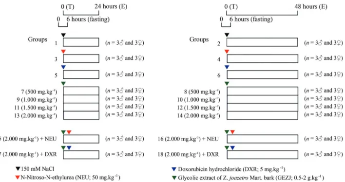
Experimental groups
The experimental groups of mice (3 males and 3 fe-males each) were assessed 24 h and 48 h after a single treat-ment administered by gavage (Figure 1). The mutagenic activity of GEZJ was assessed in mice that received doses of 0.5-2 g.kg-1(groups 7-14) and the antimutagenic activity
was assessed in mice that received NEU (50 mg.kg-1) +
GEZJ (2 g.kg-1) (groups 15 and 16) and DXR (5 mg.kg-1) +
GEZJ (2 g.kg-1) (groups 17 and 18). The doses of GEZJ
were chosen based on previous acute toxicity experiments in mice that yielded LD50values of 2.0-3.5 g/kg for several plant extracts, includingZ. joazeiro(Alvianoet al., 2008). Negative controls (groups 1 and 2: 150 mM NaCl in type 1 water) and positive controls (groups 3 and 4: 50 mg.kg-1of
NEU; groups 5 and 6: 5 mg.kg-1 of DXR) were also
in-cluded as single treatments administered by gavage (NaCl) and intraperitoneally (NEU and DXR) (OECD, 1997c).
Processing of bone marrow
MN assays using bone marrow erythrocytes were done 24 h and 48 h after treatment, using previously
de-scribed methodology (Schmid, 1976; Zambrano et al.,
430 Micronucleus test ofZ. joazeirobarks
Figure 1- Experimental protocol for assessing the mutagenic and antimutagenic activity of a glycolic extract ofZ. joazeirobark. T - treatment, E -
1982). Shortly after euthanasia, the femora were surgically and aseptically removed and the mice were appropriately discarded. Each femur was sectioned at the proximal end and the contents of the spinal canal were washed with 1.5 mL of 150 mM NaCl and transferred to a 15 mL centri-fuge tube. This material was resuspended with a Pasteur pi-pette to ensure a homogenous distribution of bone marrow cells. The suspension was then centrifuged at 1,000 rpm (Bench centrifuge, model NT 810, Nova Técnica Ind. e Com. de Equip. para Laboratório Ltda., Piracicaba, SP, Brazil) for 5 min. The supernatant was discarded and the
re-sulting pellet was resuspended in 500mL of 150 mM NaCI
solution added 4% formaldehyde. The slides (two per ani-mal) were prepared by smearing, dried at room temperature for 24 h and stained with Leishman’s eosin methylene blue dye [pure dye for 3 min followed by diluted dye in distilled water (1:6) for 15 min] to differentiate polychromatic erythrocytes (PCEs) from monochromatic erythrocytes (NCEs).
PCEs were observed by light microscopy (Nikon Eclipse E-200 microscope) at a magnification of 1000x, counted (at least 2000 anucleated polychromatic erythro-cytes per animal) with the aid of a digital cell counter (Contador Diferencial CCS02, Kacil Indústria e Comércio Ltda., PE, Brazil) and photographed using an 8.1 Mega-pixel Digital Camera (DC FWL 150). The number of PCEs, the number and frequency of MNPCEs and the ratio of polychromatic to monochromatic erythrocytes (PCE/NCE) were determined.
Statistical analysis
The data from the MN assay were analyzed by one-way analysis of variance (ANOVA) using a 9 x 2 x 2 (treat-ment x gender x time) factorial scheme followed by
multi-ple comparisons with the Tukey test (a = 0.05). All
analyses were done using SAS®version 9.2 computer soft-ware.
Results and Discussion
Ziziphus joazeiroMart. has been popularly used to treat dandruff, rheumatism, flu, fever, chronic bronchitis, gastric ulcers, indigestion, heartburn and headaches and to clean teeth (Schuhlyet al., 1999; Cartaxoet al., 2010). In addition, Z. joazeiro has potential antifungal (Cruzet al., 2007), antibacterial (Schuhlyet al., 1999; Alvianoet al., 2008; Lealet al., 2010), antioxidant (Alvianoet al., 2008) and antipyretic (Nuneset al., 1987) activities, as well as low toxicity (Alvianoet al., 2008). This information partly supports the popular use of Z. joazeiro for certain treat-ments and agrees with ethnopharmacological studies de-signed to select plants for bioactivity screening (Cruzet al., 2007). In contrast, few studies have examined the muta-genic and antimutamuta-genic effects ofZ. joazeiroMart.
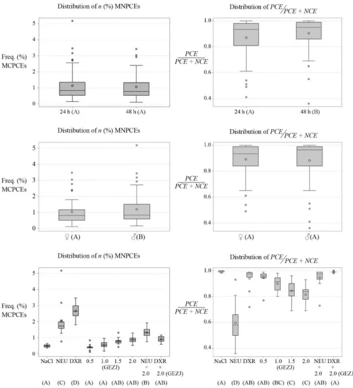
In the present study, the number and frequency of MNPCEs and the PCE/NCE ratios in mouse bone marrow were analyzed in mutagenic and antimutagenic assays of a glycolic extract ofZ. joazeirobark (Table 1 and Figure 2). Analysis of the MNPCEs revealed no significant differ-ences between the 24 h and 48 h results for the negative (NaCl) and positive (DXR and NEU) controls. However, there were significant differences (p <0.05) between the negative and positive controls at the two time intervals. There were no differences between the negative controls and the treatments with GEJZ (0.5-2 g.kg-1) or with GEJZ (2 g.kg-1) + DXR (5 mg.kg-1): these responses showed no dose or time dependence, but varied between male and
fe-male mice. Mice treated with GEJZ (2 g.kg-1) + NEU (50
mg.kg-1) had intermediate values (n and %) that differed significantly from the negative and positive controls. These results suggest absence of mutagenicity (clastogenicity and/or aneugenicity) for GEZJ, regardless of the extract dose and time interval, although the responses varied
be-tween sexes. In contrast, GEJZ (2 g.kg-1) showed
anti-mutagenic activity (anticlastogeny and/or antianeugeny)
towards the chemotherapeutic agent DXR (5 mg.kg-1) or
NEU (50 mg.kg-1), regardless of the time interval, although once again intersex variation was observed. These findings indicate that compounds in GEZJ can act against DXR-induced mutagenic effects in mouse bone marrow. Such compounds could include n-alkanes, triterpenoids [i.e., betulinic acid, alphitolic acid, ursolic acid, ester derivatives
of betulinic acid such as 7b
-(4-hydroxy-benzoylo-xy)-betulinic acid, 7b
-(4-hydro-3-methoxy-benzoyloxy)-betulinic acid and 27-(4-hydroxy-3-methoxy-benzoylo-xy)-betulinic acid] (Oliveiraet al., 2003; Schuhly et al.,
1999) and saponins (Alvianoet al., 2008). DXR has been
reported to induce micronuclei, chromatid and chromo-somal aberrations, and DNA single- and double-strand breaksin vitroand in vivo(Bean et al., 1992; Al-Harbi,
1993; Al-Shabanah, 1993; Delvaeyeet al., 1993; Jagetia
and Nayak, 1996, 2000; Shanet al., 1996; Dhawanet al., 2003; Jagetia and Aruna, 2000). In addition, the major acute toxicity induced by DXR is bone marrow suppres-sion, while the long-term clinical usefulness is limited by a cumulative, dose-dependent, irreversible, chronic cardio-toxicity that manifests itself as congestive heart failure or cardiomyopathy (Van Ackeret al., 1995, 2000).
For the PCE/NCE ratio, there were no significant dif-ferences between the negative controls (NaCl), the positive control DXR (5 mg.kg-1), the GEZJ (0.5 mg.kg-1) group, and mice treated with GEZJ (2 g.kg-1) + NEU (50 mg.kg-1) or with GEZJ (2 g.kg-1) + DXR (5 mg.kg-1) (Table 1 and Figure 1). For the treatments with GEZJ, there was a signif-icant difference between the dose of 500 mg.kg-1and the doses of 1.5 g.kg-1and 2 g.kg-1. Although there were no sig-nificant intersex differences, the responses did vary with time (24 hvs. 48 h). Lower doses of GEZJ (0.5-1 g.kg-1) were not toxic to bone marrow compared to higher doses
432
Micronucleus
test
of
Z.
joazeiro
barks
Table 1- MNPCE frequencies and PCE/NCE ratios in mouse bone marrow in mutagenic and antimutagenic assays ofZ. joazeirobark.
Treatment Number of PCEs analyzed MNPCEs* PCE / (PCE + NCE)** NCE (n)
24 h 48 h 24 h (n)A 48 h (n)A 24 h (%)A’ 48 h (%)A’ 24 hA’’ 48 hB’’ 24 h 48 h
150 mM NaCl
F1 2095 2097 7 10 0.33 0.48 1.00 1.00 5 3
F2 2094 2095 9 10 0.43 0.48 1.00 1.00 6 5
F3 2087 2089 11 8 0.53 0.38 0.99 0.99 13 11
SF S6276 S6281 S27 S28 0.43±0.10 0.45±0.05 1.00±0.00 1.00±0.00 S24 S19
M1 2095 2088 9 13 0.43 0.62 1.00 0.99 5 12
M2 2055 2088 12 11 0.58 0.53 0.98 0.99 45 12
M3 2058 2084 7 11 0.34 0.53 0.98 0.99 42 16
SM S6208 S6260 S28 S35 0.45±0.12 0.56±0.06 0.99±0.01 0.99±0.00 S92 S40
S S12484 S12541 S55A S63A 0.44±0.08A’ 0.50±0.06A’ 0.99±0.01A’’ 1.00±0.00A’’ S116 S59
N-Nitroso-N-ethylurea (NEU, 50 mg.kg-1)
F1 2148 2075 38 36 1.77 1.73 0.49 0.65 2252 1125
F2 1884 2032 32 34 1.70 1.67 0.54 0.81 1616 468
F3 2002 1948 15 31 0.75 1.59 0.61 0.93 1298 152
SF S6034 S6055 S85 S101 1.41±0.57 1.67±0.07 0.54±0.06 0.80±0.14 S5166 S1745
M1 2025 1999 64 31 3.16 1.55 0.41 0.36 2875 3501
M2 2028 1916 105 40 5.18 2.09 0.51 0.55 1972 1584
M3 2004 2069 25 38 1.25 1.84 0.67 0.65 996 1131
SM S6057 S5984 S194 S109 3.20±1.97 1.83±0.27 0.53±0.13 0.52±0.14 S5843 S6216
SM and F S12091 S12039 S279C S210C 2.30±1.66C’ 1.75±0.18C’ 0.54±0.06D’’ 0.66±0.16D’’ S11009 S7961
Doxorubicin hydrochloride (DXR, 5 mg.kg-1)
F1 2091 2017 49 36 2.34 1.78 0.72 0.96 809 83
F2 2106 2077 73 63 3.47 3.03 0.98 0.99 44 23
F3 2056 2092 57 50 2.77 2.39 0.84 0.95 394 108
SF S6253 S6186 S179 S149 2.86±0.57 2.40±0.62 0.85±0.13 0.97±0.02 1247 214
M1 2067 2086 53 61 2.56 2.92 0.98 0.95 33 114
M2 2063 2042 56 70 2.71 3.43 0.98 0.97 37 58
M3 2082 2075 46 50 2.21 2.41 0.99 0.99 18 25
SM S6212 S6203 S155 S181 2.50±0.26 2.92±0.51 0.99±0.00 0.97±0.02 88 197
SM and F S12465 S12389 S334D 330D 2.68
±0.42D’ 2.66±0.43D’ 0.92±0.07AB’’ 0.97±0.01AB’’ 1335 411
Glycolic extract ofZ. joazeiroMart. bark (0.5 mg.kg-1)
Boriollo
et
al.
433
Treatment Number of PCEs analyzed MNPCEs* PCE / (PCE + NCE)** NCE (n)
24 h 48 h 24 h (n)A 48 h (n)A 24 h (%)A’ 48 h (%)A’ 24 hA’’ 48 hB’’ 24 h 48 h
F2 2082 2035 4 8 0.19 0.39 0.94 0.97 129 65
F3 2083 2005 11 2 0.53 0.10 0.94 0.95 134 95
SFA* A** S6216 S6125 S23 S18 0.37±0.17 0.29±0.17 0.88±0.10 0.96±0.01 S883 S275
M1 2076 2060 9 7 0.43 0.34 0.99 0.98 24 40
M2 2002 2046 7 7 0.35 0.34 0.95 0.97 98 54
M3 2038 2071 7 17 0.34 0.82 0.97 0.99 62 29
SMB* A** S6116 S6177 S23 S31 0.38±0.05 0.50±0.28 0.97±0.02 0.98±0.01 S184 S123
SM and F S12332 S12302 S46A S49A 0.37±0.00A’ 0.40±0.15A’ 0.92±0.07AB’’ 0.97±0.02AB’’ S1067 S398
Glycolic extract ofZ. joazeiroMart. bark (1 g.kg-1)
F1 2062 2047 9 10 0.44 0.49 0.80 0.89 531 253
F2 2044 2053 10 9 0.49 0.44 0.91 0.93 190 147
F3 2023 2067 11 8 0.54 0.39 0.86 0.94 337 138
SFA* A** S6129 S6167 S30 S27 0.49±0.05 0.44±0.05 0.86±0.06 0.92±0.03 S1058 S538
M1 2073 2014 15 12 0.72 0.60 0 81 0 92 489 175
M2 2056 2032 3 13 0.15 0.64 0 98 0 95 37 110
M3 2101 2075 20 16 0.95 0.77 0 84 0 97 389 68
SMB* A** S6230 S6121 S38 S41 0.61±0.42 0.67±0.09 0 88±0.09 0 95±0.02 S915 S353
SM and F S12359 S12288 S68A S68A 0.55±0.09A’ 0.55±0.16A’ 0.86±0.01BC’’ 0.93±0.02BC’’ S1973 S891
Glycolic extract ofZ. joazeiroMart. bark (1.5 mg.kg-1)
F1 2082 2041 21 16 1.01 0.78 0.80 0.84 518 401
F2 2134 2107 18 15 0.84 0.71 0.89 0.80 266 528
F3 2095 2075 16 14 0.76 0.67 0.81 0.91 505 195
SFA* A** S6311 S6223 S55 S45 0.87±0.12 0.72±0.06 0.83±0.05 0.85±0.06 S1289 S1124
M1 2045 2075 12 27 0.59 1.30 0.82 0.96 455 84
M2 2125 2048 14 16 0.66 0.78 0.86 0.92 352 173
M3 2108 2022 16 8 0.76 0.40 0.85 0.69 374 928
SMB* A** S6278 S6145 S42 S51 0.67 0.09 0.83 0.45 0.84 0.02 0.84 0.15 S1181 S1185
SM and F S12589 S12368 S97AB S96AB 0.77±0.14AB’ 0.78±0.08AB’ 0.84±0.01C’’ 0.84±0.01C’’ S2470 S2309
Glycolic extract ofZ. joazeiroMart. bark (2 g.kg-1)
F1 2032 2162 21 14 1.03 0.65 0.69 0.85 922 388
F2 2173 2037 17 23 0.78 1.13 0.93 0.79 176 532
F3 2020 2070 18 18 0.89 0.87 0.84 0.85 387 378
434
Micronucleus
test
of
Z.
joazeiro
barks
Treatment Number of PCEs analyzed MNPCEs* PCE / (PCE + NCE)** NCE (n)
24 h 48 h 24 h (n)A 48 h (n)A 24 h (%)A’ 48 h (%)A’ 24 hA’’ 48 hB’’ 24 h 48 h
SFA* A** S6225 S6269 S56 S55 0.90±0.13 0.88±0.24 0.82±0.12 0.83±0.03 S1485 S1298
M1 2033 2010 17 14 0.84 0.70 0.84 0.77 383 590
M2 2058 2056 18 17 0.87 0.83 0.79 0.86 542 344
M3 2020 2037 10 26 0.50 1.28 0.70 0.88 880 277
SMB* A** S6111 S6103 S45 S57 0.74±0.21 0.93±0.30 0.78±0.07 0.83±0.06 S1805 S1211
SM and F S12336 S12372 S101AB S112AB 0.82±0.12AB’ 0.91±0.04AB’ 0.79±0.03C’’ 0.83±0.00C’’ S3290 S2509
Glycolic extract ofZ. joazeiroMart. bark (2 g.kg-1) + NEU (50 mg.kg-1)
F1 2052 2079 31 24 1.51 1.15 0.93 0.99 148 21
F2 2072 2055 26 16 1.25 0.78 0.99 0.98 28 45
F3 2071 2167 23 16 1.11 0.74 0.99 0.99 29 33
SFA* A** S6195 S6301 S80 S56 1.29±0.20 0.89±0.23 0.97±0.03 0.98±0.01 S205 S99
M1 2138 2241 32 43 1.50 1.92 0.97 0.90 62 259
M2 2144 2103 29 28 1.35 1.33 0.97 0.73 56 797
M3 2072 2076 32 27 1.54 1.30 0.94 0.99 128 24
SMB* A** S6354 S6420 S93 S98 1.46±0.10 1.53±0.35 0.96±0.02 0.86±0.13 S246 S1080
SM and F S12549 S12721 S173B S154B 1.38B’ 1.21B’ 0.97
±0.00AB’’ 0.92±0.09AB’’ S451 S1179
Glycolic extract ofZ. joazeiroMart. bark (2 g.kg-1) + DXR (5 mg.kg-1)
F1 2086 2090 23 18 1.10 0.86 0.99 0.99 14 10
F2 2080 2100 23 20 1.11 0.95 0.99 1.00 20 1
F3 2080 2075 21 17 1.01 0.82 0.99 0.99 20 24
SFA* A** S6246 S6265 S67 S55 1.07±0.05 0.88±0.07 0.99±0.00 0.99±0.01 S54 S35
M1 2086 2088 24 12 1.15 0.57 0.99 0.99 14 12
M2 2083 2076 15 18 0.72 0.87 0.99 0.99 17 24
M3 2065 2096 17 14 0.82 0.67 0.98 1.00 35 4
SMB* A** S6234 S6260 S56 S44 0.90±0.22 0.70±0.15 0.99±0.01 0.99±0.00 S66 S40
SM and F S12480 S12525 S123AB S99AB 0.99±0.12AB’ 0.79±0.12AB’ 0.99±0.00A’’ 0.99±0.00A’’ S120 S75
Means with different letters are significantly different (p <0.05). M: Male. F: Female.
(1.5-2 g.kg-1), regardless of sex, but varied between time in-tervals. Thus, the PCE/NCE ratio at higher doses was sig-nificantly lower than observed in positive the controls treated with NEU. These results suggest the absence of sys-temic toxicity at GEZJ doses of 0.5-1 g.kg-1and moderate toxicity at doses of 1.5-2 g.kg-1, regardless of mouse
gen-der, with variable responses over time (24-48 h). Whereas treatment with GEZJ (2 g.kg-1) + DXR (5 mg.kg-1) signifi-cantly reduced the MNPCEs (nand %), there was a signifi-cant increase in the PCE/NCE ratio with this same treatment, indicating that this combination was not toxic to mouse bone marrow. These results also suggest that the
Boriolloet al. 435
Figure 2- Box-plots showing the MNPCE frequencies and PCE/NCE ratios in mouse bone marrow in mutagenic and antimutagenic assays ofZ. joazeiro
bark. Means with different letters are significantly different (p <0.05). NaCl - control group treated with 150 mM NaCl, NEU - N-nitroso-N-ethylurea
(50 mg.kg-1), DXR - doxorubicin hydrochloride (5 mg.kg-1), GEZJ - Glycolic extract ofZ. joazeiroMart. bark (0.5-2 g.kg-1), GEZJ (2 g.kg-1) + NEU
phytochemical compounds responsible for the moderate toxicity (altered PCE/NCE ratio) of GEZJ (2 g.kg-1) in bone marrow may also have an important role in attenuating the
mutagenicity (nand % of MNPCE) of DRX (5 mg.kg-1).
The acute toxicity of different plant extracts, includ-ingZ. joazeiro, has previously been based on doses (1 to 4 or 5 g/kg) administered orally to different groups of mice (one dose per mouse, with each group containing eight ani-mals: four males and four females) (Alvianoet al., 2008). Behavioral parameters, including convulsion, hyperactiv-ity, sedation, grooming, loss of righting reflex, increased or decreased respiration, and changes in food and water intake were also noted. These animals were observed and weighed over a period of 14 days; no weight loss was detected. Treated mice showed no behavioral alterations and the ex-tract LD50values ranged from 2.0-3.5 g/kg. None of the ex-tracts was lethal to mice at the doses tested and the data from thein vivoassays indicated that the extracts had low toxicity (Alvianoet al., 2008). The data from the MN as-says presented here provides additional information on the
systemic toxicity of Z. joazeiro in mouse bone marrow
based on the PCE/NCE ratio that suggested moderate toxic-ity of GEZJ at doses of 1.5-2 g.kg-1that was independent of mouse gender but varied with time (24-48 h).
The PCE/NCE ratio is an indicator of the acceleration or inhibition of erythropoiesis and varies with the scoring interval. A continuous decline in the PCE/NCE ratio may reflect the inhibition of cell division, the killing of erythro-blasts, the removal of damaged cells, or dilution of the ex-isting cell pool with newly formed cells (Venkateshet al., 2007). Several mechanisms may contribute to the cytoto-xicity of DXR and MN induction (Gewirtz, 1999), includ-ing the intercalation of DXR in cellular DNA (Painter, 1978; Kiyomiya et al., 2001), stabilization of the
topoi-somerase II-DNA complex (Pommieret al., 1985; Guano
et al., 1999), free radical-mediated toxicity caused by redox cycling of the semiquinone radical (Bachuret al., 1979), or the formation of reactive oxygen species by the DXR-iron complex (Eliotet al., 1984; Myers, 1998; Konorevet al., 1999). On the other hand, chemicals such as captopril and
desferrioxamine (Al-Harbi, 1993; Al-Shabanah, 1993),b
-carotene and vitamins A, C and E (Luet al., 1996; Gulkac
et al., 2004; Costa and Nepomuceno, 2006), thiol N-acetyl-cysteine, probucol, lovastatin and hydrophilic flavonoids such as rutin and luteolin (Al-Gharably, 1996; Sadzukaet al., 1997; D’Agostiniet al., 1998; Bardelebenet al., 2002) can also reduce DXR-induced MN formation, genotoxicity and cytotoxicity. However, proponents of herbal medicine always claim that mixtures are better than pure chemicals because the dozens of biologically active compounds in plants work together to produce a greater effect than any one chemical on its own (Mackenzie, 2001).
Screening for newer pharmacological agents that can protect normal cells against DXR-induced cumulative tox-icity is essential. Many plants widely used in traditional
medicine are less toxic than pharmaceutical agents and have recently attracted the attention of researchers around the world. Plants contain many compounds and it is likely that these can provide better protection than a single mole-cule (Vidhya and Devraj, 1999). The presence of many molecules in plants may be advantageous, as some of them may counteract the toxicity of others so that the net effect may be therapeutically beneficial. For example, the effect of various concentrations (200, 250, 300, 350 and 400
mg/kg body weight) ofAegle marmeloson DXR-induced
mutagenicity in mouse bone marrow was studied (Ven-kateshet al., 2007). Mice treated with different
concentra-tions of DXR (5, 10 or 15 mg.kg-1body weight) showed a
dose-dependent elevation in the frequency of PCE and NCE in their bone marrow, and this was accompanied by a DXR-mediated dose-dependent decline in the PCE/NCE ratio. In contrast, the treatment of mice withA. marmelos
orally once a day for five consecutive days before treatment with DXR significantly reduced the frequency of DXR-induced micronuclei and significantly increased the PCE/NCE ratio at all time intervals. This chemoprotective effect may reflect the sum of interactions between different components of this complex mixture. The degree of protec-tion may depend on the individual or collective interacprotec-tion of components with the genotoxic agent. The plausible mechanisms of action ofA. marmelosin protecting against DXR-induced damage included the scavenging of O2•-,•OH and other free radicals, an increase in antioxidant status, restoration of topoisomerase II activity and inhibition of the
formation of the DXR-iron complex (Venkatesh et al.,
2007). More recently, Alves et al. (2012) evaluated the
genotoxic potential of a hydroalcoholic extract of
Copaifera lansdorffiiDesf. leaves and its influence on the genotoxicity of DXR (MN test) in peripheral blood from Swiss mice. Their finidngs demonstrated thatC. lansdorffii
Desf. was not genotoxic but that the extract significantly re-duced the number of micronuclei in DXR-treated mice. The putative antioxidant activity of one or more of the active
compounds of C. lansdorffii Desf., including two major
flavonoid heterosides (quercitrin and afzelin), may explain the effect of this plant on DXR genotoxicity.
Conclusions
This study used the MN assay to evaluate the muta-genic (clastogeny and/or aneugeny) and antimutamuta-genic
ac-tivity of an extract of Z. joazeiro bark in mouse bone
marrow. TheZ. joazeirobark extract was not mutagenic at the doses and time intervals tested, although sex-related variation was observed. The antimutagenic effect (anti-clastogeny and/or antianeugeny) ofZ. joazeirobark extract against DXR-induced genotoxicity was observed at a high dose of extract (2 g.kg-1), but was independent of the dura-tion of treatment and animal sex. Low concentradura-tions of GEZJ (0.5-1 g.kg-1) were not toxic, regardless of mouse gender and duration of treatment, whereas moderate
ity was observed at doses of 1.5-2 g.kg-1. Together, these
findings indicate that phytochemical compounds in Z.
joazeirobark can attenuate DRX-induced mutagenicity and that a high dose of extract (2 g.kg-1) showed no toxicity in the conditions tested here.
Other studies on the genotoxicity and mutagenicity of
Z. joazeiro extracts are needed to characterize the (anti)genotoxic effects and mechanisms, and to determine the potential health risks of this extract in humans. Such in-vestigations will be useful for implementing strategies re-lated to the use ofZ. joazeirobark in chemoprevention.
Acknowledgments
This research was supported by Rede Mineira de Ensaios Toxicológicos e Farmacológicos de Produtos Tera-pêuticos (REDE MINEIRA TOXIFAR - 2012) and Funda-ção de Amparo à Pesquisa do Estado de Minas Gerais (FAPEMIG).
References
Agência Nacional de Vigilância Sanitária (2010) Farmacopéia Brasileira. Fundação Oswaldo Cruz, Brasília, 545 pp. Al-Gharably NM (1996) Effect of probucol on the cytological and
biochemical changes induced by adriamycin in Swiss albino mice. Res Commun Mol Pathol Pharmacol 94:289-303. Al-Harbi MM (1993) Effect of captopril on the cytological and
biochemical changes induced by adriamycin. Food Chem Toxicol 31:209-212.
Al-Shabanah OA (1993) Inhibition of adriamycin-induced micro-nuclei by desferrioxamine in Swiss albino mice. Mutat Res 301:107-111.
Alves JM, Munari CC, Neto MABM, Furtado RA, Senedese JM, Bastos JK and Tavares DC (2012)In vivoprotective effect ofCopaifera langsdorffiihydroalcoholic extract on micro-nuclei induction by doxorubicin. J Appl Toxicol 33:854-860.
Alviano WS, Alviano DS, Diniz CG, Antoniolli AR, Alviano CS, Faria LM, Carvalho MAR, Souza MMG and Bolognese AM (2008)In vitroantioxidant potential of plant extracts and their activities against oral bacteria based on Brazilian folk medicine. Arch Oral Biol 53:545-552.
Ames BN (1983) Dietary carcinogens and anticarcinogens: Oxy-gen radicals and deOxy-generative diseases. Science 221:1256-1264.
Bachur NR, Gordon SL, Gee MV and Kon HNR (1979) NADPH cytochrome P-450 reductase activation of quinone anti-cancer agents to free radicals. Proc Natl Acad Sci USA 76:954-957.
Bardeleben RV, Dunkern T, Kaina B and Fritz G (2002) The HMG-CoA reductase inhibitor lovastatin protects cells from the antineoplastic drugs doxorubicin and etoposide. Int J Mol Med 10:473-479.
Bean CL, Armstrong MJ and Galloway SM (1992) Effect of sam-pling time on chromosome aberration yield for 7 chemicals in Chinese hamster ovary cells. Mutat Res 265:31-44. Beaver BV, Reed W, Leary S, McKiernan B, Bain F, Schultz R,
Bennett BT, Pascoe P, Shull E, Cork LC,et al.(2000)
Re-port of the American Veterinary Medical Association panel on euthanasia. J Am Vet Med Assoc 218:669-696. Braga R (1960) Plantas do Nordeste, Especialmente do Ceará.
Centro de Divulgação Universitária, Fortaleza, 540 pp. Cartaxo SL, Sousa MMA and Albuquerque UP (2010) Medicinal
plants with bioprospecting potential used in semi-arid north-eastern Brazil. J Ethnopharmacol 13:326-342.
Chandrasekaran CV, Sundarajan K, Gupta A, Srikanth HS, Edwin J and Agarwal A (2011) Evaluation of the genotoxic poten-tial of standardized extract of Glycyrrhiza glabra
(GutGardTM). Regul Toxicol Pharmacol 61:373-380. Chorilli M, Brizante AC, Rodrigues CA and Salgado HRN (2007)
Aspectos gerais em sistemas transdérmicos de liberação de fármacos. Rev Bras Farm 88:7-13.
Collaborative Study Group for the Micronucleus Test (CSGMT) (1986). Sex differences in the micronucleus test. Mutat Res 172:151-163.
Costa WF and Nepomuceno JC (2006) Protective effects of a mix-ture of antioxidant vitamins and minerals on the genoto-xicity of doxorubicin in somatic cells of Drosophila melanogaster. Environ Mol Mutagen 47:18-24.
Cruz GL (1985) Dicionário das Plantas Úteis do Brasil. Editora Civilização Brasileira S.A., Rio de Janeiro, 405 pp. Cruz MCS, Santos AM, Barbosa Jr AM, de Melo DLFM, Alviano
CS, Antoniolli AR, Alviano DS and Trindade RC (2007) Antifungal activity of Brazilian medicinal plants involved in popular treatment of mycoses. J Ethnopharmacol 111:409-412.
D’Agostini F, Bagnasco M, Giunciuglio D, Albini A and de Flora S (1998) Inhibition by oral N-acetylcysteine of doxoru-bicin-induced clastogenicity and alopecia, and prevention of primary tumors and lung micrometastases in mice. Int J Oncol 3:217-224.
Delvaeye M, Verovski V, De Neve W and Storme G (1993) DNA breakage, cytotoxicity, drug accumulation and retention in two human ovarian tumor cell lines AZ224 and AZ364 treated with adriamycin, modulated by verapamil. Anti-cancer Res 13:1533-1538.
Dhawan A, Kayani MA, Parry JM, Parry E and Anderson D (2003) Aneugenic and clastogenic effects of doxorubicin in human lymphocytes. Mutagenesis 18:487-490.
Eliot H, Gianni L and Myers C (1984) Oxidative destruction of DNA by the adriamycin-iron complex. Biochemistry 5:928-936.
Gewirtz DA (1999) A critical evaluation of the mechanisms of ac-tion proposed for the antitumor effects of the anthracycline antibiotics adriamycin and daunorubicin. Biochem Phar-macol 57:727-741.
Guano F, Pourquier P, Tinelli S, Binaschi M, Bigioni M, Animati F, Manzini S, Zunino F, Kohlhagen G, Pommier Y,et al.
(1999) Topoisomerase poisoning activity of novel disaccha-ride anthracyclines. Mol Pharmacol 56:77-84.
Gulkac MD, Akpinar G, Ustun H and Ozon KA (2004) Effects of vitamin A on doxorubicin-induced chromosomal aberra-tions in bone marrow cells of rats. Mutagenesis 19:231-236. Indart A, Viana M, Clapés S, Izquierdo L and Bonet B (2007)
Clastogenic and cytotoxic effects of lipid peroxidation prod-ucts generated in culinary oils submitted to thermal stress. Food Chem Toxicol 45:1963-1967.
Jagetia GC and Aruna R (2000) Correlation between cell survival and micronuclei-induction in HeLa cells treated with
mycin after exposure to various doses of gamma-radiation. Toxicol Lett 115:183-193.
Jagetia GC and Nayak V (1996) Micronuclei-induction and its correlation to cell survival in HeLa cells treated with differ-ent doses of adriamycin. Cancer Lett 110:123-128. Jagetia GC and Nayak V (2000) Effect of doxorubicin on cell
sur-vival and micronuclei formation in HeLa cells exposed to different doses of gamma-radiation. Strahlenther Onkol 176:422-428.
Kiyomiya K, Matsuo S and Kurebe M (2001) Differences in intracellular sites of action of adriamycin in neoplastic and normal differentiated cells. Cancer Chemother Pharmacol 47:51-56.
Konorev EA, Kennedy MC and Kalyanaraman B (1999) Cell-permeable superoxide dismutase and glutathione peroxidase mimetics afford superior protection against doxorubicin-induced cardiotoxicity: The role of reactive oxygen and ni-trogen intermediates. Arch Biochem Biophys 368:421-428. Konstantoupoulou I, Vassilopoulov L, Maviaganitsipido U and Scouras ZG (1992) Insecticidal effects of essential oils. A study of the effects of essential oils extracted from eleven Greek aromatic plants onDrosophila auraria.Experientia 48:616-619.
Leal ICR, Santos KRN, Itabaiana Jr I, Antunes OAC, Porzel A, Wessjohann L and Kuster RM (2010) Ceanothane and lu-pane type triterpenes from Zizyphus joazeiro - An anti-staphylococcal evaluation. Planta Med 76:47-52.
Lu HZ, Geng BQ, Zhu YL and Yong DG (1996) Effects of beta-carotene on doxorubicin-induced cardiotoxicity in rats. Zhongguo Yao Li Xue Bao 17:317-320.
Mackenzie D (2001) Complementary medicine, a special report. Swallow it whole. New Sci 2292:38-40.
Mateuca R, Lombaert N, Aka PV, Decordier I and Kirsch-Volders M (2006) Chromosomal changes: Induction, detection met-hods and applicability in human biomonitoring. Biochimie 88:1515-1531.
Myers C (1998) The role of iron in doxorubicin-induced cardio-myopathy. Semin Oncol 25:10-14.
Nunes PHM, Marinho LC, Nunes MLRL and Soares EO (1987) Antipyretic activity of an aqueous extract of Zizyphus
joazeiro Mart. (Rhamnaceae). Braz J Med Biol Res
20:599-601.
OECD (1997a) Guidelines for the Testing of Chemicals: Bacterial reverse mutation test. Organisation for Economic Coopera-tion and Development, Paris, Guideline 471, 11 pp. OECD (1997b) Guideline for the Testing of Chemicals:In vitro
mammalian chromosome aberration test. Organisation for Economic Cooperation and Development, Paris, Guideline 473, 14 pp.
OECD (1997c) Guideline for the Testing of Chemicals: Mamma-lian erythrocyte micronucleus test. Organisation for Eco-nomic Cooperation and Development, Paris, Guideline 474, 10 pp.
Oliveira AFM, Meirelles ST and Salatino A (2003) Epicuticular waxes from caatinga and cerrado species and their efficiency against water loss. An Acad Bras Cienc 75:431-439.
Painter RB (1978) Inhibition of DNA replicon initiation by 4-nitroquinoline 1-oxide, adriamycin, and ethyleneimine. Cancer Res 38:4445-4449.
Pommier Y, Schwartz RE, Zwelling LA and Kohn KW (1985) Ef-fects of DNA intercalating agents on topoisomerase II in-duced DNA strand cleavage in isolated mammalian cell nu-clei. Biochemistry 24:6406-6410.
Purves D, Harvey C, Tweats D and Lumley CE (1995) Genotoxity testing: Current practices and strategies used by the pharma-ceutical industry. Mutagenesis 10:297-312.
Ribeiro LR (2003) Teste de micronúcleo em medula óssea de roedorin vivo.In: Ribeiro LR, Salvadori DMF and Marques EK (eds) Mutagênese Ambiental. Ulbra, Canoas, pp 173-198.
Sadzuka Y, Sugiyama T, Shimoi K, Kinae N and Hirota S (1997) Protective effect of flavonoids on doxorubicin-induced cardiotoxicity. Toxicol Lett 92:1-7.
Schmid W (1976) Chemical mutagens. In: Hollender A (ed) The Micronucleus Test for Cytogenetic Analysis. Plenum Press, New York, pp 31-53.
Schuhly W, Heilmann J, Calis I and Sticher O (1999) New triterpenoids with antibacterial activity from Zizyphus joazeiro. Planta Med 65:740-743.
Shan K, Lincoff AM and Young JB (1996) Anthracycline-indu-ced cardiotoxicity. Ann Inter Med 125:47-58.
Silva CR, Vieira PM, Santos SC and Chen-Chen L (2011) Assess-ment ofDuguetia furfuraceagenotoxic and cytotoxic activ-ity in bacteria and mice. An Acad Bras Ciênc 84:149-156. Tavares W (1996) Manual de Antibióticos e Quimioterápicos
Anti-Infecciosos - Introdução ao Estudo dos Antimicrobia-nos. Atheneu, Rio de Janeiro, 792 pp.
UNESCO (1978) Comissão de Ética no Uso de Animais, Decla-ração Universal dos Direitos dos Animais (UNESCO), Bruxelas, Bélgica, 2 pp.
Van Acker SA, Kramer K, Grimbergen JA, Van Den Berg DJ, Van Der Vijgh WJ and Bast A (1995) Monohydroxyethyl-rutoside as protector against chronic doxorubicin-induced cardiotoxicity. Br J Pharmacol 115:1260-1264.
Van Acker FA, Van Acker SA, Kramer K, Haenen GR, Bast A and Van Der Vijgh WJ (2000) 7-Monohydroxyethylrutoside protects against chronic doxorubicin-induced cardiotoxicity when administered only once per week. Clin Cancer Res 6:1337-1341.
Varanda EA (2006) Atividade mutagênica de plantas medicinais. Rev Ciênc Farm Básica Apl 27:1-7.
Venkatesh P, Shantala B, Jagetia GC, Rao KK and Baliga MS (2007) Modulation of doxorubicin-induced genotoxicity by
Aegle marmelos in mouse bone marrow: A micronucleus
study.Integr Cancer Ther 6:42-53.
Vidhya N and Devraj SN (1999) Antioxidant effect of eugenol in rat intestine. Ind J Exp Biol 37:1192-1195.
Zambrano MA, Targa HJ and Rabello-Gay MN (1982) Physiolog-ical saline solutions as a useful tool in micronucleus and metaphase slide preparations. Stain Technol 57:48-49.
Associate Editor: Daisy Maria Fávero Salvadori
License information: This is an open-access article distributed under the terms of the Creative Commons Attribution License, which permits unrestricted use, distribution, and reproduction in any medium, provided the original work is properly cited.