M
ANDIBULAR ALLOMETRYINH
YDROCHOERUSHYDROCHAERIS(L
INNAEUS, 1766) (H
YDROCHERINAE, C
AVIIDAE)
P
EREM
IQUELP
ARÉS-C
ASANOVA¹
ABSTRACT
The mammalian masticatory apparatus is a highly plastic region of the skull and thus subjected to singular ontogenetic trajectories. Here we present the first descriptive allometric pattern
study of mandible among the capybara (Hydrochoerus hydrochaeris), based on the study
of 37 specimens. Allometric changes in shape were analyzed using geometric morphometrics techniques and the pattern of allometry was visualized. A multivariate regression of the shape component on size, estimated by the logarithm of centroid size, appeared as highly significant. Therefore, a major component of shape variation in these mandibles is related to the
attain-ment of adult size (i.e., growth).
Key-Words: Capybara; Jaw; Ontogeny; Rodentia; Scaling.
INTRODUCTION
Being the mammalian masticatory apparatus a highly plastic region of the skull, rodents are some of the most highly specialized mammals in this respect (Hautier et al., 2011). A defining characteristic of ro-dents is the grossly enlarged pair of incisors, seen in both the upper and lower jaws, which are open-rooted and continue to grow throughout life (Hautier et al.,
2011). These specializations, plus a small number of cheek teeth used for chewing, are associated with a specialized musculature (Schumacher, 1961). To cope with the demands imposed by such an unusual denti-tion and propaliny (e.g., the mandible can be moved fore and aft) (Cox et al., 2012), the masticatory mus-culature of rodents has become highly specialized. The masseter is the dominant jaw-closing muscle, forming between 60% and 80% of the masticatory musculature (Sisson et al., 1982), and is divided into
three layers in rodents: the musculus masseter (with a
pars superficialis and a pars profunda) and the musculus
zygomaticomandibularis (sometimes termed the
me-dial masseter).
Rodents have two feeding modes, gnawing at the incisors and chewing at the molars, but owing to a mismatch between the cranial and mandibular lengths, the incisors and molars cannot be in occlu-sion at the same time (Jamniczky & Hallgrímsson, 2009; Tagliaro et al., 2009). Thus, the two feeding modes are mutually exclusive, and the mandible must be moved anteriorly and posteriorly with respect to the cranium.
Variations in the masseter complex, and the as-sociated modifications of the skull, have traditionally been used as diagnostic characters to classify rodents (Parés-Casanova et al., 2015). The masseter extends its origin onto the rostrum and this can be done in one of three ways, referred to as sciuromorphy,
www.mz.usp.br/publicacoes www.revistas.usp.br/paz
ISSN impresso: 0031-1049 ISSNon-line:1807-0205
Museu de Zoologia da Universidade de São Paulo
http://dx.doi.org/10.11606/0031-1049.2017.57.35
1. Universitat de Lleida, ETSEA, Departament de Ciència Animal. Av. Alcalde Rovira Roure, 191, E-25198 Lleida, España. ORCID: 0000-0003-1440-6418. E-mail: [email protected]
hystricomorphy and myomorphy. Differences of muscle and skull morphology between the three groups confer benefits or costs on biomechanical performance (e.g., biting efficiency) (Panchetti et al.,
2008). The hystricomorphs, encompassing South American rodents plus some Old World forms such as porcupines, jerboas and capybara, have extended the zygomaticomandibularis muscle up through the orbit and anteriorly on to the rostrum through the enlarged infraorbital foramen. Its morphology produces a more effective grinding action at the molars.
Despite a recent revival of developmental stud-ies investigating the early development and patterning of the cranial musculature in mammals (Smith, 2006; Goswami, 2006; Wilson & Sánchez-Villagra, 2011) little is known about the late development and post-natal growth of the cranial system (but see, e.g., Abdala
et al., 2007), and even less about the development of
the associated musculature (Wainwright et al., 1976; Dias et al., 2011). From a functional perspective, however, such studies can provide profound insights into the selective patterns operating during early on-togeny, which ultimately determine the adult form of an organism (e.g., Herrel et al., 2008) and may help to understand the systematic position of taxa character-ized by highly derived anatomical features.
Morphometrics is defined as the quantitative description, analysis and interpretation of shape and variation of structures in biology (Richtsmeier et al.,
2002; Galan, 2016). In a fundamental area of re-search, unlike the analytical approaches, the geometric one is aimed at comparison of the shapes (Reyment, 2010). Moreover, morphometric studies have played an important role in resolving taxonomic problems (Cardini & Thorington, 2006).
By using the geometric morphometric ap-proach, variation in form can be captured and the allometric and non-allometric components can be disentangled (Zelditch et al., 2012). In this study, we analyzed patterns of allometric variation in mandible size and shape of a hystricomorph species,
Hydrochoe-rus hydrochaeris, the capybara, the largest member of
the order Rodentia.
The capybara is a semi-aquatic rodent of South America (Cueto, 1999). Adult weighs from 27 to 79 kg, and is up to 50 cm tall and 100-130 cm in length (Cueto, 1999; Chacón et al., 2013). It is found from Panama Canal through northeastern Argentina (Cueto, 1999; Ulloa, 2005). It is a selective grazer pre-ferring grasses, but also including aquatic vegetation, grains, melons and squashes (Ulloa, 2005). Three spe-cies of capybara are currently recognized:
Hydrochoe-rus hydrochaeris, the lesser capybara H. ishtmius, and
an extinct species from Argentina, H. ballesterensis
(Ojasti, 1973; Vaughan et al., 2000).
MATERIAL AND METHODS
Data collection
We examined 37 specimens of Hydrochoerus
hy-drochaeris held in the collections of the Departamento
de Biología of the Universidad del Valle in Cali
(Co-lombia) and Instituto de Ciencias Naturales of the
Uni-versidad Nacional de Colombia. Every specimen had
been taxonomically identified to the species level, and were initially collected for other studies. As sex infor-mation was not available for all specimens studied, we performed all our analyses irrespective of sex.
Mandible landmarks obtention
Digital images of left lateral hemimandibles were taken with a Nikon D1500 digital camera equipped with an 18-105 mm Nikon DX telephoto lens. Each mandible was placed in the center of the optical field, with body oriented parallel to the image plane. The 13 landmarks on the left hemimandible (lateral as-pect) were digitized by using TpsDig ver. 2.26 software (Rohlf, 2016). The landmarks chosen were present on all specimens and were considered to sufficiently sum-marize the morphology of the lateral aspect of hemi-mandible – alveolus, tips of processes, and point of maximum curvature of structures – (Cardini & Slice, 2004) (Fig. 1). Moreover, they can be used as the carri-er of biological hypotheses of diffcarri-erent morphogenetic mandibular units. Since the mandible is constituted by a unique dentary bone of relatively simple shape, most of the landmarks taken were of type 2 (e.g., it is sup-ported only by geometric, not histological evidence; for instance, the maxima of curvature) (Bookstein, 1991).
Statistical analyses
To obtain information on shape with differenc-es related to size, position and orientation removed (Rohlf, 2005). The data were first superimposed on Bookstein’s shape coordinates by IMP CoordGen8 (Sheets, 1998). We used centroid size (CS), the square root of the summed squared distances of each land-mark from the centroid of the landland-mark configuration as a geometric measure of mandible size (Rohlf, 2005). Subsequently, mandibular form of each specimen was Parés-Casanova, P.M.: Allometry in HYDROCHOERUSHYDROCHAERIS mandible
represented by CS, and by multidimensional shape vector in linearized Bookstein’s shape space.
For the smallest shape variation around the point of tangency, the best point of tangency is the sam-ple mean form. TpsSmall ver. 1.33 software (Rohlf, 2015b) was used to assess this correlation between the 2D distances to the Euclidean distances in that tan-gent space. The correlation was very close to linear for all of the data (r = 1.000; slope, b = 0.999), suggesting that tangent space was an adequate approximation to Kendall’s shape space (e.g., the mathematical proper-ties of the shape space for landmark configurations) and that no specimens deviated appreciably from the linear regression line (Rohlf, 2005). Thus, although the mandible is not a perfect flat object, we consid-ered that the two-dimensional approach implied quite a limited loss of information, and we proceeded with the morphometric analyses.
Size and shape variation
To explore variation in mandibular shape among specimens and to visualize its changes, we conducted a Principal Component Analysis (PCA) on the covari-ance matrix of the shape variables. The PCA analysis was performed using PAST Package ver. 2.17c
(Ham-mer et al., 2001).
Analysis of allometry and removing the effect of size
To assess the effect of size on shape, we re-gressed the shape of the collection of specimens (cap-tured as coordinates of landmarks) onto size (CS, log transformed) as independent variable using TpsRegr
ver. 1.36 software (Rohlf, 2015a).
RESULTS
The results of multivariate regression of shape variables (20 coordinates) on log CS revealed that shape variation could be clearly explained by allometry (Generalized Goodall F-test: F = 7.4662, df = 22, 770,
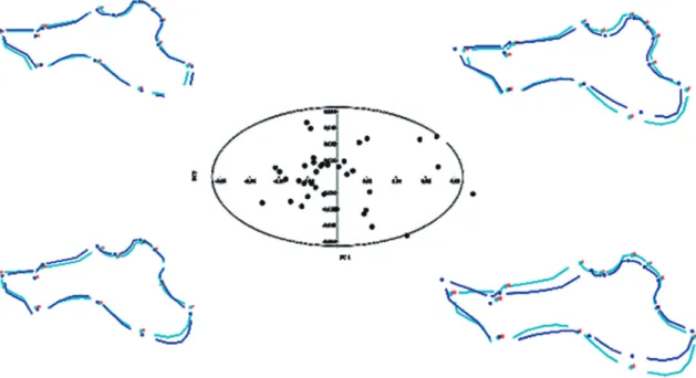
p = 0.0000). Thus, our analyses indicated significant shape changes according to size changes. The position of the specimens in the morphospace defined by the first two axes obtained by PCA analysis of mandible shape variables and visualization of related, mandibu-lar shape changes are presented in Fig. 2. The first two axes explained 54.8% (PC1 + PC2 = 33.5 + 21.3%) of the total observed variance. An additional 42.3% of variance was spread across PC3-PC23. Jolliffe cut-off was 8.734E-05. Both axes tended to distinguish between small specimens (understood as young ani-mals) and bigger ones (understood as old aniani-mals). This suggests that most of the variation is driven by shape changes during growth.
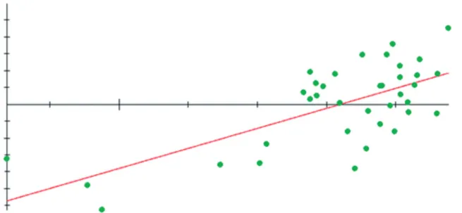
Individual regressions of shape on centroid size display the pattern of allometric shape variation with-in the size range observed (Fig. 3). As such, allometry therefore explains a substantial part of shape variation and plays an important role in determining that main modifications are located in the ramus (landmarks 4, 5, 6 and 8) and in ventral part of the corpus (land-marks 9 and 10) and diastema (land(land-marks 12 and 13) (Fig. 4).
DISCUSSION
For H. hydrochaeris there are papers concerning
gastrointestinal parasites, blood parasites, anatomical studies on miology, habitats, diets and potential use of food, morphophysiology of urogenital system… but none is concerning (at least to author’s knowledge) to allometry. This is the first study of allometry of capyba-ra focused on mandible using geometric morphometric methods. These methods can be defined as the quanti-tative description, analysis and interpretation of shape and variation of structures in biology. In a fundamental area of research, unlike the analytical approaches, the geometric one is aimed at comparison of the shapes.
In the sample studied of H. hydrochaeris there appeared a positive allometry, e.g., mandibular shape did vary according to mandibular size. Our analyses determined that mandibles exhibit positive allometry, whereby the relative size of mandibles becomes greater with increasing body size, indicating a functional disso-ciation with age (if the shape were the same throughout the life of animals, mechanical considerations, such the relative engulfment capacity and the specific physical forces at play during the feeding process, it would not increase allometrically with body size). A major com-ponent of shape variation in the mandibles of
H. hy-drochaeris is thus related to the attainment of adult size
(i.e., postnatal growth). As an integral component of
the craniomandibular apparatus, mandibles play an
FIGURE 2: Principal Component Analysis scatter plot for first two axes, which explained 54.8% (PC1 + PC2 = 33.5 + 21.3%) of the total observed variance, for Hydrochoerus hydrochaeris mandibles (n = 37). Ellipse is 95% confidence. Each dot represents one specimen. The major patterns of morphological variation in the pooled sample are reflected. Jolliffe cut-off was 8.734E-05.
important role in feeding mechanics. So the detected growth pattern seems to be related to dental eruption: the body must become elongated along its whole length to provide space for the additional teeth developed in this part, and the depth of the body must then increase, owing to increased growth of the alveolar part, to afford room for the roots of the teeth. Bony parts which enable the jaw to withstand the powerful action of the mastica-tory muscles (muscular attachment points) change less. Thus, the forces produced by the action of the mastica-tory muscle are not affected by mandibular size.
These robust scaling relationships also would al-low to predict mandible shape from fragmentary re-mains in the capybara. Furthermore, if the evolution of morphology is arguably the evolution of allometry, its changes in different sloth species could also con-tribute to a better understanding of Hydrocherus evo-lutionary relationships.
In conclusion, mandibular muscle mass in-creases during postnatal development and growth, as does the force-generating capacity of the jaw adductor muscles (notably the masseter complex).
FIGURE 4: Loadings for Principal Component 1 for the 13 mandibular landmarks analyzed (PC1 = 33.5% of the total observed variance). Landmarks that contributed less to variation are located in the ramus (landmarks 4, 5, 6 and 8) and in ventral part of the corpus (landmarks 9 and 10) and diastema (landmarks 12 and 13).
RESUMEN
El aparato masticatorio de los mamíferos es una región altamente plástica del cráneo y, por ello, sujeta a trayec-torias ontogénicas singulares. En este estudio presentamos el primer estudio descriptivo del patrón alométrico de la
mandíbula en el capibara (Hydrochoerus
hydrochae-ris), basado en el estudio de 37 especímenes. Los cambios
alométricos en la forma pura fueron analizados median-te técnicas de morfometría geométrica, permitiendo la visualización del patrón de alometría. Una regresión multivariada de la forma pura sobre el tamaño, estima-do por el logaritmo del tamaño del centroide, apareció como altamente significativo. De ello, deducimos que la variación de la forma pura en la mandíbula está rela-cionada con la llegada al tamaño adulto, es decir, con el crecimiento.
Palabras-Clave: Capibara; Mandíbula; Ontogenia;
Rodentia; Escalado.
ACKNOWLEDGEMENTS
We thank Catalina Cárdenas and Hugo Fer-nando López for access to the Instituto de Ciencias
Naturales of the Universidad Nacional de Colombia
collection, and Óscar Murillo, to the Departamento
de Biología of the Universidad del Valle collection. We
thank anonymous reviewers for their comments and suggestions.
REFERENCES
Abdala, F.; Flores, D.A. & Giannini, N.P. 2007. Postweaning Ontogeny of the Skull of Didelphis albiventris. Journal of Mammalogy, 82(1):190-200.
Bookstein, F.L. 1991. Morphometric tools for landmark data. Geometry and biology. New York, Cambridge University Press. Cardini, A. & Slice, D.E. 2004. Mandibular shape in the genus
Marmota (Rodentia, Sciuridae): a preliminary analysis using outlines. Italian Journal of Zoology, 71(1):17-25.
Cardini, A. & Thorington, R.W. 2006. Postnatal Ontogeny of Marmot (Rodentia, Sciuridae) Crania: Allometric Trajectories and Species Divergence. Journal of Mammalogy, 87(2):201-215. Chacón, J.P.; Linares, J.A.; Carrascal, J.V. & Ballesteros, J.C.
2013. Área de acción del chigüiro (Hydrochoerus isthmius) en un sistem agropecuario en Córdoba, Colombia. Revista Colom-biana de Ciencia Animal, 5(2):270-281.
Cox, P.G.; Rayfield, E.J.; Fagan, M.J.; Herrel, A.; Pataky, T.C. & Jeffery, N. 2012. Functional evolution of the feeding system in rodents. PLoS ONE, 7(4):1-11. e32699. http://doi. org/10.1371/journal.pone.0036299.
Cueto, G.R. 1999. Biología reproductiva y crecimiento del car-pincho (Hydrochoerus hydrochaeris) en cautiverio: una interpre-tación de las estrategias poblacionales. Universidad de Buenos Aires.
Dias, G.J.; Cook, R.B. & Mirhosseini, M. 2011. Influence of food consistency on growth and morphology of the mandibular condyle. Clinical Anatomy, 24(5):590-598.
Galan, A.L. 2016. Morfometría geométrica: el estudio de la for-ma y su aplicación en biología. Temas de Ciencia y Tecnología,
19(55):53-59.
Goswami, A. 2006. Notes and Comments. Cranial Modularity Shifts during Mammalian Evolution. The American Naturalist,
168(2):270-280.
Hammer, Ø.; Harper, D.A.T. & Ryan, P.D. 2001. Past: Paleon-tological Statistics Software Package for Education and Data Analysis. Palaeontologia Electronica, 4(1).
Hautier, L.; Lebrun, R.; Saksiri, S.; Michaux, J.; Vianey-Liaud, M. & Marivaux, L. 2011. Hystricognathy vs. sciurognathy in the rodent jaw: a new morphometric assessment of hystricog-nathy applied to living fossil Laonastes (Rodentia, Diatomyi-dae). PLoS ONE, 6(4)1-11. e18698. http://doi.org/10.1371/ journal.pone.0018698.
Herrel, A.; De Smet, A; Aguirre, L.F. & Aerts, P. 2008. Morphological and mechanical determinants of bite force in bats: do muscles matter? The Journal of Experimental Biology,
211:86-91.
Jamniczky, H.A. & Hallgrímsson, B. 2009. A comparison of covariance structure in wild and laboratory Muroid crania. Evolution, 63(6):1540-1556.
Ojasti, J. 1973. Estudio biológico del chiguire o capibara. Ca-racas, Ediciones del Fondo Nacional de Investigaciones Agropecuarias.
Panchetti, F.; Scalici, M.; Carpaneto, G.M. & Gibertini, G. 2008. Shape and size variations in the cranium of elephant-shrews: a morphometric contribution to a phylogenetic debate. Zoomorphology, 127(2):69-82.
Parés-Casanova, P.M.; Samuel, O.M. & Olopade, J.O. 2015. Non-functional sexually dimorphic mandibular differences in the African rodent Thryonomys swinderianus (Temminck, 1827). Annals of Biological Research, 6(10):26-31. Available at: http://scholarsresearchlibrary.com/archive.html.
Reyment, R.A. 2010. Morphometrics: An historical essay. In:
Morphometric for nonmorphometricians. New York, Springer. p. 9-25. (Lecture notes in earth sciences, 124). Available at: www.springerlink.com/index/10.1007/978-3-540-95853-6. Richtsmeier, J.T.; DeLeon, V.B. & Lele, S.R. 2002. The promise
of geometric morphometrics. American Journal of Physical Anthropology, Suppl. 35:63-91.
Rohlf, F.J. 2005. Geometric morphometrics simplified. Trends in Ecology & Evolution, 20(1):13-14.
Rohlf, F.J. 2015a. The tps series of software. Hystrix, The Italian Journal of Mammalogy, 26(1):9-12. www.italian-journal-of-mammalogy.it/article/download/11264/pdf_11264. Rohlf, F.J. 2016. tpsDig v. 2.26. New York, Stone Brook. Available
at: http://life.bio.sunysb.edu/ee/rohlf/software.html. Schumacher, G.H. 1961. Funktionelle morphologie der
kaumusku-latur. Jena, Gustav Fisher.
Sheets, H.D. 1998. IMP: CoordGen8-Coordinate Generation Utility.
Available at: www3.canisius.edu/~sheets/CoordGenManual. htm.
Sisson, S., Grossman, J.D. & Getty, R. 1982. Anatomía de los Animales Domésticos. Barcelona, Salvat Editores.
Smith, K.K. 2006. Craniofacial development in marsupial mammals: developmental origins of evolutionary change. De-velopmental Dynamics, 235(5):1181-1193.
Tagliaro, M.L.; de Oliveira, R.M.; Callegari-Jac-ques, S.M. & Jeckel-Neto, F.A. 2009. Morphological changes in the mandible of male mice associated with aging and biomechanical stimulus. Anatomical Record,
292(3):431-438.
Ulloa, A.R. 2005. Distribución del hábitat del chigüire (Hydrochaeris hydrochaeris Linne 1766) en sabanas inundables de la Estación Biológica el Frío, Venezuela. Mérida-Venezuela, Universidad de Los Andes.
Vaughan, T.A.; Ryan, J.M. & Zaplewski, N.J.C. 2000.
Mammalogy. Fort Worth, Texas, S. C. Publishing.
Wainwright, S.A.; Biggs, W.D.; Currey, J.D. & Gosline, J.M. 1976. Mechanical design in organisms. Princeton, NJ, Princeton University Press.
Wilson, L.A.B. & Sánchez-Villagra, M.R. 2011. Evolution and phylogenetic signal of growth trajectories: The case of chelid turtles. Journal of Experimental Zoology Part B: Molecular and Developmental Evolution, 316(1):50-60.
Zelditch, M.L.; Swiderski, D.L. & Sheets, D.H. 2012.
Geometric morphometrics for biologists: A Primer. 2.ed. San Diego, Elsevier Academic Press.
Aceito em: 29/11/2017 Publicado em: 20/12/2017 Editor Responsável: Marcelo Duarte
Publicado com o apoio financeiro do Programa de
Apoio às Publicações
Científicas Periódicas da USP
Produzido e diagramado na Seção de Publicações do
Museu de Zoologia da
Universidade de São Paulo
Os periódicos Papéis
A
vulsos de Zoologia
e
Arquivos de Zoologia estão licenciados sob uma Licença CC-BY