Inês Isabel Gomes Ferreira
Licenciada em Bioquímica
Study of glycosidic changes in lung
cancer: potential effectors in
hematogenous metastasis
Dissertação para obtenção do Grau de Mestre em
Bioquímica para a Saúde
Orientador: Doutora Paula Alexandra Quintela Videira, Professora Auxiliar, Faculdade de Ciências e Tecnologia da Universidade Nova de Lisboa
Co-orientador: Doutor António Bugalho, Professor Auxiliar, Faculdade de Ciências Médicas, Universidade Nova de Lisboa
Júri: A definir
Nova Medical School - Faculdade de Ciências Médicas da Universidade Nova de Lisboa
The work developed until the present date has originated:
- One Poster:
I. Ferreira; M. Carrascal; A. Bugalho; A. Fernandes; P. Baptista; P. Videira. Role of fucosyltransferase 3 in hematogenous metastasis of lung cancer. iMED 7.0 Conference
Acknowledgements
- Ao grupo de Glicoimunologia do Centro de Estudos de Doenças Crónicas (CEDOC) da Nova Medical School, por me receber no seu laboratório;
- À Professora Doutora Paula Videira, a minha orientadora, pela oportunidade oferecida, orientação prestada e por todo o apoio e ensinamentos durante esta dissertação. Ao Professor Doutor António Bugalho, o meu co-orientador, pela disponibilidade e por toda a ajuda prestada, principalmente a nível clínico;
- À Mylène Carrascal pela amizade, ajuda constante, paciência e disponibilidade para me guiar no decorrer desta tese. Um agradecimento especial por me ter ensinado a gostar “tanto”de fazer Western Blotting’s. Sem ela, tudo teria sido mais difícil;
- Ao Doutor Dário Ligeiro por me ter recebido no seu laboratório de Genética Molecular do Centro de Histocompatibilidade do Sul e pela preciosa ajuda em todo o processo de extracção de RNA das amostras e no processo de Real-Time PCR;
- À Graça Marques, Inês Iria, Leonor Rodrigues e Lili pela amizade, conversas, boa disposição, bons momentos e ajuda prestada. Um obrigado especial à Lili pela sua tentativa falhada em tentar cuidar da minha saúde e por me acordar logo de manhã com luz no laboratório;
- À minha italiana preferida Silvia Achilli pela amizade e todos os bons momentos proporcionados durante a sua estadia em Lisboa;
- A todos os restantes membros do grupo de Glicoimunologia por todos os preciosos conselhos, bons momentos e ajuda prestada no decorrer deste trabalho;
- A todos os amigos que me apoiaram no decorrer desta dissertação, em especial à minha Rainha Susy pela amizade, ajuda, apoio, conversas e por ser das pessoas mais incríveis que já conheci. À Princesa Rita por apesar de longe continuar a ser uma grande amiga e um apoio fundamental durante todo este ano. À minha afilhada Raquel por ser a melhor afilhada de sempre e por ser um Exemplo de força e coragem;
- Ao meu melhor amigo e companheiro de vida Luís Garcez por me aturar durante estes últimos anos, apoiar em todos os momentos e nunca deixar de acreditar em mim;
Abstract
Lung cancer (LC), one of the major causes of mortality related to cancer in Portugal, may lead to hematogenous metastasis. Adhesion of cancer cells to endothelium is considered one of the crucial steps involved in metastasis. In blood cells, this adhesion is initiated by endothelial selectin ligands (E-SL) that are glycoproteins or glycolipids decorated mostly with sialyl-Lewis x (sLex) and sialyl-Lewis a (sLea).
While LC has been described as expressing these sialyl Lewis antigens, its functional role in allowing LC adhesion to endothelium is still poorly understood.
We analyzed paired tumor and normal tissues samples from non-small cell lung cancer (NSCLC) patients and three LC cell lines. Immunoblotting assays with anti-sLex/sLea and E-selectin chimera demonstrated that LC tumor tissues significantly overexpress E-SL and flow cytometry results indicated that E-SL are also abundantly expressed in LC cell lines.
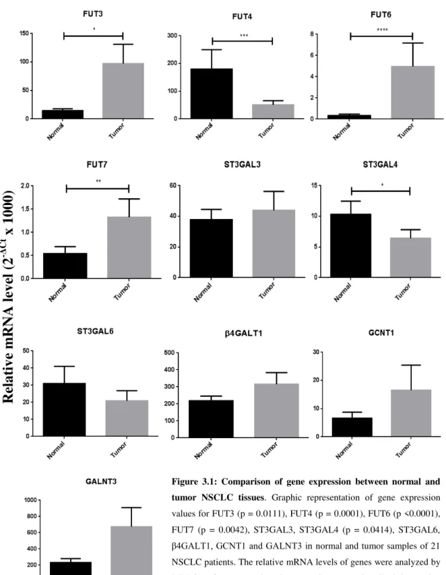
To understand the mechanism behind the overexpression of E-SL in LC tissues and cell lines, we analyzed the expression of genes involved in its biosynthesis, namely
FUT3, FUT4, FUT6, FUT7, ST3GAL3, ST3GAL4, ST3GAL6, β4GALT1, GCNT1 and
GALNT3. It was observed the overexpression of fucosyltransferases FUT3, FUT6 and FUT7 in LC tumor tissues and FUT3 in LC cell lines, being this last one correlated with an increased reactivity of the LC cells to endothelial selectins. It was described that low expression of FUT4 in tumor tissues is correlated with early stages of NSCLC. We also analyzed scaffolds proteins of sLex/sLea and it was identified the carcinoembryonic antigen as an E-SL in NSCLC.
Resumo
O cancro do pulmão (LC), uma das principais causas de mortalidade relacionada com cancro em Portugal, pode levar à formação de metástases hematogénicas. A adesão das células tumorais ao endotélio é considerada um dos passos fundamentais envolvidos na metástase. Em células sanguíneas, esta adesão é mediada por ligandos de E-selectina (E-SL), glicoproteínas ou glicolípidos decorados principalmente com sialyl-Lewis x (sLex) e sialyl-Lewis a (sLea).
Tem sido descrito a expressão destes antigénios em LC, contudo o seu papel funcional em permitir a adesão das células de LC ao endotélio é ainda pouco compreendido.
Foram analisadas amostras emparelhadas normais e tumorais de pacientes com cancro de pulmão de não-pequenas células (NSCLC) e três linhas celulares de LC.
Immunoblotting assays com anti-sLex/sLea e molécula quimérica de E-selectina
demonstraram que tecidos tumorais de LC sobreexpressam significativamente E-SL e resultados de citometria de fluxo demonstraram uma expressão elevada de E-SL nas linhas celulares.
Para compreender o mecanismo da sobreexpressão de E-SL em tecidos tumorais e linhas celulares de LC, foi analisada a expressão de genes envolvidos na biossíntese de E-SL, nomeadamente FUT3, FUT4, FUT6, FUT7, ST3GAL3, ST3GAL4,
ST3GAL6, β4GALT1, GCNT1 e GALNT3. Observou-se a sobreexpressão das
fucosiltransferases FUT3, FUT6 e FUT7 em tecidos tumorais de LC e FUT3 em linhas celulares de LC, sendo que neste último, esta expressão é correlacionada com um aumento da adesão das células de LC às selectinas endoteliais. Foi observado que uma baixa expressão de FUT4 em tecidos tumorais está associada com estadios menos avançados de NSCLC. Foram analisadas ainda proteínas decoradas com sLex/sLea, tendo-se identificado como E-SL o antigénio carcinoembrionário em NSCLC.
Index
1. Introduction ... 1
1.1 Carbohydrates ... 1
1.1.1 Glycosylation & Glycoproteins ... 2
1.1.1.1 N-glycans ... 5
1.1.1.2 O-glycans ... 6
1.1.2 Altered glycosylation in cancer ... 7
1.1.2.1 Sialyl-Lewis x and a antigens... 8
1.1.2.1.1 Role in metastasis ... 10
1.2 Lung cancer ... 12
1.2.1 Non-small cell lung cancer ... 13
1.2.1.1 Etiology ... 13
1.2.1.2 Diagnosis and staging ... 14
1.2.1.3 Metastasis in NSCLC ... 15
1.2.1.4 Treatment ... 15
1.3 Context and objectives of the work developed for this thesis ... 16
2. Material and methods ... 19
2.1 Culture and maintenance of cell lines ... 19
2.2 Biological samples ... 20
2.2.1 Tissues ... 20
2.3 Techniques ... 20
2.3.1 Real-Time Polymerase Chain Reaction (RT- PCR) ... 20
2.3.2 Blotting techniques: western blotting and dot blotting ... 22
2.3.3 Flow cytometry ... 23
2.3.4 Adhesion Assays ... 24
2.3.5 Immunoprecipitation ... 24
2.4 Glycosidic characterization of lung cancer cells and NSCLC tissues ... 25
2.4.1 Analysis of gene expression by RT-PCR ... 25
2.4.1.1 Total RNA extraction and reverse transcription to cDNA ... 25
2.4.1.2 Real Time-PCR ... 26
2.4.2 Phenotypic analysis ... 27
2.4.2.1 Expression of sLex and sLea antigens in tissues samples by dot blotting ... 27
2.4.2.2 Expression of E-SL, sLex/sLea antigen, mucin 1 (MUC1), CD44 and carcinoembryonic antigen (CEA) in tissues samples by western blotting ... 28
2.4.2.3 Immunoprecipitation of CEA in tissue samples ... 31
2.4.2.4 Cell lines staining with antibodies by flow cytometry ... 33
2.4.3. Functional analysis of E-selectin ligands in LC cell lines by adhesion assay ... 34
3. Results ... 36
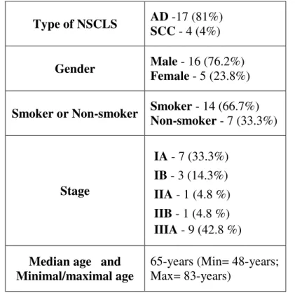
3.1. Patient’s characteristics... 36
3.2 Glycosidic characterization of normal and tumor tissues from NSCLC patients ... 37
3.2.1 Analysis of gene expression by RT-PCR ... 37
3.2.2 Expression of sLex/sLea antigens in tissues samples by dot blotting ... 43
3.2.3 Expression of sLex/sLea antigens and E-SL in tissues samples by western blotting 46 3.2.4 Expression of glycoproteins MUC1 and CD44 in tissues samples by western blotting ... 49
3.2.5 Expression of carcinoembryonic antigen in tissues samples by western blotting ... 51
3.2.5.1 Confirmation of CEA as E-SL in patient 7 by immunoprecipitation of CEA and western blotting ... 54
3.3 Glycosidic characterization of lung cancer cell lines ... 57
3.3.1 Analysis of gene expression by RT-PCR ... 57
3.3.2 Phenotypic analysis by flow cytometry ... 58
3.3.3 Functional analysis of E-selectin ligands by adhesion assay ... 60
4. Discussion and conclusions ... 62
4.1 FUT3, FUT6 and FUT7 are upregulated, while FUT4 and ST3GAL4 are downregulated in NSCLC tumor tissues compared with normal NSCLC tissues. ... 63
4.2 Increased expression of FUT4 is associated to a more advanced stage of NSCLC.... 64
4.3 sLex/sLea are more expressed in NSCLC tumor tissues than normal tissues. ... 65
4.4 Increased expression of ST3GAL3 and GCNT1 is related with enhanced expression of sLex/sLea antigens in tumor NSCLC tissues. ... 65
4.5 E-selectin ligands in lung cancer: CEA is an N-glycan E-SL in NSCLC while CD44 and MUC1 don’t seem to be. ... 66
4.6 Enhanced expression of sLex/sLea on lung cancer cells which may be attributed to elevated levels of FUT3 correlates with increased ability to adhere to E-selectin. ... 68
4.7 Future perspectives ... 69
4.8 General conclusions ... 71
5. References ... 72
Index of figures
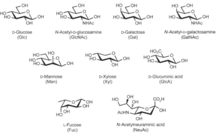
Figure 1.1: Common monosaccharides found in mammals.. ... 1
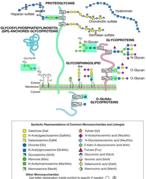
Figure 1.2: Common classes of animal glycans. ... 3
Figure 1.3: Biosynthesis of N-glycans. ... 6
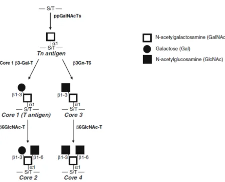
Figure 1.4: Common O-GalNAc glycan core structures biosynthetic pathways.. ... 7
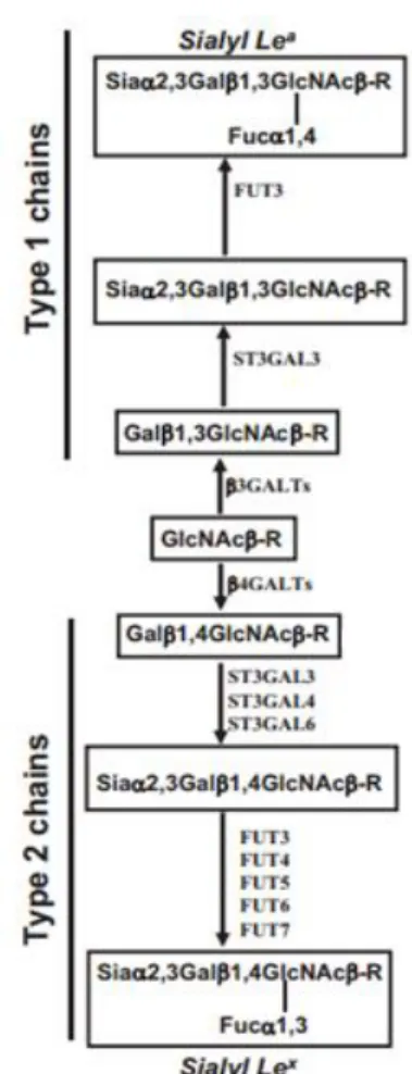
Figure 1.5: Structures and enzymes involved on biosynthesis of sialyl-Lewis x and sialyl-Lewis a antigens. ... 9
Figure 1.6: Schematic representation of the multi-step process of metastasis in cancer... ... 12
Figure 3.1: Comparison of gene expression between normal and tumor NSCLC tissues...39
Figure 3.2: Comparison of the expression of fucosyltransferases and sialyltransferasse in lung tumor tissues. ... 40
Figure 3.3: Comparison of fucosyltransferases and sialyltransferases expression in tumors, according to smoke habits and age. ... 41
Figure 3.4: Comparison of the fucosyltransferases and sialyltransferases expression in tumors, according to tumor stage. ... 42
Figure 3.5: Expression of sLex and sLea in NSCLC patient’s matched normal/tumor tissue pairs.. ... 44
Figure 3.6: Comparison of ST3GAL3 and GCNT1 expression in tumors, according expression of sLex/sLea. ... 46
Figure 3.7: Western Blotting analysis of E-SL proteins present in tumor lysates from eight NSCLC patients. ... 47
Figure 3.8: Western blotting analysis of CD44 present in tumor lysates from eight NSCLC patients. ... 51
Figure 3.9: Western blotting analysis of CEA proteins present in tumor lysates from eight NSCLC patients ... 52
Figure 3.10: Comparison of western blotting analysis with HECA-452 staining, E-Ig staining and CEA staining in tumor lysates from four NSCLC patients. ... 53
Figure 3.11: Western blotting analysis of CEA proteins present in tumor lysates from eight NSCLC patients. ... 54
Figure 3.12: Western blotting analysis of CEA immunoprecipitate (IP-CEA) from tumor lysate of patient number 7. ... 55
Index of tables
Table 1.1: The most common glycosyltransferases and nucleotide donors in animal cells ... 4 Table 2.1: List of genes analyzed in tissue samples and LC cell lines by RT-PCR………26 Table 2.2: List of molecules/antibodies and respective dilutions used in western blotting experiences ... 30 Table 3.1: Clinical characteristics of patients enrolled in this study: type of NSCLC, gender,
Index of appendixes
Appendix 6.1: TNM staging of Lung Cancer...81
Appendix 6.2: Constitution of buffer solutions used in this work...82
Appendix 6.3:Patient’s characteristics…...83
Appendix 6.4: Data sets from cBioPortal database...84
Appendix 6.5: Statistical analysis of Spearman correlations...87
Appendix 6.6: Values of NSCLC lysates quantification...88
Abbreviations
AD - Adenocarcinoma
ALK - Anaplastic lymphoma kinase Asn - Asparagine
β4GALT1 - UDP-Gal:betaGlcNAc beta 1,4- galactosyltransferase, polypeptide 1 CD62E - CD62 antigen-like family member E, also known as E-selectin
CD62L - CD62 antigen-like family member L, also known as L-selectin CD62P - CD62 antigen-like family member P, also known as P-selectin CEA - Carcinoembryonic antigen
CLA - Cutaneous Lymphocyte-associated Antigen CMP - Cytosine Monophosphate
Ct - Threshold cycle
DNA -Deoxyribonucleic acid
EDTA - Ethylenediaminetetraacetic acid EGF - Epidermal growth factor
EGFR - Epidermal growth factor receptor
E-Ig - Recombinant mouse E-Selectin/CD62E Fc chimera ER -Endoplasmic reticulum
E-SL - E-selectin ligands FBS - Fetal Bovine Serum FSC - Forward scatter FUT - Fucosyltransferase
FUT3 -Fucosyltransferase 3 (galactoside 3(4)-L-fucosyltransferase, Lewis blood group) FUT4 -Fucosyltransferase 4 (alpha (1,3) fucosyltransferase, myeloid-specific)
FUT6 -Fucosyltransferase 6 (alpha (1,3) fucosyltransferase) FUT7 -Fucosyltransferase 7 (alpha (1,3) fucosyltransferase) GalNAc - N-Acetyl- D- galactosamine
GlcNAc - N-Acetyl-D-glucosamine GT - Glycosyltransferase
HCELL - Hematopoietic Cell E-selectin/L-selectin Ligand IP - Immunoprecipitation
IP-CEA - CEA immunoprecipitate LC - Lung cancer
MFI - Median Intensity Fluorescence mRNA - Messenger RNA
MUC1 - Mucin 1
NSCLC - Non-small cell lung cancer NST - Nucleotide Sugar Transporter PNGase F - Peptide-N-glycosidase F PSGL-1 - P-selectin glycoprotein ligand-1 RNA - Ribonucleic acid
RT - Reverse transcription
RT-PCR - Real-time polymerase chain reaction SCC - Squamous cell lung cancer
SCLC - Small cell lung cancer
SDS-PAGE - Sodium Dodecyl Sulfate Polyacrylamide Gel Electrophoresis Ser - Serine
Sia - Sialic acid sLea -Sialyl-Lewis a sLex -Sialyl-Lewisx SSC - Side scatter ST - Sialyltransferase
ST3GAL3 - ST3 beta-galactoside alpha-2,3-sialyltransferase 3 ST3GAL4 - ST3 beta-galactoside alpha-2,3-sialyltransferase 4 ST3GAL6 - ST3 beta-galactoside alpha-2,3-sialyltransferase 6 Thr - Threonine
TNM - Tumor-node-metastasis
1. Introduction
1.1 Carbohydrates
Carbohydrates are one of the four main classes of organic molecules in living systems, besides the lipids, nucleic acids and proteins (Ghazarian, Idoni and Oppenheimer, 2011). The basic structural units of carbohydrates are monosaccharides. There are nine monosaccharides commonly found in mammals (Figure 1.1).
Monosaccharides are linked together to form oligosaccharides (2-20 monosaccharides residues linked by glycosidic linkages) or polysaccharides (long chains of monosaccharides linked by glycosidic linkages). In most cases, people use the term “glycan” instead of carbohydrate to refer any form of mono-, oligo- or polysaccharide free or covalently attached to another molecule, forming a glycoconjugate.
The glycome, analogous to genome, transcriptome and proteome, involves all the glycans that can be synthesized by an organism. Different types of cells, tissues and organisms, express different types of glycans and the glycome also varies during stages of development/differentiation and during malign transformation (Tateno et al., 2007). The glycome is estimated to be 10-104 times bigger than the proteome, depending on species (Freeze, 2006).
Figure 1.1: Common monosaccharides found in mammals. The abbreviation of
The structural diversity of glycans is enormous and results of several carbohydrate characteristics: the number of available monosaccharides building blocks, glycosidic bond position, anomeric configuration (α or β glycosidic linkage), the capacity of glycans to form branched or linear scores structures and carbohydrates modifications (Ghazarian, Idoni and Oppenheimer, 2011).
The glycan structures are not encoded directly in the genome and for that reason, they are called secondary gene products. There are several genes in the genome that after transcription and translation, generate transporters and enzymes (like glycosidases and glycosyltransferases) responsible for the biosynthesis and the assembly of glycans (Varki et al., 2009; Taylor and Drickamer, 2011) .
Glycans are involved in the regulation of multiple cellular mechanisms like: differentiation, cell adhesion, contact inhibition, receptor activation, endocytosis, cell-cell recognition, cell-cell growth and development, anticoagulation, host immune response, metastasis, molecular trafficking and clearance, signal transduction, membrane rigidity, host-pathogen interaction during infection and disease development (Varki et al., 2009; Ghazarian, Idoni and Oppenheimer, 2011; Ohtsubo and Marth, 2006; Raman et al., 2005). Glycans can also affect the intrinsic properties of proteins which they are linked to, such as their solubility in water, their proper protein folding and functional group orientation and protection from proteases (Dall’Olio, 1996).
1.1.1 Glycosylation & Glycoproteins
Glycosylation is a process of the enzymatic addition of glycans to a noncarbohydrate moiety, such as proteins, lipids or other organic compound. It is the most complex form of post-translational modification of proteins. It is important not confuse glycosylation with glycation which is a nonenzymatic and irreversible process of adding glycans, that is elevated in several diseases (Ohtsubo and Marth, 2006).
(Hadley et al., 2014). Glycoproteins are also found at the extracellular matrix that surrounds cells and can be secreted into biological fluids, for example the serum (Taylor and Drickamer, 2011).
The biosynthesis of glycoproteins requires activation of monosaccharides to nucleotide-sugars donors. First of all, monosaccharides are obtained by the cell from dietary sources, salvage processes from glycoconjugates degraded within cells and endogenous conversion from other monosaccharides (Yarema and Bertozzi, 2001). The conversion of monosaccharides into nucleotide-sugars donors involves normally the phosphorylation of one or more hydroxyl groups of the monosaccharides. The
nucleotide-sugar donor contains the energy required for the transference of a monosaccharide residue from a nucleotide-sugar donor to the hydroxyl group of an acceptor substrate (in this case, oligosaccharide, monosaccharide or protein), by action of a large group of enzymes called glycosyltransferases (GTs) (Palcic, 1994). GTs are specific for a nucleotide-sugar donor but may recognize more than one different acceptor (Varki et al., 2009). GTs act sequentially, so the product of one GT may yields an acceptor substrate for the action of another GT. They are classified according to the sugar they transfer (Table 1.1).
Acceptor + Glycosyl DonorGlycosyltransferase→ Glycosylated acceptor + Nucleotide
Table 1.1:The most common glycosyltransferases and nucleotide donors in animal cells. UDP - Uridine
Diphosphate; CMP - Cytosine Monophosphate; GDP - Guanine Diphosphate.
Besides GTs, glycosidases are also involved in the biosynthesis of glycans. These enzymes are responsible for the hydrolysis of glycosidic linkages to remove sugars from proteins and play an important role in glycan processing in the Golgi apparatus and endoplasmic reticulum (ER), as well as in the modulation of the glycan landscape in the extracellular milieu.
In eukaryotes, most glycosylation reactions occur predominantly in Golgi apparatus and ER lumen. On the other hand, nucleotide-sugars are synthetized in cytosol with exception for CMP-sialic acid synthetized in nucleus (Hadley et al., 2014).
Monosaccharide Glycosyltransferase family Nucleotide donor
Glucose Glucosyltransferase UDP
Galactose Galactosyltransferase UDP
N-acetylglucosamine N-acetylglucosaminyltransferase UDP
N-acetylgalactosamine N-acetylgalactosaminyltransferase UDP
Xylose Xylosyltransferase UDP
Glucuronic acid Glucuroniltransferase UDP
Sialic acid Sialyltransferase CMP
Mannose Manosyltransferase GDP
For this reason, the nucleotide-sugars must be transported into the ER lumen or Golgi. This transport is performed by proteins known as nucleotide sugar transporters (NSTs). NSTs are very hydrophobic proteins that function as antiporters: they exchange cytosolic nucleotide sugars into the lumen of the organelles for the corresponding luminal nucleoside monophosphate, such as CMP for CMP-sugars, UMP for UDP-sugars and GMP for GDP-UDP-sugars (Hadley et al., 2014).
As previously mentioned, the most common types of glycoproteins are N -glycans and O-glycans, which will be described below.
1.1.1.1 N-glycans
In eukaryotic organisms, N-glycans are the most studied form of glycoproteins (90% of glycoproteins are N-glycosylated).
N-glycans are covalently attached to proteins at amide nitrogen of asparagine
(Asn) side chains. There is a minimal consensus sequence that can accept an N-glycan: Asn-X-Ser/Thr (starts with Asn, followed by any amino acid except proline (Pro) and ends with serine (Ser) or threonine (Thr)). The most common sugar linked to Asn is N-acetylglucosamine (GlcNAc), with a β configuration (GlcNAcβ1-Asn).
N-glycans biosynthesis (figure 1.3) begins with the formation of a lipid-linked
oligosaccharide (fourteen sugars covalently attached to a lipid dolichol) on the cytoplasmic and luminal face of ER. After that, the entire oligosaccharide is transferred
en bloc to the Asn residue of a nascent polypeptide by a multi-enzyme complex named
Figure 1.3: Biosynthesis of N-glycans. OST, oligosaccharyltransferase; Glcase, glucosidase; UGGT, UDP– glucose-glycoprotein glucosyltransferase; Hmt1p, HnRNP methyltransferase 1; Mnl1p, mannosidase-like protein 1; N-Gase, N-glucosidase; Man, mannosidase; GnT, N-acetyl glucosaminyltransferase; CNX, calnexin. Adapted from Wolfert and Boons, 2013.
1.1.1.2 O-glycans
Several types of O-glycans have been identified but the most common form is the mucin-type O-linked glycans, also called O-GalNAc glycans. Mucins are heavily O -glycosylated glycoproteins (molecular weight > 200kDa) that can be soluble, secreted or expressed in the membrane. They are present at many epithelial surfaces such as respiratory, reproductive and gastro-intestinal tracts and play an important role in the protection against pathogens.
O-glycans biosynthesis begins in cis Golgi apparatus with the binding of
1.1.2 Altered glycosylation in cancer
Alterations in glycosylation are one of the main characteristics associated with
malignant transformation and tumor progression. Cancer progression involves many steps summarize as follows: alterations in extracellular and intercellular signaling pathways, cell growth, detachment of the tumor cells from the primary tumor into the circulation, extravasation of surrounding tissues, cell proliferation and colonization at distant sites (metastasis) (Häuselmann and Borsig, 2014; Reymond, D'Água and Ridley, 2013).
In cancer cells, changes in glycans can take a variety of forms: loss or excessive expression of certain structures, incomplete or truncated glycans and less frequent, the appearance of new antigens. Typically, there are modifications in the expression levels of GTs that lead to the expression of those altered glycans. Besides GTs, modifications in other molecules like transporters and sugar-nucleotides can also alter the phenotype of cancer cells (Kobata and Amano, 2005).
Altered glycosylation in N-glycans is normally associated with an increase in
β1,6-branching caused by the enhanced expression of β1– 6-N-acetylglucosaminyltransferase-5 (GlcNAcT-V) (Dennis, Granovsky and Warren, 1999;
Varki et al., 2009). In cancers with epithelial origin, mucins are the major carriers of incomplete glycosylation in the O-linked pathways, that results in the expression of antigens in cell’s surface such as sialyl-Tn (sTn, addiction of sialic acid to Tn antigen), sialyl-T (sT, addiction of sialic acid to T antigen), Tn and T antigens, considered tumor-associated carbohydrate antigens (TACA) (Tarp and Clausen, 2008).
This master thesis focuses on one of the most important families of tumor antigens, which include sialyl-Lewis x (sLex) and sialyl-Lewis a (sLea) glycans.
1.1.2.1 Sialyl-Lewis x and a antigens
Lewis epitopes are synthesized by a series of GTs including N-acetilglucosaminyltransferases, galactosyltransferases, sialyltransferases (STs) and fucosyltransferases (FUTs). The sLea and sLex antigens result from mono-fucosylated substitution of the α-2,3- linked sialic acid containing type 1 or type 2 chains (Dall’Olio
et al., 2012; Dall’Olio et al., 2014), respectively (Figure 1.5). These two structures are
found at terminal ends mainly on the β-1,6 branching of N-glycans or O-linked glycans
attached to glycoproteins and sometimes on glycolipids. Besides cell surface glycoconjugates, these antigens can also be detected in serum.
Clinical studies show an overexpression of sLex and sLea antigens on the surface of tumor cells including colon, gastric, pancreatic and lung cancer (Häuselmann and Borsig, 2014; Mizuguchi et al., 2007; Nakamori et al., 1993; Tozawa et al., 2005) and sometimes their overexpression is correlated with a poor prognosis. Nowadays, sLea is used as a tumor biomarker in pancreatic and colon cancer (Ugorski and Laskowska, 2002).
The terminal steps of the biosynthesis of sLex and sLea involve α 1 3/4 - fucosyltransferases and α 2 3 - sialyltransferases.
FUTs catalyze the transfer of a fucose residue from GDP-α-L-fucose to an acceptor, normally galactose or GlcNAc. This family is composed by three main sub-families: α 1 2 FUT, α 1 3 FUT and α 1 6 FUT. 1 3 FUTs (composed by FUT3, FUT4, FUT5, FUT6, FUT7 and FUT9) represent the most important and critical enzymes involved in the last step of the biosynthesis of sialyl Lewis antigens (Chen, 2011).
Typically, one GT only produces one specific glycosidic linkage but FUT3 can produce two glycosidic linkages: α 1 3 in sLex and α 1 4 in sLea. Importantly, FUT3 is the only enzyme able to synthetize sLea epitope (Dall’Olio et al., 2012).
Although FUT4 can synthetize sLex antigen with a weak activity, it is generally involved in the formation of nonsialyl Lewis antigens, as is FUT9 (Mollicone, Cailleau
Figure 1.5:Structures and enzymes involved on biosynthesis of sialyl-Lewis x and sialyl-Lewis a antigens. In the figure, sialyl-Lewis x is denominated by Sialyl Lex and sialyl-Lewis a is denominated
and Oriol, 1995). FUT6 is an important enzyme involved in the synthesis of type 2 chain structures and FUT7 is highly expressed on leukocytes and is responsible for the sLex increase in leukemia cell surfaces and other types of tumors (Chen, 2011).
Related to ST family, ST3GALs transfer sialyl group from CMP-Sia to galactose, forming an α 2 3 linkage in the final product. ST3GAL family is divided into 6 subtypes. ST3GAL3 uses as substrate type 1 and type 2 chains although it prefers the former. ST3GAL4 and ST3GAL6 prefer type 2 chain as substrate (Chen, 2011).
Increased levels of FUTs and STs have been found in various types of tumors and this expression is correlated with the level of sLex and sLea antigens (Carvalho et
al., 2010; Pérez-Garay et al., 2013; Vajaria et al., 2014) .
1.1.2.1.1 Role in metastasis
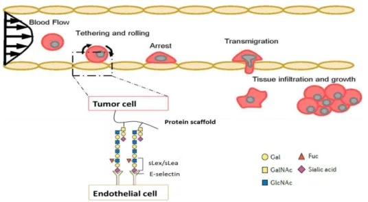
Tumor cells mimic the process of extravasation used mainly by leukocytes that migrate from the blood to reach sites of injury, infection or inflammation (Strell and Entschladen, 2008). There are four main steps in the extravasation process: tethering and rolling, integrin activation, firm adhesion and transendothelial migration (also called diapedesis). Tethering is characterized by the first contact of the leukocytes with activated endothelium, resulting in rolling of the cells along the surface of the endothelium. Tethering and rolling of leukocytes are mediated by selectins and their glycoprotein ligands. After that, rolling leukocytes in response to specificchemokines and cytokines activate their integrins that leads to a firm adhesion to the endothelium and a following transendothelial migration to the injury site (Ley et al., 2007; Reymond, D'Água and Ridley, 2013; Strell and Entschladen, 2008).
stored in Weibel-Palade bodies and α-granules, respectively. In response to inflammatory cytokines, P-selectin is translocated to cell surface in endothelial cells. L-selectin is expressed in the most of leukocytes and E-L-selectin is expressed by activated endothelial cells.
The minimal recognition motif for all three selectins is mainly the sLex and/or sLea antigens, so the interaction between cancer cells and selectins is possible because of the presence of these sialylated and fucosylated structures known as selectin ligands on the cell surface of tumor cells (figure 1.6) (Barthel et al., 2007; Gout, Tremblay and Huot, 2008; Läubli and Borsig, 2010) . Of all selectins, E-selectin (107 - 115kDa cell surface glycoprotein) is the major receptor expressed on activated endothelial cells that are involved in adhesion events during metastasis although P-selectin and L-selectin can also contribute for the process (Gout, Tremblay and Huot, 2008; Kannagi et al., 2004). The presence of E-selectin ligands (E-SL) on tumor cells is correlated with high adhesion to activated endothelium. Several E-SL carriers have been identified on tumor cells such as P-selectin glycoprotein ligand-1 (PSGL-1), a glycoform of CD44 known as hematopoietic cell E-selectin/L-selectin (HCELL), several mucins, dead receptor-3 and CD24 among others (Barthel et al., 2007; Burdick et al., 2012; Häuselmann and Borsig, 2014; Läubli and Borsig, 2010).
1.2 Lung cancer
Lung cancer (LC) is one of the most common forms of cancer (approximately 1.6 million new cases of cancer annually) and the leading cause of death related to cancer in the world, with highest overall prevalence in industrialized regions, such as Europe and North America (Alberg, Brock and Samet, 2005; Cruz, Tanoue and Matthay, 2011). In Portugal it is responsible for 4.200 deaths/year (first cause of cancer related mortality). It occurs predominately in persons between 50-70 years and is more common in men than in women. The global situation is not encouraging, with an overall 5-years survival rate estimated at only 17% in developing countries (American Cancer Society, 2014).
It is characterized by the growth of abnormal cells that starts in the lungs. Over time, these cells continue to grow and divide, forming a mass of tissue called tumor. LC cells can spread to other parts of the body, through blood vessels or lymph vessels and can form new tumors in other tissues (metastasis).
Historically, it was divided in two major types of LC: Small cell lung cancer (SCLC) and Non-small cell lung cancer (NSCLC).
SCLC, as the name indicates, is characterized by small cells that normally spread quickly through the body. It represents 15% of all LC cases, almost always
Figure 1.6: Schematic representation of the multi-step process of metastasis in cancer. Adapted from
caused by smoking and typically starts in the bronchi close to the center of the chest. The prognosis of this type of cancer is very poor and there are only two stages in SCLC: limited (tumor only on one side of the chest) and extensive stage (Kitamura et al., 2008).
1.2.1 Non-small cell lung cancer
It is the most common type of LC comprising 85% of all LC cases and is divided in three major subtypes: adenocarcinoma (AD), squamous cell carcinoma (SCC) and large cell carcinoma. These cancer cells tend to grow and spread more slowly than SCLC.
AD represents 40% of LC cases and usually occurs in a peripheral location within the lung. This subtype of cancer arises from mucus-producing cells in bronchial mucosal glands and it is the most common type of cancer in non-smokers. Analysis by immunohistochemistry shows 89% positive for thyroid transcription factor (TTF-1), being a useful marker in the diagnosis of tumors with lung origin (Teh and Belcher, 2014).
SCC usually occurs in the central parts of the lungs, representing 25% of LC cases. It develops in squamous cells (flat cells that line the airways). These cells have an irregular surface, characterized by the presence of keratin pearls. Unlike AD, immunohistochemistry of SCC shows negative for TTF-1 (Teh and Belcher, 2014).
Finally, large cell carcinoma (10% of LC cases) manifests as a peripheral lesion that has a high tendency to metastasize. The remaining 10% of the NSCLC cases correspond to other subtypes much less common or cases when a more specific diagnosis cannot be made (Teh and Belcher, 2014).
1.2.1.1 Etiology
secondhand smoke (passive smoke inhalation) can increase the risk of developing LC by 30% (Cruz, Tanoue and Matthay, 2011; Groot and Munden, 2012).
Other features can also play an important role in LC such as inherited factors (responsible for 8% of LC cases), atmospheric pollution and exposure to asbestos, radon gas or other chemicals. People with non-malignant lung diseases have a high risk of developing LC.
1.2.1.2 Diagnosis and staging
The symptoms of NSCLC may take a long time to appear and in general, LC is often confused with other less serious conditions. Normally, this type of cancer only produces symptoms in an advanced stage of the disease, so more than 50% of the patients are diagnosed at an advanced stage, which leads to a poor prognosis and survival.
After a comprehensive history and physical exam, a chest x-ray or computed tomography are initially performed. To confirm a diagnosis of cancer, the cells or tissues have to be sampled, normally by minimally invasive methods such as bronchoscopy, transthoracic needle biopsy or thoracentesis (depending on the primary lesion location and potential metastasis).
1.2.1.3 Metastasis in NSCLC
Metastatic LC occurs when tumor cells escape from the lung and travel to other parts of the body through blood (hematogenous metastasis) or lymph system. It can be metastatic at the time of diagnosis or during/following the treatment.
LC is the most common cause of brain metastases. Up to 33% of patients with NSCLC develop symptomatic brain metastases during the disease. Besides the brain, LC cells frequently metastasize to the other lung, adrenal glands, bone and liver (Riihimäki et al., 2014; Stenbygaard et al., 1999).
Patients in stage IV are often diagnosed with multiple disseminated metastases (Schuchert e Luketich, 2003).
Metastatic patterns varied depending on histological subtype, gender and age at the time of diagnosis of the disease, however it was reported that SCC is less predisposed to metastasize than AD and the last one prefers spreading to the bone. Survival in metastatic LC is worst in patients with liver or bone metastases (Riihimäki
et al., 2014).
According to literature, there were alterations in some tumor suppressor proteins such as p16 that occurs only in metastatic NSCLC and not in primary NSCLC. Besides that, mutations on oncogenes also affect the pattern of LC spread. NSCLC patients with mutations in anaplastic lymphoma kinase (ALK) were more likely to have pericardial or pleural metastases whereas mutations in ALK and epidermal growth factor receptor (EGFR) were more likely to develop liver metastases (Aisner and Marshall, 2012; Doebele et al., 2012).
1.2.1.4 Treatment
Overall, there are three main treatments available for NSCLC that can be used separately or in combination: surgery, radiotherapy and chemotherapy. Depending on the stage of disease, the patients are treated in different ways.
Chemotherapy is characterized by anti-cancer drugs that are delivery into the body. This therapy is used in almost 80% of the patients at some point of the disease: before surgery as a neo-adjuvant therapy, after surgery as adjuvant therapy trying to kill any tumor cells that may have been left behind and for more advanced stages as the main treatment. There are several drugs used in NSCLC treatment but American Society for Clinical Oncology (ASCO) recommends a platinum combination for first-line treatment (Reck et al., 2013).
For advanced stages (stage III and IV), tumors are rarely cured and the main aims are improve and extend the quality of life.
Nowadays, clinical trials are focused on targeted therapy, blocking specific genes or proteins that contribute to cancer survival. This strategy may reduce treatment side effects and is less harmful to healthy cells. For example, AD patients have EGFR mutations in 25% of the cases. Mutations in this receptor result in tumor cells that proliferate uncontrollably. In this case, a target therapy can be used including EGFR inhibition mainly by whole ErbB family blockers (Langer, 2004). Target therapy against angiogenesis is another strategy to stop cancer. Angiogenesis which is characterized by the development of new blood vessels used by tumor cells depends on the interaction of growth factors like vascular endothelial growth factor (VEGF) or fibroblast growth factor (FGF) and respective receptors (VEGFR and FGFR). Inhibition of these molecules may offer a direct anti-tumor effect (Alberg et al., 2005).
1.3 Context and objectives of the work developed for this thesis
Currently, LC is the major cause of mortality related to cancer in Portugal. The critical pathophysiological pathways to the development of NSCLC are barely known. The high incidence of mortality along with the need for an early diagnosis and a proper staging of the disease, determines the urgency to find mechanisms associated with LC and with the development of LC metastasis. A major clinical hurdle that contributes to LC mortality is the metastatic spread of primary tumor cells. Nevertheless the mechanisms triggering hematogenous metastasis of LC are unclear.
have the potential of binding to E-selectin expressed on activated endothelial cells. However, its functional role in allowing LC adhesion to endothelium is still poorly understood.
This thesis had as main aim the identification of new glycan-based biomarkers, in particular sLex and sLea, in NSCLC. It envisaged that a better understanding of their pathophysiological role in tumor cell adhesion to E-selectin, may give us hints to predict tumor progression and metastasis formation. A better understanding of glycosidic changes taking place throughout the tumor progression will help the identification of new diagnostic/prognosis biomarkers and potential therapeutic targets. Thus, this work was outlined in two specific topics:
1-Identification of glycosidic antigens (sLea and sLex) expressed in normal and NSCLC tissues. Normal and tumor matched samples were collected from 48
patients submitted to thoracic surgery based on suspicion of LC. This topic had the following secondary objectives:
a) Phenotypic analysis of these glycosidic antigens by Dot Blotting to analyze the expression of E-SL in matched normal and tumor LC tissues.
b) Expression analysis of several genes involved in the biosynthesis of sLex and sLea antigens in matched normal and tumor tissues of NSCLC patients by Real-Time Polymerase Chain Reaction (RT-PCR), then deducting the altered biosynthetic pathways. We also analyzed the correlation between the gene expression and the expression of sLex and sLea.
2 -Identification of sLex and sLea in NSCLC cell lines, having the following
secondary objectives:
a) Analysis by RT-PCR the expression of genes involved in sLex and sLea biosynthesis in commercial NSCLC cell lines (H292, H1299 and A549).
b) Phenotypic analysis of E-SL by flow cytometry.
2. Material and methods
2.1 Culture and maintenance of cell lines
For this work, were used three cell lines derived from non-small cell lung cancer (A549, H1299 and H292) kindly provided by professor Pi-Wang Cheng, from University of Nebraska Medical Center, United States of America. We also used a cell line of colorectal cancer (LS 174T) provided by Professor Fabio Dall’Olio from University of Bologna, Italy. All the cell lines are adherent to plastic surfaces charged negatively and grew up in culture flasks in an incubator with a humidified atmosphere of 5% CO2 at 37ºC.
The cell line A549 (ATCC® CLL-185™) is derived from a 58 years old Caucasian male lung carcinoma. The cell line H292 (ATCC® CRL-1848™) is derived from a cervical node metastasis of a pulmonary mucoepidermoid carcinoma in a 32 years old Negro female. The cell line H1299 (ATCC® CRL-5803™) is derived from a lymph node metastasis of NSCLC in a 43 years old Caucasian male. The cell line LS 174T (ATCC® CL-188™) was established from a Duke’s type B adenocarcinoma of colon in a 58 years old Caucasian female.
H1299 and H292 cell lines were cultivated in Roswell Park Memorial Institute medium-1640 (RPMI-1640), A549 in Dulbecco’s Modified Eagle’s Medium (DMEM) and LS 1474T in Minimum Essential Medium (MEM), all from Sigma-Aldrich. The basal medium were supplemented with 10% of heat inactivated fetal bovine serum (FBS) from Gibco™, 2mM of L-glutamine (Gibco™) and 100µg/mL of
Penicillin/Streptomycin (Gibco™). RPMI-1640 medium was also supplemented with
1mM of sodium pyruvate (Gibco™) and 0.1mM of MEM non-essential amino acids solution (Gibco™).
LC cells lines were cultivated in T-25cm2 flasks and LS 1474T in T-75cm2 flasks. The culture medium of the cell lines was renovated each two/three days and the cells were normally detached from the flask at a confluence about 70-80% and passed into new flasks, according to the following protocol:
0.25% Trypsin- Ethylenediaminetetraacetic acid solution (Trypsin-EDTA solution)
from Gibco™, acting for 5min in the incubator, at 37ºC. Then we put complete medium
into the T-flask with the released cells, to stop the action of Trypsin-EDTA and collected all the content of T-flask to a 15mL falcon. The cell’s pellet was obtained by a centrifugation at 1200rpm for 5min and it was ressuspended in new medium (6mL for T-25cm2 and 15mL for T-75cm2). Cells were split in new T-flasks, normally LC cell lines in a proportion of 1:3 and the LS 174T cell line 1:4.
These cell lines were also stored in cryogenic tubes at -80ºC and in liquid nitrogen, using a freezing solution constituted by simple RPMI-1640 medium supplemented with 5% of dimethyl sulfoxide (DMSO) from Merck and 20% of FBS.
2.2 Biological samples
2.2.1 Tissues
Samples collected from 48 patients submitted to thoracic surgery based on suspicion of LC were performed at Unidade de Técnicas Invasivas Pneumológicas in Hospital Pulido Valente (Lisboa), under coordination of Dr. António Bugalho.
For each patient, fragments of tumor tissue and normal pulmonary tissue (local as far as possible of the tumor, likely in a different pulmonary lobule) with a size above 0.5 cm3 were collected in duplicate for sterile cryogenic tubes, immediately placed in liquid nitrogen.
For this master thesis, normal and tumor tissue samples were divided in two pieces of approximately 40mg: one for RNA extraction and another one to be used in blotting techniques.
2.3 Techniques
2.3.1 Real-Time Polymerase Chain Reaction (RT- PCR)
sensitive, specific and reproducible method widely used to study the gene expression levels, since combine the process of amplification and detection into a single step.
In my thesis, the fluorescent technology used was TaqMan type probe. This probe consists in a specific sequence of oligonucleotides complementary to our gene of interest, with one molecule reporter fluorescent at one end and one molecule quencher at the other. When the reporter is excited by an appropriate laser, it absorbs the light and emits it at a specific wavelength. While the quencher remains near the reporter, it will absorb the emitted light. During the reaction of RT-PCR, the TaqMan probe hybridizes with the target sequence and Taq DNA polymerase begins the extension of the primers. This polymerase has exonuclease activity 5’3’, which involves the hydrolysis of TaqMan probes. During the extension, the Taq enzyme encounters the probe that will be
degraded. Degrading the probe, the quencher is no longer in close proximity to the reporter and consequently the light emitted by the reporter can be detected. Thus, in each cycle of PCR, the fluorescence increases exponentially, in a way proportional to the amount of the PCR product formed ( Wong and Medrano, 2005).
In this work, the quantification strategy used was the relative quantification based on the expression levels of a target gene compared with a control gene, normally a housekeeping gene which is usually abundant and constitutively expressed. This is an essential step to normalize the amount of DNA in all the samples, errors in reverse transcription and also efficiencies variations.
To calculate the relative changes in gene expression, the method used was 2-ΔΔCt method, a mathematical model developed by Livak and Schmittgen (Livak and Schmittgen, 2001; Schmittgen and Livak, 2008), with the following arithmetic formula:
Fold change (RQ) = 2-ΔΔCt
, wherein the threshold cycle (Ct) is the cycle number at which the fluorescent signal of
the reaction crosses the threshold and ΔCt corresponds to the difference between the value of Ct for the gene of interest and for the endogenous control. Thus, ΔΔCt is the
gene of interest and the control gene is approximately equal, and it is approximately 100%. If we do not want to compare two conditions (for example: tumor vs. normal or treated vs. non-treated), the relative expression of the gene of interest can be calculated with the expression 2-ΔCt × 1000, that gives us the number of copies of the interest gene for each 1000 molecules of endogenous control. The mRNA expression was normalized using the geometric mean of the expression of two endogenous controls, beta actin and glyceraldehyde-3-phosphate dehydrogenase (GAPDH).
During this master thesis, RT-PCR was performed in 7500 Fast Real-Time PCR System (Applied Biosystems) and the results were analyzed using Sequence Detection Software, version 1.3.
2.3.2 Blotting techniques: western blotting and dot blotting
Western blotting, also called immunoblotting, is a common analytical technique for detecting proteins in a given sample of tissue homogenate or extract. This method usually consists of five main steps: gel electrophoresis, transfer of proteins to a membrane, blocking, detection and visualization.
First of all, the proteins are separated according to their molecular weight by gel electrophoresis on a Sodium Dodecyl Sulfate Polyacrylamide Gel Electrophoresis (SDS-PAGE) and after that migrated proteins are transferred to a membrane. The membrane is then incubated and blocked to prevent non-specific background binding of the detection antibodies. The blocked membrane is incubated with a primary antibody (1º antibody) that recognizes a specific protein of interest. To remove unbound antibodies, the membrane is washed and after that, it is incubated with a secondary antibody (2º antibody) that recognizes specific portions of the 1º antibody. The 2º antibody is conjugated with a reporter enzyme, normally horseradish peroxidase (HRP) or alkaline phosphatase, able to catalyze a chemical substrate to produce either chemiluminescence light that can be detected with photographic film, color that can be visualized on the membrane or fluorescence detected by a camera that captures a digital image of the western blotting. The thickness of the band is reflective of the amount of antibodies bound, thus the amount of the protein of interest as well.
electrophoresis. Instead, the lysate that contains the protein of interest is applied directly onto a membrane as a circular dot. This is followed by the steps of blocking, detection and visualization as described previously for western blotting. Dot blotting is a less time consuming method but can only confirm the presence or absence of a protein, there is no information about the size of the protein of interest.
2.3.3 Flow cytometry
This technique allows analyzing the physical and chemical characteristics of particles or cells like relative size, granularity and/or fluorescence intensity. A flow cytometer is constituted mainly by three systems: fluidics, optics and electronics.
When a sample in solution enters into a flow cytometer, the cells are focused into a stream of single cells by the fluidics system. After that, cells enter in the optics system that consists of lasers, lenses that collect the emission light and optical filters which direct the resulting light signals to the appropriate detectors. Thus, each cell passes focused laser beam and the light can be refracted (Side Scatter - SSC) or scattered (Forward Scatter -FSC) in all directions. SSC is indicative of the granularity inside the cells and FSC provides information about cell’s size. The detected light signals are converted into electronic signals by photodetectors that receive light pulses and the amplifiers associated to them will convert the received signal into a voltage value. The processors will combine all measurements of a particle, making it an event, which can be processed by the computer - electronic system (Radcliff and Jaroszeski, 1998).
One of the most common ways to identify the expression of a particular surface marker in the cells includes the use of fluorophores-labeled antibodies. When a fluorescent molecule conjugated with a monoclonal or polyclonal antibody is added to the cells, the antibody binds to a specific antigen on the cell surface or inside the cell. After the laser light strikes the fluorophore, a fluorescent signal is emitted and can be detected by the flow cytometer. This signal is measured as Median Fluorescence Intensity (MFI), an estimative of the amount of antibodies that bind specific to the target of interest.
(488nm) and one red laser (638nm) able to detect six different fluorescence. Blue laser detects four fluorescence’s BL1 (green), BL2 (yellow), BL3 (red) and BL4 (red) and the red laser detects RL1 (far red) and RL2 (near IR red). The results were analyzed using the program FlowJo v10.0.7.
2.3.4 Adhesion Assays
These adhesion assays were performed based on the Stamper- Woodruff assay (Stamper and Woodruff, 1976), an in vitro model to study the interaction between lymphocytes and high-endothelial venules of lymph nodes.
In my work, there were slight modifications of the previous method. A recombinant mouse E-Selectin/CD62E Fc chimera (E-Ig) was immobilized on coated slides and then was blocked to reduce the non-specific interactions. This molecule will recognize all the E-selectin ligands, sialylated and fucosylated molecules which bind to the lectin domain of E-selectin.
2.3.5 Immunoprecipitation
Immunoprecipitation (IP) is one of the most common processes that enable the affinity purification of antigens from a complex mixture on a small scale using a specific antibody.
Normally, the antibody for the protein of interest and sample (usually cell lysate) are incubated, so the antibody can bind to the protein in solution. This step is followed by the addition of affinity beads to capture the antigen-antibody complex. In my master thesis, we used agarose beads, that have attached proteins G, immunoglobulins (Ig) binding proteins recognizing and binding preferentially for the heavy chains on Fc region of the antibodies. After binding the antigen-antibody complex to the beads, non-bound components are washed away and the antigen is eluted with an appropriate elution buffer (typically reducing SDS-PAGE sample buffer) that will disrupt the affinity interactions.
At the end, the purified antigen obtained by IP can be analyzed by western blotting or enzyme-linked immunosorbent assay (ELISA).
2.4 Glycosidic characterization of lung cancer cells and NSCLC tissues
2.4.1 Analysis of gene expression by RT-PCR
2.4.1.1 Total RNA extraction and reverse transcription to cDNA
Total RNA extraction was performed in tissues samples and three LC cell lines. Before the extraction, tissue samples were cut in small pieces and homogenized in 300µL of Mili-Q water, for 20s inside a sterile 2mL tube in a homogenizer (Heidolph DIAX900). Regarding to LC cell lines, the cells were removed from the bottom of the flask as described in section 2.1 of Material and Methods and for each cell line, a pellet between 1- 3 × 10⁶ cells were stored into a 1.5mL tube at -80ºC, until RNA extraction. Tissue samples homogenates and thawed pellets of cell lines were used to proceed to RNA extraction, following the instructions of the NZY Total RNA Isolation kit from Nzytech.
RNA was quantified by spectrometry using a spectrophotometer (Bio-Rad SmartSpec™ Plus) and the purity of the samples was analyzed by the ratio of absorbance (Abs) at 260nm and 280nm (Abs260/Abs280). The concentration of the samples was determined using the following relation: one unit of Abs at 260nm corresponds to 40µg of RNA/mL in solution.
Conversion to cDNA was performed using the High-Capacity cDNA Reverse Transcription Kit (Applied Biosystems). A mixture of 50µL was made by reaction
containing 3.5μL MultiScribe™ reverse transcriptase (50U/μL), 10µL reverse
2.4.1.2 Real Time-PCR
Each reaction of PCR was performed in a final volume of 15µL, constituted by
7.5μL of TaqMan® Universal Master Mix II no AmpErase® uracil-N-glycosylase
(UNG) 2× (Applied Biosystems), 1.7µL cDNA, 2.8µL mili-Q water and 3µL of Taqman probes and primers (TaqMan® Pré-Developed Assay Reagents 20× - Applied Biosystems) specific for each gene of interest. The genes analyzed by RT-PCR are described in table 2.1. For tissue samples, all the genes in table 2.1 were analyzed except for the CEACAM5 gene (this gene was analyzed only in LC cell lines).
Table 2.1:List of genes analyzed in tissue samples and LC cell lines by RT-PCR: ACTB and GAPDH were used as endogenous controls.
*Assay identification is the identification given for Applied Biosystems that refers to a set of probes and corresponding primers for each gene. The prefix “Hs” is an abbreviation for Homo sapiens, specie in which the assay was formulated. The suffix “m1” indicates an assay whose probes and primers were designed across an exon-exon junction and “s1” indicates an assay whose probes and primers were designed within a single exon. ** UniGene ID refers to the identification number for each gene in UniGene NCBI database (http://www.ncbi.nlm.nih.gov/unigene).
Gene symbol Gene name Assay identification*
UniGene
ID**
ACTB Beta Actin Hs99999903_m1 60 GAPDH Glyceraldehyde-3-phosphate
dehydrogenase Hs99999905_m1 2597 β4GALT1 UDP-Gal:betaGlcNAc beta 1,4-
galactosyltransferase, polypeptide 1 Hs00995149_m1 2683
CEACAM5 Carcinoembryonic antigen-related cell
adhesion molecule 5 Hs00944025_m1 1048
FUT3 Fucosyltransferase 3 (galactoside
3(4)-L-fucosyltransferase, Lewis blood
group) Hs00356857_m1 2525
FUT4 Fucosyltransferase 4 (alpha (1,3)
fucosyltransferase, myeloid-specific) Hs01106466_s1 2526
FUT6 Fucosyltransferase 6 (alpha (1,3)
fucosyltransferase) Hs00173404_m1 2528
FUT7 Fucosyltransferase 7 (alpha (1,3)
fucosyltransferase) Hs00237083_m1 2529
GALNT3 Polypeptide
N-acetylgalactosaminyltransferase 3 Hs00237084_m1 2591
GCNT1 Glucosaminyl (N-acetyl) transferase 1,
core 2 Hs00946933_m1 2650
ST3GAL3 ST3 beta-galactoside
alpha-2,3-sialyltransferase 3 Hs00196718_m1 6487
ST3GAL4 ST3 beta-galactoside
alpha-2,3-sialyltransferase 4 Hs00272170_m1 6484
ST3GAL6 ST3 beta-galactoside
The mixture of all the components was made in a 96-well microplate (Fast Optical 96-Well Reaction, Applied Biosystems). After sealed, the plate was centrifuged at 2000rpm during 5min. The thermal cycling conditions were 1 cycle of 2min at 50ºC, 1 cycle of 10min at 95ºC and 50 cycles of 15s at 95ºC and 1min at 60ºC. Each reaction was made in duplicate and to normalize the values of each sample was used the mean of Ct values of two endogenous controls (β-actin and GAPDH).
The analysis of gene expression was performed using 2-ΔΔCt method, as explain in topic 2.3.1 of this master thesis.
2.4.2 Phenotypic analysis
2.4.2.1 Expression of sLex and sLea antigens in tissues samples by dot
blotting
Almost all the tissue samples analyzed by RT-PCR were also studied by dot blotting, except samples from patient number 15, 20 and 21.
In order to release the proteins of interest, tissues samples needed to be lysed. For each sample, the piece of tissue kept for blotting techniques was cut and placed in a 2mL round bottom microfuge tube and homogenized in 330µl of lysis buffer (see Appendix 6.2) for 20s at homogenizer. Then the homogenates were maintained in constant agitation for 2 days at 4ºC. After the lysis, the samples were centrifuged at 17000xg for 2min and the supernatant was saved and used for all dot blotting and western blotting experiences.
In order to know the amount of protein to place in each dot, lysates were quantified using the protocol recommended by Pierce® BCA Protein Assay Kit (Thermo Scientific).
(1:1000; Biolegend) diluted in PBS 1×-Tween 0.1% for 1h at room temperature under agitation. After the incubation, the antibody solution was removed and the membrane washed three times (5min each) with PBS 1×-Tween 0.1%. After that, the membrane was incubated with secondary antibody Anti-Rat IgM-HRP (1:2500; SouthernBiotech) diluted in PBS 1×-Tween 0.1% for 1h at room temperature under agitation. At the end of the incubation period, the antibody solution was removed and the membrane was washed three times (5min each) with PBS 1×-Tween 0.1%. Finally, it was added to the membrane a solution with two reagents of the detection kit Lumi-Light Western Blotting Substrate (Roche) in a ratio of 1:1 in order to cover the entire surface of the membrane for 1min. The excess liquid was removed and the membrane inserted in a plastic sleeve into a cassette. In dark room, a photographic film (Amersham Hyperfilms™ ECL) was placed on the membrane for 2min. Then, the film was passed in revelation solution (Sigma-Aldrich), washed in distilled water, passed in fixer solution (Sigma-Aldrich) and washed again in distilled water. Depending on the quality of revelation, the membrane was exposed at different exposure times (2min and 5min).
Image’s analysis was performed using ImageJ 1.48v software.
2.4.2.2 Expression of E-SL, sLex/sLea antigen, mucin 1 (MUC1),
CD44 and carcinoembryonic antigen (CEA) in tissues samples by western
blotting
The tumor tissues samples that showed a high expression of sLex and sLea by dot blotting were chosen to be analyzed by western blotting. Also the matched normal tissues samples were analyzed.
Lysate samples were ressuspended in a solution containing loading buffer (5×
SDS-PAGE sample loading buffer; Nzytech) with β-mercaptoetanol (Sigma-Aldrich) at
a final concentration of 20µg/10µL and boiled during 7min at 95-100ºC in order to denature the proteins.
Nzytech) were loaded onto gel. Initially, the gel was run at low voltage (100V) and after the samples entered on the resolving gel, the voltage was increased to 150V to separate the proteins until the end of the gel. When the dye molecule (bromophenol blue dye present in loading buffer) reached the bottom of gel, the power was turned off.
After that, it was prepared the cassette of the transfer system (Mini-PROTEAN Tetra System; Bio-Rad). First of all, two fiber pads and two filter papers were soaked in transfer buffer 1× (see Appendix 6.2) and then one fiber pad was placed on the gray side of the cassette. Then, a sheet of filter paper was place on the fiber pad and the gel was put gently on it. A membrane of PVDF (Sequi-Blot™ PVDF, membrane roll, 0.2µm, Bio-Rad; activated previously 5min in methanol- VWR) was placed on the gel, being careful to remove any air bubbles which may have formed. The cassette was completed by placing a piece of filter paper on the membrane and the last fiber pad.
The cassette was placed inside the transfer system with a frozen blue cooling
unit and filled to the “blotting” mark on the tank with transfer buffer 1×. At the power
supply, it was selected a time of 90min and amperage of 400mA.
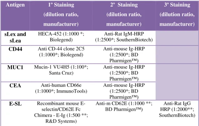
Table 2.2: List of molecules/antibodies and respective dilutions used in western blotting experiences: * Diluted
in TBS 1×-Tween 0.1%; ** Diluted in TBS 1×-Tween 0.1% + 2mM CaCl2.
The washing buffer used was TBS 1× -Tween 0.1%, except for E-SL staining that was TBS 1×-Tween 0.1%. + 2mM CaCl2.
In the case of MUC1 staining, it was used the membrane previously used for sLex/sLea staining. After the final step of revelation in sLex/sLea staining, the membrane was washed two times (10min each) with TBS 1×-Tween 0.1% to remove chemiluminescent substrate. Then, the membrane was incubated in Western Blotting Stripping Buffer (Thermo Scientific) during 15min under agitation to remove rests of primary and secondary antibodies without removing or damaging the immobilized antigen. At the end of incubation, it was washed two times (10min each) with TBS 1×-Tween 0.1%. Finally, the membrane was blocked with non-fat milk 10% diluted in TBS 1×-Tween 0.1% during the night at 4ºC under agitation and the following steps were the same as described previously.
Relatively to E-SL staining, there was an extra step: the addiction of a third antibody. At the end of the incubation period, the secondary antibody solution was removed and the membrane washed three times (5min each) with TBS 1×-Tween 0.1% + 2mM CaCl2. After that, the membrane was incubated with third antibody for 1h at
Antigen 1º Staining
(dilution ratio,
manufacturer)
2º Staining
(dilution ratio, manufacturer) 3º Staining (dilution ratio, manufacturer) sLex and sLea
HECA-452 (1:1000 *; Biolegend)
Anti-Rat IgM-HRP (1:2500*; SouthernBiotech) CD44 Anti CD-44 clone 2C5
(1:1000*; Biolegend)
Anti-mouse Ig-HRP (1:2500*; BD
Pharmigen™)
MUC1 Mucin-1 VU4H5 (1:100*; Santa Cruz)
Anti-mouse Ig-HRP (1:2500*; BD
Pharmigen™)
CEA Anti-human CD66e (1:1000*; ImmunoTools)
Anti-mouse Ig-HRP (1:2500*; BD
Pharmigen™)
E-SL Recombinant mouse E-selectin/CD62E Fc Chimera - E-Ig (1:500 **;
R&D Systems)
Anti-m CD62E (1:1000 **;
room temperature under agitation. The next steps were the same as described previously.
Depending on the quality of revelation, the membrane was exposed at different exposure times (30s, 1min, 2min, 10min, 30min).
2.4.2.3 Immunoprecipitation of CEA in tissue samples
First of all, it was added 80µl of beads-protein G in a tube denominated clearing and 60µL in a tube denominated IP. After that, it was added 1mL of lysis buffer to wash the beads and the tubes were inverted and centrifuged at 12000×g for 1min at 4ºC. The beads were washed three more times with 1mL of lysis buffer (centrifuge to pellet the beads between each wash).Supernatant was carefully removed after each wash to not let the beads dry.
To blocking non-specific interactions, each tube was incubated with 500µL of lysis buffer with bovine serum albumin (BSA from Sigma Aldrich - 0.04g for each 4mL of lysis buffer) for 1h at 4ºC under constant agitation. After that the beads were centrifuged at 12000×g for 1min at 4ºC and washed three times with 1mL of lysis buffer (centrifuge to pellet the beads between each wash). The lysate of the sample of interest (100µg/300µL in lysis buffer) was added to the clearing tube and incubated for 2h at 4ºC under constant agitation. With this step (called pre-clearing), the proteins in lysate that bind non-specific to the beads will be removed. After pre-clearing, the lysate was completely removed and transferred to a new 1.5mL tube, where it was added 3µL of anti-human CD66E (1µg/1µL; ImmunoTools) and incubated for 2h at 4ºC under constant agitation. Finally, the mix of lysate and antibody was added to the IP tube and incubated overnight at 4ºC under constant agitation.