Comparative molecular analysis of
Herbaspirillum
strains by RAPD,
RFLP, and 16S rDNA sequencing
Juliana R.L. Soares-Ramos, Humberto J.O. Ramos, Leonardo M. Cruz, Leda S. Chubatsu,
Fábio O. Pedrosa, Liu U. Rigo and Emanuel M. Souza
Universidade Federal do Paraná, Departamento de Bioquímica e Biologia Molecular, Curitiba, PR, Brazil.
Abstract
Herbaspirillum spp. are endophytic diazotrophic bacteria associated with important agricultural crops. In this work, we analyzed six strains of H. seropedicae (Z78, M2, ZA69, ZA95, Z152, and Z67) and one strain of H. rubrisubalbicans (M4) by restriction fragment length polymorphism (RFLP) using HindIII or DraI restriction endonucleases, random amplified polymorphic DNA (RAPD), and partial sequencing of 16S rDNA. The results of these analyses ascribed the strains studied to three distinct groups: group I, consisting of M2 and M4; group II, of ZA69; and group III, of ZA95, Z78, Z67, and Z152. RAPD fingerprinting showed a higher variability than the other methods, and each strain had a unique electrophoretic pattern with five of the six primers used. Interestingly,H. seropedicae M2 was found by all analyses to be genetically very close to H. rubrisubalbicans M4. Our results show that RAPD can distinguish between allHerbaspirillum strains tested.
Key words: Herbaspirillum, RAPD, RFLP, Phylogeny, 16S rDNA.
Received: December 9, 2002; Accepted: August 10, 2003.
Introduction
Herbaspirillumspp. are a group ofβ-Proteobacteria (Baldaniet al., 1986; Gilliset al., 1990) comprising three species: H. seropedicae (Baldani et al., 1986), H. rubrisubalbicans(Baldaniet al., 1996) andH. frisingense (Kirchhof et al., 2001). These bacteria are endophytic diazotrophs capable of colonizing plant tissues of gramineae such as rice, maize, sugar cane, sorghum, ba-nana, and pineapple (Olivareset al., 1993; Baldaniet al., 1986, 1992; Cruz et al., 2001). Inoculation with Herbaspirillumspp. can improve plant growth and produc-tivity (Baldaniet al., 1995). These effects are attributed in part to the nitrogen fixed by the bacteria (Döbereiner and Pedrosa, 1987; Döbereiner 1991; Döbereineret al., 1995; Boddeyet al., 1995; Olivareset al., 1996) and also to the production of phytohormones, such as auxins and gibberillins, which can induce plant growth (Bastiánet al., 1998). However, evaluation of the bacterial contribution to the associated plant under field conditions is complicated by the presence, in both soil and seeds, of other bacteria which can associate with the crop (Barraquioet al., 1997). The recovery and identification of inoculated strains is
es-sential to correlate gains in productivity with successful colonization.
In recent years, much progress has been made in the development of molecular tools to identify bacteria, such as restriction fragment length polymorphism (RFLP), 16S ri-bosomal DNA (rDNA) sequencing, and random amplified polymorphic DNA (RAPD).
In this work, we analyzed six strains of Herbaspirillum seropedicae(Z78, M2, ZA69, ZA95, Z152 and Z67) and one strain of Herbaspirillum rubrisubalbicans(M4) by a combination of methods for genetic differentiation, including RFLP, RAPD, and com-parison of partial sequences of the 16S rRNA genes.
Materials and Methods
Strains and culture conditions
TheHerbaspirillum seropedicae strains M2, ZA69, ZA95, Z152, Z78 and Z67, and the H. rubrisubalbicans strain M4 used in this study were grown overnight in 50 mL NFbHP-malate (Klassenet al., 1997) at 30 °C in a rotary shaker.
RAPD fingerprinting
Genomic DNA ofHerbaspirillumstrains was puri-fied as follows: 5 mL of the overnight cultures were centri-fuged, and the pellet was suspended in 500µL TES buffer
www.sbg.org.br
Send correspondence to Emanuel M. Souza. Universidade Fed-eral do Paraná, Departamento de Bioquímica e Biologia Molecular, Caixa Postal 19046, 81531-990 Curitiba, PR, Brazil. E-mail: [email protected].
(50 mmol/L Tris-HCL, 20 mmol/L EDTA, and 100 mmol/L NaCl, pH 8.0), treated with 100 µg of lysozyme, and incubated at 30 °C for 3 h. Cells lysis was completed by adding 50µL SDS (10%), followed by incu-bation at room temperature for 5 min and treatment with proteinase K (100 µg/mL) at 37 °C overnight. Purified DNA was obtained by three successive extractions with equilibrated phenol (1 vol) and one with chloroform: isoamyl alcohol (24:1). The genomic DNA was precipi-tated with 2 volumes of absolute ethanol, washed with 70% ethanol, vacuum-dried, and suspended in 200µL of T10E1 (10 mM Tris, 1 mM EDTA, pH 8.0).
Ten ng of genomicHerbaspirillumDNA were sub-mitted to random amplification using the Ready.To.GoTM RAPD Analysis Beads Kit (Amersham Biosciences) ac-cording to the manufacturer’s instructions. Six different primers, previously described for characterization of bacte-rial strains (Amersham Biosciences), were used in the RAPD reactions (Table 1). The electrophoretic profiles were scored according to the presence (1) or absence (0) of a particular band, generating a binary matrix. Similarity of all pair-wise combinations of the numerical profiles was determined by Dice’s coefficient (Dice, 1945) and clus-tered by unweighted pair-group analysis using arithmetical averages (UPGMA) (Sokal and Michener, 1958). A dendrogram was constructed from the patterns, using the NTSYS pc 2.0 program (Exeter Software - scientific soft-ware for teaching and research - http://www. exetersoftware.com/index.html). All samples and a nega-tive control without DNA were run in parallel in the same thermocycler. At least three independently amplified frag-ment patterns of each sample were obtained to confirm the result.
RFLP fingerprinting
Genomic DNA isolated as described above was di-gested withHindIII orDraI (Invitrogen) and submitted to agarose gel (0.7%) electrophoresis. Analyses were per-formed as described for RAPD fingerprinting.
DNA sequencing of 16S rDNA
The 16S rDNA of theHerbaspirillumstrains was am-plified using primers Y1 and Y3 as previously described (Cruz et al., 2001). Briefly, diluted overnight cultures (1:10) were boiled for 10 min and cooled on ice. Reaction mixtures (25µL) contained 10µL of boiled cultures,Taq DNA polymerase buffer (50 mmol/L KCl, 1.5 mmol/L MgCl2, 10 mmol/L Tris-HCl, pH 9.0), 200µmol/L of each deoxynucleotide, 0.4µmol/L of each primer, and 1U ofTaq DNA polymerase (Invitrogen). The mixtures were incu-bated at 93 °C for 2 min, followed by 34 amplification cy-cles of 93 °C for 45 s, 62 °C for 30 s, and 72 °C for 2 min each. Conserved regions of 16S rDNA from subclasses al-pha, beta and gamma of the Proteobacteria were identified from the alignment of 65 sequences from the GenBank da-tabase, and used to design the primers 16S362f, 16S786f, 16S1203f, 16S1110r, and 16S805r (Table 1). DNA se-quences of the amplified products were determined using dye terminator chemistry in an ABI Prism 377 automated DNA sequencer (Applied Biosystems). The 16S rDNA se-quences obtained were deposited in the GenBank database, and their accession numbers are in parentheses: H. seropedicaestrains M2 (AY191276), ZA69 (AY191272), ZA95 (AY191274), Z152 (AY191273), Z78 (AY191275).
DNA Sequencing analysis
DNA sequences were assembled and edited using the BioEdit package (Hall, 1999) and aligned by the Contig
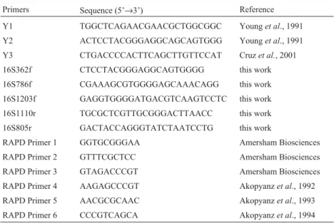
As-Table 1- Sequences of the primers used.
Primers Sequence (5’→3’) Reference
Y1 TGGCTCAGAACGAACGCTGGCGGC Younget al., 1991
Y2 ACTCCTACGGGAGGCAGCAGTGGG Younget al., 1991
Y3 CTGACCCCACTTCAGCTTGTTCCAT Cruzet al., 2001
16S362f CTCCTACGGGAGGCAGTGGGG this work
16S786f CGAAAGCGTGGGGAGCAAACAGG this work
16S1203f GAGGTGGGGATGACGTCAAGTCCTC this work
16S1110r TGCGCTCGTTGCGGGACTTAACC this work
16S805r GACTACCAGGGTATCTAATCCTG this work
RAPD Primer 1 GGTGCGGGAA Amersham Biosciences
RAPD Primer 2 GTTTCGCTCC Amersham Biosciences
RAPD Primer 3 GTAGACCCGT Amersham Biosciences
RAPD Primer 4 AAGAGCCCGT Akopyanzet al., 1992
RAPD Primer 5 AACGCGCAAC Akopyanzet al., 1993
sembly Program (CAP) (Huang, 1992). The phylogenetic tree was obtained by a neighbor-joining method, using the MEGA 2 software (Kumaret al., 2001).
Results and Discussion
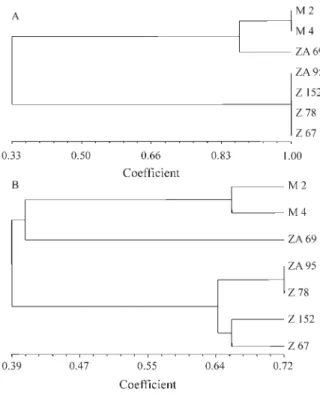
The total DNA restriction patterns of the Herbaspirillum strains were used to construct a dendrogram (Figure 1A), which clustered them into three groups (Figure 1A and Table 2). TheHindIII orDraI re-striction profiles of M2 and M4 were identical, as were those ofH. seropedicae Z67 and Z78, thus defining two electrophoretic types. Strains ZA95 and Z152 had identical patterns with DraI, but HindIII produced two different bands. However, analyses of their electrophoretic profiles positioned these strains together with Z67 and Z78, form-ing a cluster with a similarity coefficient of 100%. H. seropedicae ZA69 had a distinct electrophoretic pattern and was allocated into a separate group, closer to the M2/M4 cluster. These results indicate that, as far as the sep-aration of theHerbaspirillumstrains is concerned, restric-tion length polymorphism has a limited potential.
Genomic diversity of theHerbaspirillumstrains was also investigated by random amplified polymorphic DNA (RAPD) analysis. Each of the six primers used generated electrophoretic DNA patterns for the strains studied
(Table 2 and Figure 2). The analysis of these patterns pro-duced highly congruent DNA fingerprint clustering, in overall agreement with the RFLP results (Figure 1A). With the exception of primer 1, all primers produced unique pat-terns for M2 and M4, allowing unequivocal differentiation of these strains. As with RFLP, analysis of the RAPD fin-gerprinting patterns revealed three main clusters of strains (Figure 1B), with a similarity level of approximately 39%. Cluster I was formed by M2 and M4, with a similarity level of 65%. Strain ZA69 was located in a separate branch, closer to M2 and M4, but with a similarity level of 40%. Strains ZA95, Z78, Z152, and Z67 were located in a sepa-rate cluster, occupying distinct positions in the dendrogram and forming two sub-groups, comprising strains ZA95 and Z78 (72% similarity), and Z152 and Z67 (65% similarity), respectively. Cluster analysis of RAPD profiles supported the differences noted by visual observation of the electro-phoretic profiles (data not shown). Furthermore, the pro-files obtained by RAPD showed a higher level of variability than those obtained by RFLP, since, using primers 2 to 6, all strains could be distinguished by at least one band (Ta-ble 2). These results show that RAPD is the most sensitive and convenient method tested to unequivocally identify Herbaspirillumstrains using whole genomic DNA. How-ever, since low reproducibility has been attributed to RAPD profiling, this may reduce its potential application (Penner et al., 1993; Wanget al., 1993). To minimize this effect, we used the same procedure for DNA purification from all strains and a commercially available RAPD system with standardized components and conditions, which was reli-able and reproducible in this study. The Ready-To-Go (Amersham Biosciences) beads format also significantly reduced the number of pipetting steps, thereby increasing the reproducibility of the RAPD technique.
Figure 1-Panel A– Dendrogram inferred from the RFLP profiles ob-tained by digestion of total DNA ofHerbaspirillumstrains with restriction endonucleasesHindIII orDraI.Panel B– Dendrogram inferred from RAPD profiles of theHerbaspirillumstrains. Similarities were calculated using Dice’s coefficient, and clustering was achieved by UPGMA (Sokal and Michener, 1995).
Table 2- Genotypic characterization ofHerbaspirillumspp., as revealed by RAPD and RFLP profiling and 16S rDNA sequence analysis.
Organism RAPD patterna RFLP patternb 16S rDNAc
H. seropedicae
M2 aaaaaa aa a
ZA69 cccccc bb b
ZA95 dddddd cc c
Z152 deeeee cd c
Z67 efffff ce c
Z78 fggggg ce c
H. rubrisubalbicans
M4 bbbbbb aa a
aRAPD patterns obtained with different primers. Each letter defines a
common pattern for primers 1 to 6, respectively (see Table 1).
bRFLP patterns obtained with restriction endonucleasesHindIII andDraI, respectively. Each letter defines a common pattern.
c
Lasker (2002) recently reported that repeated runs of the same or different DNA preparations of the same strain ofAspergillus fumigatusproduced highly reproducible re-sults using RFLP, RAPD, sequence-specific DNA primer (SSDP) analysis or polymorphic microsatellite markers (PMM) analysis (Lasker, 2002). Coenye et al. (2002) reached similar conclusions by analyzing Burkholderia cepacia Genomovar III isolates, using pulsed-field gel electrophoresis (PFGE), BOX-PCR fingerprinting and ran-dom amplified polymorphic DNA (RAPD) typing.
Zhanget al. (2002) comparing typing methods to dif-ferentiateStreptococciGroup B showed that RAPD was a useful assay for the rapid characterization of these strains and was more discriminatory than conventional serological assays. These authors also reported that the RAPD assay was faster, more convenient and easier to perform than al-ternative DNA analytical procedures such as pulsed-field gel electrophoresis. In addition, van den Braaket al. (2002) obtained highly congruent DNA fingerprint clustering of vancomycin-resistant enterococci (VRE), employing well-standardized RAPD or PFGE protocols.
The 16S rDNA from allHerbaspirillumstrains used in this work was amplified using primers Y1 (Younget al., 1991) and Y3 (Cruzet al., 2001), which allowed the ampli-fication of the nearly complete 16S rRNA genes. The PCR products were thoroughly sequenced in both orientations, yielding the complete sequence of the Y1-Y3 fragment with a three-times redundancy. The partial sequence of the 16S rRNA gene of theH. seropedicaestrain Z67, deposited in the GenBank database (Kirchhofet al., 2001), differed from the sequence obtained in this study by three bases. The 16S rDNA sequence of theH. rubrisubalbicansstrain M4
deposited in GenBank (Kirchhofet al., 2001) was identical to that determined in this work.
Ribosomal operons are of great relevance for the study of bacterial evolution and phylogeny (Woese, 1987), and sequencing of 16S rDNA has been widely used to re-construct phylogenetic relationships of microorganisms (Gutellet al., 1994; Luzet al., 1998).
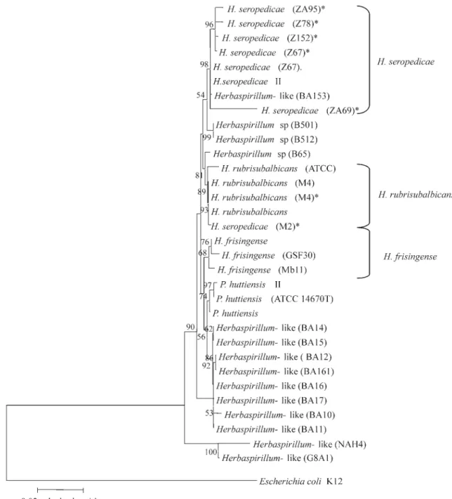
Phylogenetic analysis of the partial 16S rDNA se-quences from theHerbaspirillumstrains studied here, to-gether with related sequences deposited in GenBank (Figure 3), positioned the Herbaspirillum species (H. seropedicae, H. rubrisubalbicans and H. frisingensis), Herbapirillum-like isolates, and the related species Pseu-domonas huttiensisin separate branches, well supported by the bootstrap values (see Figure 3). Within the H. seropedicae branch, strains Z67, Z78, ZA95, and Z152 formed a tight cluster with a bootstrap value of 96. Strain ZA69 formed a separate cluster in the same branch, consis-tent with its taxonomic position. StrainsH. seropedicaeM2 andH. rubrisubalbicans M4 had very similar 16S rDNA sequences, and grouped in theH. rubrisubalbicansbranch with a bootstrap value of 89. Although strains M2 and M4 belong to different species, as determined by physiological assays (Baldaniet al., 1996), the analyses carried out in this work showed very close genetic proximity.
The genetic proximity between strains of the species H. seropedicaeandH rubrisubalbicanswas also observed by Cruzet al. (2001), using ARDRA (amplified rDNA re-striction analysis) and sequencing of approximately 300pb of the 16S rDNA, thereby highlighting the low level of vari-ability of these organisms. Kirchhofet al.(2001) and Ca-ballero-Mellado and Martinez-Romero (1994) evaluating
Figure 2- Agarose gel (2.0%) electrophoresis of RAPD profiles of theHerbaspirillum seropedicae(M2, Z69, ZA95, ZA152, Z67 and Z78) and
Herbaspirillum frisingense and Gluconacetobacter diazotrophicusstrains, also concluded thatHerbaspirillum strains formed a group of tight genetic relationships and low variability.
Herbaspirillumspp. are endophytic diazotrophs with low survival in plant-free soils (Baldaniet al., 1992), sug-gesting a strong adaptation of these bacteria to the endophytic state. McArthur et al. (1988) suggested that close genetic proximity may be accounted for by adaptation to very stable habitats for long periods.
In summary, molecular tools evaluating either the 16S rRNA gene or the entire genome (RAPD and RFLP) al-lowed distinction of the sevenHerbaspirillumstrains stud-ied. RAPD profiling using a commercially available system unequivocally differentiated all the strains tested and proved very reliable. This finding agrees with the observa-tion that the 16S rDNA sequences are highly conserved among the species and potentially useful to distinguish be-tween distantly related organisms (Woese, 1987). In con-trast, RAPD utilizes fragment amplification of the whole
genome, therefore being well suited to detect differences between closely related organisms.
Acknowledgments
We thank Roseli Prado, Valter Baura and Julieta Pie for technical assistance. CNPq/PRONEX/Fundação Arau-cária/Paraná Tecnologia supported this work.
References
Baldani JI, Baldani VLD, Seldin L and Döbereiner J (1986) Char-acterization of Herbaspirillum seropedicaegen. nov., sp. nov., a root-associated nitrogen fixing bacterium. Int J Syst Bacteriol 36:86-93.
Baldani VLD, Baldani JI, Olivares F and Döbereiner J (1992) Identification and ecology of Herbaspirillum seropedicae and the closely related Pseudomonas rubrisubalbicans. Symbiosis 13:65-73.
Baldani VLD, Olivares FL and Döbereiner J (1995) Selection of Herbaspirillum spp. strains associated with rice seedlings amended with15N-labeled fertilizer. 202. International
Sym-posium on Sustainable Agriculture for the tropics - The role of Biological Nitrogen Fixation, Angra dos Reis, Brazil. Baldani JI, Pot B, Kirchhof G, Falsen E, Baldani VLD, Olivares
FL, Hoste B, Kersters K, Hartmann A, Gillis M and Döbereiner J (1996) Emended description of Herbaspirillum; inclusion of [Pseudomonas] rubrisubalbicans, a mild plant pathogen, asHerbaspirillum rubrisubalbicanscomb. nov.; and classification of clinical isolates (EF group 1) asHerbaspirillumspecies 3. Int J Syst Bacteriol 46:802-810.
Barraquio WL, Revilla L and Ladha JK (1997) Isolation of endophytic diazotrophic bacteria from wetland rice. Plant Soil 194:15-24.
Bastián F, Cohen A, Piccoli P, Luna V, Baraldi R and Bottini R (1998) Production of indole-3-acetic acid and gibberellins A1 and A3 by Acetobacter diazotrophicus and Herbaspirillum seropedicaein chemically-defined culture media. Plant Growth Regul 24:7-11.
Boddey RM, de Oliveira OC, Urquiaga S, Reis VM, Olivares FL, Baldani VLD and Döbereiner, J (1995) Biological nitrogen fixation associated with sugar cane and rice: contributions and prospects for improvement. Plant Soil 174:195-209. Caballero-Mellado J and Martinez-Romero E (1994) Limited
genetic diversity in the endophytic sugarcane bacterium Acetobacter diazotrophicus. Appl Environ Microbiol 60:1532-1537.
Coenye T, Spilker T, Martin A and LiPuma JJ (2002) Compara-tive assessment of genotyping methods for epidemiologic study of Burkholderia cepacia Genomovar III. J Clin Microbiol 40:3300-3307.
Cruz LM, Souza EM, Weber OB, Baldani JI, Döbereiner J and Pedrosa FO (2001) 16S ribosomal DNA characterization of nitrogen-fixing bacteria isolated from banana (Musaspp.) and pineapple (Ananas comosus(L.) Merril). Appl Environ Microbiol 67:2375-2379.
Dice LR (1945) Measures of the amount of ecologic association between species. Ecology 26:297-302.
Döbereiner J and Pedrosa FO (1987) The genusAzospirillum. In: Nitrogen-fixing bacteria in non-leguminous crop plants. Madison: Science Tech. Publishers, 155 pp.
Döbereiner J (1991) The generaAzospirillumandHerbaspirillum. In: Ballows A, Triiper HG, Dowrkin M, Harder W (eds) The Prokaryotes. Berlin: Springer-Verlag, pp 2236-2253. Döbereiner J, Baldani VLD and Reis VM (1995) Endophytic
oc-currence of diazotrophic bacteria in non-leguminous crops. In: Fendrik I, del Gallo M, Vanderleyden J, de Zamaroczy M (eds)AzospirillumVI and related microorganisms. Berlin, Heidelberg: Springer-Verlag, pp 15-30.
Gillis M, Döbereiner J, Pot B, Goor M, Falsen E, Hoste B, Reinhold B and Kersters K (1990) Taxonomic relationships between [Pseudomonas] rubrisubalbicans, some clinical isolates (EF GROUP 1), Herbaspirillum seropedicaeand [Aquaspirillum] authrotrophicum. In: Polsinelli M, Materassi R, Vincenzini M (eds) Nitrogen Fixation. Dordrecht: kluver Acad. Publish. pp 293-294.
Gutell RR, Larsen N and Woese CR (1994) Lessons from an evolving rRNA: 16S and 23S rRNA structures from a com-parative perspective. Microbiol Rev 58:10-26.
Hall TA (1999) BioEdit: a user-friendly biological sequence alignment editor and analysis program for Windows 95/98/NT. Nucleic Acids Symp Ser 41:95-98.
Huang X (1992) A contig assembly program based on sensitive detection of fragment overlaps. Genomics 14:18-25. Kirchhof G, Eckert B, Stoffels M, Baldani JI, Reis VM and
Hartmann A (2001)Herbaspirillum frisingensesp. nov., a new nitrogen-fixing bacterial species that occurs in C4-fibre plants. Int J Syst Evol Microbiol 1:157-168.
Klassen G, Pedrosa FO, Souza EM, Funayama S and Rigo LU (1997) Effect of nitrogen compounds on nitrogenase activity in Herbaspirillum seropedicae SMR1. Can J Microbiol 43:887-891.
Kumar S, Tamura K, Jakobsen IB and Nei M (2001) Mega 2: mo-lecular evolutionary genetic analysis software. Bioinformatics 17:1244-1245.
Lasker BA (2002) Evaluation of performance of four genotypic methods for studying the genetic epidemiology of Aspergillus fumigatus isolates. J Clin Microbiol 40:2886-2892.
Luz SP, Rodriguez-Valera F, Lan R and Reeves PR (1998) Varia-tion of the ribosomal operon 16S-23S gene spacer region in representatives of Salmonella enterica subspecies. J Bacteriol 180:2144-2151.
McArthur JV, Kovacic DA and Smith MH (1988) Genetic diver-sity in natural populations of a soil bacterium across a land-scape gradient. Proc Natl Acad Sci 85:9621-9624.
Olivares FL, James EK, Reis VM, Baldani VLD, Baldani JI and Döbereiner J (1993) Colonização do tecido vascular por Herbaspirillum spp. em sorgo e cana-de-açúcar. Fitopat Bras 18:313.
Olivares FL, James EK, Baldani JI and Döbereiner J (1996) Infec-tion of mottled stripe disease-susceptible and resistant sugar cane varieties by the endophytic diazotrophHerbaspirillum. New Phytol 135:723-737.
Sokal RR and Michener CD (1958) A statistical method for evalu-ating systematic relationships. Univ Kans Sci Bull 38:1409-1438.
van den Braak N, Power E, Anthony R, Endtz HP, Verbrugh HA and van Belkum A (2000) Random amplification of poly-morphic DNA versus pulsed field gel electrophoresis of SmaI DNA macrorestriction fragments for typing strains of vancomycin-resistant enterococci. FEMS Microbiol Lett 192:45-52.
Wang D, Wayne MMY, Taricani M, Buckingam K and Sandham HJ (1993) Artifactual variation in randomly amplified
polymorphic DNA banding patterns. Biotechniques 14:214-218.
Woese CR (1987) Bacterial evolution. Microbiol Rev 512:221-71.
Young JPW, Downer HL and Eardly BD (1991) Phylogeny of the phototrophicRhizobiumstrains BTAil by polymerase chain reaction-based sequencing of a 16S rRNA gene segment. J Bacteriol 173:2271-2277.
Zhang GW, Kotiw M and Daggard G (2002) A RAPD-PCR geno-typing assay which correlates with serotypes of group B streptococci. Lett Appl Microbiol 35:247 250.