VITRO CONDITION
1Farzad Aala, 2Umi Kalsom Yusuf, 3Farida Jamal, 4Sassan Rezaie*
1
Department of Medical Mycology & Parasitology, Faculty of Medicine, Kurdistan University of Medical Sciences, Sanandaj,
Kurdistan, Iran; 2Department of Biology, Faculty of Science; Universiti Putra Malaysia, 43400 Serdang, Selangor, Malaysia;
3
Department of Medical Microbiology and Parasitology, Faculty of Medicine and Health Sciences; Universiti Putra Malaysia,
43400 Serdang, Selangor, Malaysia; 4Department of Medical Biotechnology, School of Advanced Technologies in Medicine,
Tehran University of Medical Sciences, Tehran, Iran.
Submitted: May 04, 2011; Approved: January 16, 2012.
ABSTRACT
Dermatophytosis is caused by a group of pathogenic fungi namely, dermatophytes, is among the most
prevalent infectious diseases worldwide. Azole drugs are widely used in the treatment of dermatomycosis,
but can cause various side effects and drug resistance to the patients. Hence, for solving this problem can be
used from the plant extract as alternative for chemical drugs. Allicin is a pure bioactive compound isolated
from garlic was tested for its potential as a treatment of dermatomycosis in this study. This study evaluated
the in vitro efficacy of pure allicin against ten isolates of Trichophyton rubrum and the MIC50 and MIC90
ranged from 0.78-12.5 µg/ml for allicin. The results revealed that the order of efficacy based on the MICs
values, all isolates showed almost comparable response to allicin and ketoconazole except for some isolates,
at 28 ºC for both 7 and 10 days incubation. Mann-Whitney test indicate that MICs at 7 days incubation was
not observed a significant difference between the effects of allicin and ketoconazole (p > 0.05), but MICs at
10 days incubation, a significant difference was observed (p ≤ 0.05). On the other side, time kill studies
revealed that allicin used its fungicidal activity within 12-24 h of management in vitro as well as
ketoconazole. In conclusion, allicin showed very good potential as an antifungal compound against
mycoses-causing dermatophytes, almost the same as the synthetic drug ketoconazole. Therefore, this
antifungal agent appears to be effective, safe and suitable alternative for the treatment of dermatomycosis.
Key words: Allicin, Antifungal drugs, Dermatophytes, MIC (minimal inhibitory concentration.
INTRODUCTION
Dermatophytosis is among the most popular and prevalent
infectious disease worldwide that caused by dermatophytes.
Dermatophytes are the main cause of superficial fungal
infections of the skin, hair and nails of human and other
animals (2, 12). Among the various types of dermatophytes,
Trichophyton rubrum is clinically the most common genus of
human pathogenic dermatophytes and has been the most
frequently isolated dermatophytes (20). The azoles group of
antifungal drugs has been used to cure dermatomycosis.
Ketoconazole and fluoconazole are effective in treating
dermatomycosis but may cause undesirables side effects and
toxicity to patients. It has been reported that the increased in
the use of these drugs may cause drug resistance to all agents in
the azoles group (1, 10). Hence, this study was aimed at the
investigation on the use of a plant extracts as one of the
alternatives to treat dermatomycosis. One of the most common
plant extracts being used for medicinal purposes is garlic
extracts (Allium sativum). The therapeutic effect of garlic is
because of sulfur-containing compounds and thiosulfinates
(17). The interaction of sulfur compounds (such as allicin) with
sulphur (thiol-containing enzymes) groups of microbial
enzymes (such as trypsin and other proteases) which caused
inhibition of microbial growth (14). Allicin (diallyl
thiosulphinate) is a pure bioactive compound isolated from
garlic. It is manufactured via an enzymatic response from
freshly crushed garlic (5). Allicin is made with a mixture of
allinase (the enzyme that is stocked in an isolated section in
garlic) and alliin (a compound in fresh garlic) (14). Allicin has
been reported to show antibacterial properties (3) and
antifungal activities (5, 10, 14, 17). Ajoene is the ingredient
from allicin that has antifungal action against Aspergillus niger
and Candida albicans (19). Currently, pure grade allicin is
available commercially. Pyun and Shin (10) found out that
oil fractions of Allium plants are weaker compared to allicin
against Trichophyton spp. The growth of new antifungal agents
as well as the incidence of fungal infections in human
highlighted the antifungal susceptibility testing of
dermatophytes (6, 13). The National Committee for Clinical
Laboratory Standards (NCCLS) (new name Clinical and
Laboratory Standards Institue (CLSI) M38-A established in
2002 has currently issued a standardized in vitro protocol for
susceptibility testing. Barros et al. (2) have reported that the
susceptibility tests to antifungal agents were suitable and
reliable. This method is based on the calculation of the MICs.
Pyun and Shin (10) investigated the activity of allicin alone and
the combined effects of Allium oils with ketoconazole against
Trichophyton spp. They used the broth microdilution method
which is based on the calculation of the MIC with further
calculation of the FICI for drug combination.
This study was aimed at the evaluation of in vitro
antifungal activity of allicin against 10 isolates of T. rubrum,
which is determined by broth microdilution.
MATERIALS AND METHODS
Study design
Ten isolates of T. rubrum were examined for their
interactions with natural product; allicin, and drug;
ketoconazole respectively. Protocol outlined in Clinical and
Laboratory Standards Institue (CLSI, formerly the NCCLS)
M38-A, susceptibility testing guidelines for filamentous fungi,
was employed (8). These interactions were evaluated in two
incubation periods of 7 and 10 days respectively to compare
the effectiveness of the treatment.
Isolates
Nine clinical isolates of T. rubrum from culture collection
of clinical isolates preserved at the laboratory of Medical
Mycology Department in Tehran University of Medical
Sciences, Iran were selected. T. rubrum (ATCC-10218) was
used as a control strain. All isolates were kept in sterile saline
(0.85%) v/v NaCl at 4 ºC until required for bioassays.
Media
The standard RPMI 1640 (Sigma chemicals Co, USA)
medium based on CLSI guidelines M38-A was prepared.
Antifungal compounds and dilutions
Two antifungal agents namely, allicin (Alexis-
Biochemicals Co, San Diego, USA) was dissolved in 10 mg/ml
in methanol/water/formic acid (60:40:0.1), then stored at -20 to
-70 °C. Ketoconazole (Sigma chemicals Co, USA), was
dissolved in 100% dimethyl sulfoxide (DMSO) at 1.5 mg/ml
stock solution of 1000 µg/ml. The stock was 2Χ diluted in
RPMI 1640 when the antifungal mixture was examined
individually to give the same final strength required for the test
(13). A series of twofold dilutions were prepared at 100 times
higher than the highest expected final test concentration
(intermediate concentration). The DMSO solutions were
diluted 1:50 in RPMI 1640 medium to make twice the final
intensity required for the test (final concentration). The stock
solutions were kept at -70 °C until needed. Allicin and
ketoconazole drug dillutions ranged from 0.05 to 25.0 µg/ml
and 0.03 to 16.0 µg/ml, correspondingly, as outlined in the
CLSI guidelines M38-A.
Inoculum preparation
All isolates were transfered from sterile saline (0.85%) v/v
NaCl solution and subcultured to potato dextrose agar (Difco
Laboratories, Detroit, Michigan) and kept at 28 ºC for 7 days to
allow for sporulation. The seven-day-old colonies were
immersed in 5 ml of sterile saline (0.85%) v/v NaCl solution,
and the culture exterior was smoothly probed with the tip of a
Pasteur pipette to remove the spores. The resulting mixture of
conidial and hyphal portions was filtrated with a Whatman’s
filter no. 40 (pore size:8 µm), which filtered the hyphal
fragments but allowing the passage of microconidia into the
collecting flask below as suggested by Santos and Hamdan
(12); Santos et al. (13); Barros et al. (2). The conidial
suspensions’s densities in cuvettes were determined using
spectrophotometer (U-1900-UV/VIS, Model: BJO-0003,
Hitachi, Tokyo-Japan) at 520 nm wavelength. The conidial
suspensions’s densities varied from 0.15 to 0.17 (70% to 72%
transmittance). The inoculum concentrations ranged from
2-4×10 conidia/ ml. The inoculum quantifications were
determined by calculating number of microconidia using a
hematocytometer, and by plating 0.01 ml of each inoculum
suspensions on sabouraud dextrose agar (Difco Laboratories,
Detroit, Michigan). The plates were kept at 28 ºC and
monitored daily for the existence of colony from units (CFU).
The CFU were calculated as soon as growth becomes
observable. These suspensions were diluted 1:50 in RPMI 1640
medium, which matched with 2x, which was the density
needed (about 2-4×10 conidia/ ml). The test inoculums were
prepared in enough volume to inoculate each well with 100 µl
of the corresponding diluted inoculum suspensions.
Test procedure
U-bottomed 96 well microdilution plates (Brand 781660,
Wertheim, Germany) were used.
Antifungal susceptibility testing
To each well, 100 µl of the corresponding fungal
inoculum suspension prepared earlier was pipetted and 100 µl
of the twofold dilution of antifungal compounds. Each test
plate included two drug-free controls, one with only RPMI
1640 medium (negative control) and another with 100 µl of
RPMI 1640 with 100 µl of the inoculum suspension (positive
control). This medium was prepared by adding powdered
RPMI 1640 medium in distilled water, then buffered to PH 7.0
with 0.165 mol/L 3-[N-morpholino] propane sulfonic acid
(MOPS).
Incubation time and temperature
Microdilution trays were incubated at 28 ºC without
agitation and were visually examined after 7 days to observe
the growth of colonies as recommended by Santos and
Hamdan (12), Santos et al. (13), and Barros et al. (2). After the
visual reading, the density of fungal growing on the plates were
read with a spectrophotometer (Benchmark plus Microplate
Spectrophotometer-Model; Bio-Rad 170-6930-Japan) set at
520 nm wave length. The absorbance ranged from 0.15 to 0.17
(70 to 72% transmittance) after 7 and 10 days of incubation.
Evaluation of the MIC
The lowest concentration of an antifungal agent that
or broth dilution susceptibility test is called MIC which is an
acronym for the Minimal Inhibitory Concentration. The results
of MIC are recorded in micrograms per milliliter. All growths
in the microdilution wells were compared to the growth control
to determine the endpoint in microdilution procedures. To
compare the actions of the tested drug, the MIC of all drugs
were determined. This included the readings of the MIC50 and
MIC90; namely, the lowest concentration of the drugs at which
50 or 90% of the growth of a microorganism was inhibited (9,
11, 12, 13, and 15).
Time-kill studies
Ten isolates of T. rubrum were grown as mentioned above
in the presence of 12.5 µg/ml allicin and 4 µg/ml ketoconazole
to determin the time-kill activities. The reason behind the
selection of these concentrations was because they were
twofold above the MICs set for these isolates in the
microdilution broth method explained above. Conidia in
cultures were dislodged after 1, 2, 4, 8, 12 and 24 h and by
scraping and then diluted serially in twofold dilutions which
were later poured over sabouraud dextrose agar. Colonies were
counted after an interval of 7 days incubation at 28 ºC. The
calculation of the MFC (Minimal Fungicidal Concentration) as
the drug concentration responsible for 99.9% reduction of
viability was made. Such high percentage of reduction took
place after 1-24 h in the presence of allicin and ketoconazole as
compared with untreated viable spore counts at 0 h (14).
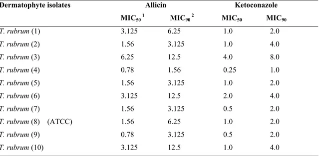
RESULTS
The MICs of allicin was obtained at 0.78-12.5 µg/ml and
ketoconazole 0.25-8.0 µg/ml in the study. Results showed that
the order of efficacy based on the MIC50 and MIC90 values, all
isolates showed almost comparable response to allicin and
ketoconazole except for some isolates at 28 ºC at both 7 and 10
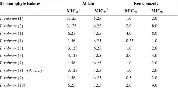
days incubation. Values for MICs at 10 days incubation were
higher than for 7 days incubation for allicin and ketoconazole
(Table 1 and 2).
Time–kill study outcomes reveal that conidia grown from
T. rubrum exhibited a reduction in viability after 12-24 h of
incubation in the presence of 12.5 µg/ml allicin and 4 µg/ml
ketoconazole (Figure 1). It also shows that allicin decreased the
growth of T. rubrum almost as well as ketoconazole.
Table 1. Effects of Allicin and Ketoconazole on Trichophyton rubrum at 28 ºC at 7 days incubation.
Allicin Ketoconazole Dermatophyte isolates
MIC50 1 MIC90 2 MIC50 MIC90 T. rubrum (1) 3.125 6.25 1.0 2.0
T. rubrum (2) 1.56 3.125 1.0 4.0
T. rubrum (3) 6.25 12.5 4.0 8.0
T. rubrum (4) 0.78 1.56 0.25 1.0
T. rubrum (5) 1.56 3.125 1.0 2.0
T. rubrum (6) 3.125 12.5 2.0 4.0
T. rubrum (7) 1.56 3.125 0.5 2.0
T. rubrum (8) (ATCC) 1.56 6.25 1.0 2.0
T. rubrum (9) 0.78 3.125 0.5 2.0
T. rubrum (10) 3.125 12.5 1.0 4.0
1 MIC
50 is the MIC at which 50% of the growth of a microorganism was inhibited (µg/ml). 2 MIC
Table 2. Effects of Allicin and Ketoconazole on Trichophyton rubrum at 28 ºC at 10 days incubation.
Allicin Ketoconazole Dermatophyte isolates
MIC50 1 MIC90 2 MIC50 MIC90 T. rubrum (1) 3.125 6.25 1.0 2.0
T. rubrum (2) 3.125 6.25 2.0 8.0
T. rubrum (3) 6.25 12.5 4.0 8.0
T. rubrum (4) 1.56 6.25 0.25 1.0
T. rubrum (5) 3.125 6.25 1.0 2.0
T. rubrum (6) 3.125 12.5 2.0 4.0
T. rubrum (7) 1.56 6.25 1.0 2.0
T. rubrum (8) (ATCC) 3.125 12.5 1.0 2.0
T. rubrum (9) 1.56 6.25 0.5 2.0
T. rubrum (10) 6.25 12.5 2.0 4.0
1 MIC
50 is the MIC at which 50% of the growth of a microorganism was inhibited (µg/ml). 2 MIC
90 is the MIC at which 90% of the growth of a microorganism was inhibited (µg/ml).
Figure 1.Time-kill curve of T. rubrum (ATCC-10218) conidia incubated in the presence of 12.5 µg/ml allicin and 4µg/ml keteconazole. The outcomes are characteristic of two independent examinations carried out in triplicate.
Data analysis
A series of Mann-Whitney tests were used to compare the
effects of allicin and ketoconazole on T. rubrum after 7 days
incubation. In summary, there is no significant differences
between allicin and ketoconazole (Mann-Whitney U=28.00,
p>0.05).
Another series of Mann-Whitney tests were used to
compare the effects of allicin and ketoconazole on T. rubrum
after 10 days incubation. Briefly, there is significant difference
p≤0.05).
Determination of all the MICs were performed in
triplicate.
DISCUSSION
Dermatophytes are a group of fungi able to invade
keratinized tissues of human and animals, causing
dermatomycosis (2). Dermatophytosis is one of the common
infectious diseases worldwide (2, 12). The azole antifungal
drugs such as imidazole (e.g. ketoconazole) are commonly
used in the treatment of dermatomycosis. They are synthetic
antifungal drugs and although effective, but, because of
increased use of these medications, an incidence of drug
resistance to all agents has been reported. They are also known
generally to cause side effects such as itching, allergic rash,
hepatotoxicity (for ketoconazole) (1). Therefore we investigate
the use of a plant-based, biodegradable natural product as an
alternative namely allicin. Allicin is a pure, bioactive and the
most powerful medicinal compound isolated from garlic and it
has different biological properties such as antimicrobial and
antifungal activities. Several studies showed that it could be
used as the treatment of fungal infections (3, 14). This study
used allicin as an antifungal agent against 10 isolates of T.
rubrum in comparison with ketoconazole. The MICs of the
allicin and ketoconazole against T. rubrum isolates assessed by
broth microdilution assay are listed in Table1 and 2.
Yamada and Azuma (18) proved that the MICs of allicin
obtained at 28 ºC for 5 days incubation are 0.78-6.25 µg/ml. In
our investigation, the MICs of allicin obtained at 28 ºC for 7
days incubation are MIC50 with 0.78-6.25 µg/ml and MIC90
with 1.56-12.5 µg/ml. Furthermore the MICs of ketoconazole
attained at 28 ºC for 7 days incubation are equal to MIC50 and
MIC90 of ketoconazole with 0.25-4.0 µg/ml and 0.5-8.0 µg/ml
respectively. The results were in line with Santos and Hamdan
(12) and Korting et al. (7), but proved to be different from the
study of Fernandez-Torres et al. (4).
The results of this study revealed that the MICs for 10
days incubation increased 1 dilutions for drugs tested
separately in comparison with the MICs for 7 days incubation
except for ketoconazole. The results are supported with Santos
and Hamdan (12) who proved that an increased incubation time
of 10 days compared to 7 days increases MICs from 1 to 2
dilutions with the same medium, 7 days incubation time
compared to 4 days proved to generate similar results.
Therese et al. (16) showed that the isolates were resistant
in vitro to ketoconazole, with the MICs>0.8 μg/ml. Our results
displayed that the isolates were all resistance to ketoconazole
in MICexcept for T. rubrum 4, 7 and 9, thus this data confirm
the resistance of T. rubrum isolatesto ketoconazole.
Time-kill study outcomes reveal that in the presence of
12.5 µg/ml allicin and 4 µg/ml ketoconazole, conidia grown
from T. rubrum exhibited a reduction in viability after 12-24 h
of incubation. It also shows that allicin decreased the growth of
T. rubrum almost as well as ketoconazole. Our results are in
agreement with the previously study by Shadkchan et al. (14)
who demonstrated that conidia grown from strains A. fumigatus
13, A. niger 1, A. terreus 3 and A. flavus 2 displayed a loss in
viability after 8-12 h of incubation in the presence of 16 µg/ml
allicin. Time-kill study results is also confirmed our MICs
results in this study, because both of these results showed that
allicin decreased growth of T. rubrum almost as well as
ketoconazole.
In conclusion, allicin is potentially as good as antifungal
compound against dermatophytes, almost as good as
ketoconazole. Therefore, it can be considered as an effective,
safe and suitable alternative in the treatment of
dermatomycosis. However, further research is required
including “in vivo” studies, as there is a lack of data in this
field.
REFERENCES
present and future. Ann Saudi Med. 18 (1), 28-38.
2. Barros, M.; Santos, D.; Hamdan, J. (2007). Evaluation of susceptibility of Trichophyton mentagrophytes and Trichophyton rubrum clinical isolates to antifungal drugs using a modified CLSI microdilution method (M38-A). J Med Microbiol. 56,514-518.
3. Cai, Y.; Wang, R.; Pei, F.; Liang, B.-B. (2007). Antibacterial Activity of Allicin Alone and in Combination with b -Lactams against
Staphylococcus spp. and Pseudomonas aeruginosa. J Antibio. 60 (5), 335-338.
4. Ferna´ndez-Torres, B.; Inza, I.; Guarro, J. (2003). Comparison of in vitro antifungal susceptibilities of conidia and hyphae of dermatophytes with thick-wall macroconidia. Antimicrob Agents Chemother. 47,3371-3372. 5. Gardner, CD.; Lawson, LD.; Block, E.; Chatterjee, LM.; Kiazand, A.;
Balise, RR.; Kraemer, HC. (2007). Effect of raw garlic vs. commercial garlic supplements on plasma lipid concentrations in adults with moderate hypercholesterolemia: a randomized clinical trial. Arch Intern Med. 167 (4), 346-353.
6. Ghannoum, M. A.; Chaturvedi, V.; Espinel-Ingroff, A.; Pfaller, M. A.; Rinaldi, M. G.; Lee-Yang, W.; Warnock, D. W. (2004). Intra- and interlaboratory study of a method for testing antifungal susceptibilities of dermatophytes. J Clin Microbiol. 42,2977-2979.
7. Korting, H. C.; Ollert, M.; Abeck, D.; the German Collaborative Dermatophyte Drug Susceptibility Study Group. (1995). Results of German multicenter study of antimicrobial susceptibilities of
Trichophyton rubrum and Trichophyton mentagrophytes strains causing tinea unguium. Antimicrob Agents Chemother. 39,1206-1208.
8. National Committee For Clinical Laboratory Standards. (2002). Reference method for broth dilution antifungal susceptibility testing of filamentous fungi. Approved standard M38-A. National Committee for Clinical Laboratory Standards, Wayne, Pa.
9. Pelletier, R.; Loranger, L.; Marcotte, H.; Carolis, E. (2002). Voriconazole and fluconazole susceptibility of Candida isolates. J Med Microbiol. 51, 479-483.
10. Pyun, M.; Shin, S. (2006). Antifungal effects of the volatile oils from Allium plants against Trichophyton species and synergism of the oils with ketoconazole. Phytomedicine. 13,394-400.
11. Rex, JH.; Pfaller, MA.; Walsh, TJ. (2001). Antifungal susceptibility testing: practical aspects and current challenges. Clin Microbiol Rev. 14, 643-658.
12. Santos, D. A.; Hamdan, J. S. (2005). Evaluation of broth microdilution antifungal susceptibility testing conditions for Trichophyton rubrum. J Clin Mirobiol. 43, 1917-1920.
13. Santos, D. A.; Barros, M. E. S.; Hamdan, J. S. (2006). Establishing a Method of Inoculum Preparation for Susceptibility Testing of
Trichophyton rubrum and Trichophyton mentagrophytes. J Clin Mirobiol. 44 (1), 98-101.
14. Shadkchan, Y.; Shemesh, E.; Mirelman, D.; Miron, T.; Rabinkov, A.; Wilchek, M.; Osherov, N. (2004). Efficacy of allicin, the reactive molecule of garlic, in inhibiting Aspergillus spp. in vitro, and in a murine model of disseminated aspergillosis. J Antimicrobial Chemother. 53, 832-836.
15. Swinne, D.; Watelle, M.; Nolard, N. (2005). In vitro activities of voriconazole, fluconazole, itraconazole and amphotericin B against non
Candida albicans yeast isolates. Rev Iberoam Micol. 22, 24-28. 16. Therese, K. L.; Bagyalakshmi, R.; Madhavan, H. N.; Deepa, P. (2006).
Invitro Susceptibility Testing by Agar Dilution Method to Determine The Minimum Inhibitory Concentration of Amphotericin B, Fluconazole and Ketoconazole Against Ocular Fungal Isolates. Indian J Med Microbiol. 24 (4), 273-279.
17. Woods-Panzaru, S.; Nelson, D.; McCollum, G.; Ballard, L. M.; Millar, C. (2009). An examination of antibacterial and antifungal properties of constituents described in traditional Ulster cures and remedies. Ulster Med J. 78 (1), 13-15.
18. Yamada, Y.; Azuma, K. (1977). Evaluation of the In Vitro Antifungal Activity of Allicin. Antimicrob Agents and Chemother. 11 (4), 743-749. 19. Yoshida, S.; Kasuga, S.; Hayashi, N.; Ushiroguchi, T.; Matsuura, H.;
Shizutoshi, N. (1987). Antifungal Activity of Ajoene Derived from Garlic. Appl Environ Microbiol. 53 (3), 615-617.
20. Zaugg, C.; Monod, M.; Weber, J.; Harshman, K.; Pradervand, S.; Thomas, J.; Bueno, M.; Giddey, K.; Staib, P. (2009). Gene expression profiling in the human pathogenic dermatophyte Trichophyton rubrum
during gowth on proteins. Eukaryotic cell. 8 (2), 241-250.