NEOFORMANS, CRYPTOCOCCUS ALBIDUS AND CRYPTOCOCCUS LAURENTII COMPLEX
Reginaldo dos Santos Pedroso1*, Joseane Cristina Ferreira2, Karen Regina Carim da Costa3, Regina Celia Candido2
1
Escola Técnica de Saúde, Universidade Federal de Uberlândia, Uberlândia, MG, Brasil; 2Faculdade de Ciências Farmacêuticas de Ribeirão Preto, Universidade de São Paulo, Ribeirão Preto, SP, Brasil; 3Faculdade de Ciências Farmacêuticas, Universidade
Federal do Amazonas, Manaus, AM, Brasil.
Submitted: June 07, 2011; Returned to authors for corrections: November 08, 2011; Approved: June 07, 2012.
ABSTRACT
Various organisms have been characterized by molecular methods, including fungi of the genus
Cryptococcus. The purposes of this study were: to determine the discriminatory potential of the RAPD
(Random Amplified Polymorphic DNA) primers, the pattern of similarity of the Cryptococcus species, and
discuss their useful application in epidemiological studies. We analyzed 10 isolates of each specie/group: C.
albidus, C. laurentii complex, C. neoformans var. grubii, all from environmental source, and two ATCC
strains, C. neoformans var. grubii ATCC 90112, and C. neoformans var. neoformans ATCC 28957 by
RAPD-PCR using the primers CAV1, CAV2, ZAP19, ZAP20, OPB11 and SEQ6. The primers showed a
good discriminatory power, revealing important differences between them and between species; the SEQ6
primer discriminated a larger number of isolates of three species. Isolates of C. laurentii showed greater
genetic diversity than other species revealed by all six primers. Isolates of C. neoformans were more
homogeneous. Only the primer CAV2 showed no amplification of DNA bands for C. albidus. It was
concluded that the use of limited number of carefully selected primers allowed the discrimination of different
isolates, and some primers (e.g., CAV2 for C. albidus) may not to be applied to some species.
Key words: Cryptococcus albidus; Cryptococcus laurentii; RAPD; Molecular Markers.
INTRODUCTION
Fungi of the genus Cryptococcus are important agents of
infections in immunocompromised individuals, especially
those with AIDS. C. neoformans and C. gattii are the main
species involved in cryptococcosis (9, 16), but other species, as
C. laurentii, C. albidus, C. uniguttulatus, C. luteolus, C.
adeliensis have been encountered in human and animal
infections in recent decades (15).
Typing of Cryptococcus isolates, especially C.
neoformans complex (wich includes C. neoformans and C.
gattii), relies on well established phenotypic characteristics,
such as exoenzyme production, serotyping, morphotyping,
antifungal susceptibility, killer toxin sensitivity patterns,
among other (5, 6, 13). However, more sensitive and specific
tools, as molecular typing methods, that are able to distinguish
subpopulations of the same species of organisms have been
developed in recent years. These tools have allowed
distinguishing isolates with different profiles of virulence
factors, susceptibility to antifungal drugs, and to discriminate
strains from distinct geographic areas (12, 28).
Several molecular typing methods, as reliable and
practical options, have been studied and improved, although
not all methods are equally discriminatory. Randomly
amplified polymorphic DNA (RAPD), pulsed-field gel
electrophoresis (PFGE), restriction fragment length
polymorphism (RFLP), amplified fragment length
polymorphism (AFLP), and more recently, multigene sequence
analysis to multilocus sequence typing (MLST) are the most
commonly used for the C. neoformans complex analysis.
Actually, MLST has been purposed as the standardized method
for strain typing of C. neoformans isolates (17, 21). On the
other hand, the RAPD has been one of the most used for typing
different microorganisms, such as Candida albicans (3, 22).
These markers have been applied in detecting polymorphisms,
identification and isolation of specific DNA fragments, and
have application in genomic analysis of a wide variety of
species, with a relatively low cost (27). However, a good
typability and accuracy of the method is desirable, so it is
important to choose primers that allow a high discriminatory
power of the isolates.
The purposes of the present study were to determine the
discriminatory potential of the RAPD primers, to analyze the
molecular profile of three species of Cryptococcus by
RAPD-PCR, and to discuss their possible usefulness in
epidemiological studies.
MATERIALS AND METHODS
Microorganisms
Ten isolates of each specie/group, C. albidus (identified as
CRA01, CRA03, CRA04, CRA06, CRA07, CRA08, CRA10,
CRA11, CRA12 and CRA15), C. laurentii complex (CRL01,
CRL02, CRL04, CRL05, CRL08, CRL09, CRL10, CRL11,
CRL12 and CRL13) and C. neoformans var. grubii (CN13,
CN17, CN18, CN19, CN20, CN22, CN23, CN24, CN25,
CN26), were obtained from air and avian droppings in public
urban areas inhabited by pigeons at the city of Ribeirão Preto,
State of São Paulo, Brazil (18). Identification of the isolates
was done by classical methods and confirmed by ID32C
system (Bio-Merieux, Marcy I'Etoile, France). C. neoformans
var. grubii ATCC 90112 and C. neoformans var. neoformans
ATCC 28957 were included in this study. The strains were
stored in both at –20ºC in BHI-glycerol and at 22-28°C in
Sabouraud dextrose agar (SDA) subcultured bimonthly.
Genomic DNA extraction and RAPD-PCR
Genomic DNA was extracted as described by Bolano et
al. (7), with some modifications (19) and stored at –20°C. The
reactions were performed using the primers described in Table
1, in a final volume of 25 μL, containing 2.5 µL of 10x enzyme
buffer with KCl (Fermentas, Glen Burnie, MD, USA), 3
mmol/L of MgCl2, 0.2 mmol/L of dNTPs, 50 ng of DNA, 0.4
mmol/L of each primer and 1U of Taq DNA polymerase
(Fermentas, Glen Burnie, MD, USA). PCR was performed in a
Mastercycler thermocycler (Eppendorf, Hamburg, Germany).
The reactions were as follows: initial denaturation at 94°C for 5
min, 40 cycles of 1 min at 94°C, 1 min at 36°C, 2 min at 72°C,
followed by final extension for 10 min at 72°C. Control tubes
without genomic DNA were included in each reaction as a
control. Amplification products were submitted electrophoresis
on 1% agarose gel in TBE buffer 0.5x (89 mmol/L of
Tris-borate and 2 mmol/L of EDTA), at 80V for 3 h.
The processed gels were stained with 1 mg/L of ethidium
bromide (Invitrogen, São Paulo, SP, Brazil), visualized with
UV transilluminator and the images captured by a capture
system (Alpha-Innotech, San Leandro, CA, USA). Molecular
USA) were used as reference to band sizes. Only intense and
reproducible fragments were considered for analysis.
Reproducibility was assessed by PCR-amplification on two
different occasions.
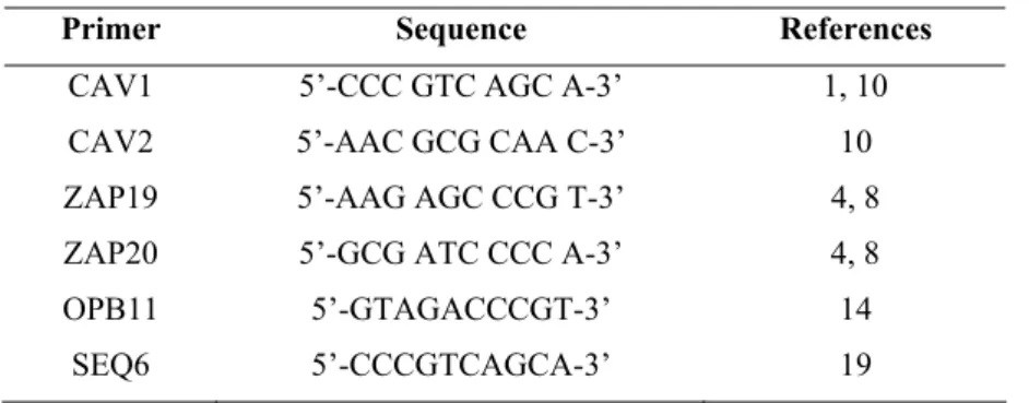
Table 1. Primer sequences for RAPD-PCR.
Primer Sequence References
CAV1 5’-CCC GTC AGC A-3’ 1, 10
CAV2 5’-AAC GCG CAA C-3’ 10
ZAP19 5’-AAG AGC CCG T-3’ 4, 8
ZAP20 5’-GCG ATC CCC A-3’ 4, 8
OPB11 5’-GTAGACCCGT-3’ 14
SEQ6 5’-CCCGTCAGCA-3’ 19
The Jaccard coefficient (Sj) was used to calculate the
distance between the isolates according to comparison of the
bands and their sizes (3). The Multi-variate Statistical Package
3.0 (MVSP 3.0, Kovach Computing Services, Pentraeth,
Wales, UK) software was used to analyze the differences. A
dendrogram was generated from the data obtained by the
cluster method UPGMA (Unweighted Pair Group Method
using Arithmetic Averages). A Sj value of 1.00 indicated that
the isolates were identical; values between 0.81 and 0.99
represented high similarity, but not identity, and suggest the
occurrence of microevolution on a given isolate; values below
0.80 represented unrelated isolates (3, 22).
RESULTS
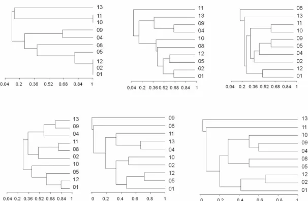
RAPD patterns for isolates in this study have been
translated into the dendrograms (Figures 1 to 3).
C. laurentii complex isolates had the greatest genetic
diversity. Identical patterns (Sj = 1.00) were found only with
primer CAV1 in a few isolates; three isolates (CRL01, CRL02
and CRL12) constituted one cluster and two isolates (CRL10
and CRL11) another one. All isolates were separated from each
other by some other primers, that separated all isolates with
similarities ranging from 0.00 to 0.800. The primer OPB11
showed that CRL09 is not related with any other isolates.
Figure 1 shows the dendrogram of C. laurentii complex
isolates.
C. albidus isolates, in general, showed higher genetic
similarity than C. laurentii isolates. Only the primer CAV2 did
not amplify any DNA bands. Primers ZAP19 and CAV1
formed similar clusters, that had at least seven identical
isolates; six groups (Sj = 0.135 to 0.800) were clustered by
SEQ6, four of them with a pair of identical isolates (Sj = 1.00).
OPB11 grouped six identical isolates (CRA01, CRA06,
CRA07, CRA08, CRA10 and CRA11) highly related to two
other isolates (CRA03 with Sj = 0.842 and CRA12, Sj = 0.875).
C. neoformans isolates, including ATCC strains, formed
three unrelated cluster (Sj ≤ 0.80), with the primers CAV1,
ZAP20 and OPB11, all of them grouping the same isolates,
distributed as follows: one cluster with eight identical isolates,
the other with three isolates and another one composed by
28957. Primer CAV2 formed two cluster, one formed by 28957
(var. neoformans) and the other grouping all identical isolates
(all of var. grubbi). The primers ZAP19 and SEQ6 (see Figure
4) had higher discriminatory power between the primers tested,
Figure 1. Dendrograms showing the relationships of C. laurentii isolates generated from the RAPD-PCR profiles obtained with
primers (from left to right) CAV1, CAV2 and ZAP19 (above); ZAP20, OPB11 and SEQ6 (below).
Figure 2. Dendrograms showing the relationships of C. albidus isolates generated from the RAPD-PCR profiles obtained with
Figure 3. Dendrograms showing the relationships of C. neoformans isolates generated from the RAPD-PCR profiles obtained with
primers (from left to right) CAV1, CAV2, ZAP19 (above); ZAP20, OPB11 and SEQ6 (below).
Figure 4. Examples of amplification obtained with the primer SEQ6. Line 1: molecular size marker (Lambda DNA/Hind III;
Fermentas); lines 2 to 13 represent C. neoformans isolates 13, 17, 18, 19, 20, 22, 23, 24, 25, 26, ATCC 90112 and ATCC 28957,
DISCUSSION
Molecular techniques have been widely used to study
genetic variations in uni- and multicellular organisms (1-4, 28),
and RAPD-PCR has been used in studies of human pathogenic
fungi as well as those causing diseases in animals and plants (1,
28). Molecular studies of microorganisms contribute to the
epidemiological studies and are also useful in the relationship
between molecular profile and phenotypic characteristics (12).
In this study, we observed high genetic variability among
isolates of C. laurentii complex with all the primers used, as
demonstrated in previous studies (19, 23, 25). The greatest
differences among isolates of C. laurentii complex (minor Sj)
were observed with the primers CAV2, ZAP19, OPB11 and
SEQ6. That heterogeneity is probably caused by different
species and subspecies that constitute the C. laurentii complex,
which can only be identified by more specific methods, such as
rDNA sequencing (D1/D2 and/or ITS regions).
In clinical or medical laboratory, especially in Brazil, the
laboratories are able to identify the C. laurentii complex, but
not the accurate specie or subspecie. In this line, the use of
molecular methods such as RAPD-PCR associated with the
phenotypic tests can help identify the species or subspecies of
the microorganisms and also their typing.
C. albidus is another specie that is reported as a species
complex. In some isolates have been observed a high genetic
heterogeneity, revealed by different molecular methods (1, 19,
24). However, in this study we demonstrated that most isolates
had high similarity or were identicals (Figure 2). Among the
primer used, we observed that CAV2 did not amplify any
bands. Despite the small number of C. albidus isolates studied,
the CAV2 primer seems inappropriate and ZAP20, OPB11 and
SEQ6 were the primers that formed more clusters and are most
promising for use in studies envolving C. albidus.
Isolates of C. neoformans were best discriminated by
primer SEQ6, that formed eight groups, one of them grouping
five isolates, including strain ATCC 90112, a strain that
belongs to the var. grubii isolated from the cerebrospinal fluid
in the United States. The identity revealed by most isolates of
C. neoformans suggests that new primers should be researched
to increase the discriminatory power. Among the primers
utilized, we noted that C. neoformans ATCC 28957, a strain of
the var. neoformans, formed an evident separate group with all
the primers. It is interesting to note that the primer CAV2
grouped all isolates except 28957, as shown in Figure 3.
The study of molecular markers is promising for the
typing of microorganisms. These studies are interesting to
study the relationship between molecular profile and
phenotypic characteristics, such as in vitro virulence factors
production (laccases, phospholipases, proteases), phenotypic
switching, resistance to antifungal drugs, or associations
between geographic origin of the isolates and molecular
profiles. Certain molecular profiles may be related to specific
features of the phenotype, as has been shown for C.
neoformans and another species isolates from different parts of
the world (1, 3, 4, 10, 14, 26). Recently the evidence of the
relationship between susceptibility to antifungal agents and
different genotypes of C. neoformans/ C. gattii complex has
increased (26).
The use of a single molecular marker is usually not
sufficient to discriminate a large number of isolates. The
establishment of a rational screening protocol, with primers
carefully chosen and carried out in sequential reactions, or even
the sequential analysis of results can allow discrimination of
isolates or even increase the discriminatory power of the
method, expanding the field of application of molecular
markers. More studies with larger numbers of isolates and
primers, available for practical application in clinical and
research laboratories, will facilitate the work of researchers,
will reduce cost and time consuming.
In conclusion, we observed that the use of limited number
of carefully selected primers allowed the discrimination of
different isolates, and not every primers (e.g., CAV2 for C.
C. laurentii complex will be required to detail the identification
at the level of the species or subspecies, for example, by
comparison with a validated method such as rDNA sequencing
(D1/D2 domain or ITS). More studies, however, are necessary
to determine the effectiveness of this tool and primers studied
here and other in clinical isolates, as well as in veterinary and
environmental isolates.
ACKNOWLEDGEMENTS
This study was supported in part by CNPq (Conselho
Nacional de Desenvolvimento Científico e Tecnológico),
Brazil.
REFERENCES
1. Almeida, A.M.F.; Matsumoto, M.T.; Baeza, L.C.; Oliveira e Silva, R.B.; Kleiner, A.A.; Melhem, M.D.E.S.; Mendes-Giannini, M.J.; Laboratory Group on Cryptococcosis. (2007). Molecular typing and antifungal susceptibility of clinical sequential isolates of Cryptococcusneoformans
from São Paulo State, Brazil. FEMS Yeast Res. 7 (1), 152-164.
2. Arif, I.A.; Bakir, M.A.; Khan, H.A.; Al Farhan, A.H.; Al Homaidan, A.A.; Bahkali, A.L.; Al Sadoon, M.; Shobrak, M. (2010). A brief review of molecular techniques to assess plant diversity. Int. J. Mol. Sci. 11 (5), 2079-2096.
3. Bacelo, K.L.; Costa, K.R.C.; Ferreira, J.C.; Candido, R.C. (2010). Biotype stability of Candida albicans isolates after culture storage determined by randomly amplified polymorphic DNA and phenotypical methods. Mycoses. 53 (6), 468-474.
4. Blasi, E.; Brozzetti, A.; Francisci, D.; Neglia, R.; Cardinali, G.; Bistoni, F.; Vidotto, V.; Baldelli, F. (2001). Evidence of microevolution in a clinical case of recurrent Cryptococcus neoformans meningoencephalitis.
Eur. J. Clin. Microbiol. Infect. Dis. 20 (8), 535-543.
5. Boekhout, T.; Belkun, A.; Leenders, A.C.A.; Verbrugh, H.A.; Mukamurangwa, P.; Swinne, D.; Scheffers, W.A. (1997). Molecular typing of Cryptococcus neoformans: taxonomic and epidemiological aspects. Int. J. Syst. Bacteriol. 47 (2), 432-442.
6. Boekhout, T.; Scorzetti, G. (1997). Differential killer toxin sensitivity patterns of varieties of Cryptococcus neoformans. J. Med. Vet. Mycol. 35 (2), 147-149.
7. Bolano, A.; Stinchi, S.; Preziosi, R.; Bistoni, F.; Allegrucci, M.; Baldelli, F.; Martini, A.; Cardinali, G. (2001). Rapid methods to extract DNA and RNA from Cryptococcus neoformans. FEMS Yeast Res. 1 (3), 221-224.
8. Cardinali, G.; Martini, A.; Preziosi, R.; Bistoni, F.; Baldelli; F. (2002). Multicenter comparison of three different analytical systems for evaluation of DNA banding patterns from Cryptococcus neoformans. J. Clin. Microbiol. 40 (6), 2095-2100.
9. Casadevall, A.; Perfect, J.R. (1998). Cryptococcus neoformans. ASM Press, Washington, D.C.
10. Cavalcante, S.C.; Freitas, R.S.; Vidal, M.S.; Dantas, K.C.; Levi, J.E.; Martins, J.E. (2007). Evaluation of phenotypic and genotypic alterations induced by long periods of subculturing of Cryptococcus neoformans
strains. Mem. Inst. Oswaldo Cruz. 102 (1), 11-17.
11. Fonseca, A.; Scorzetti, G.; Fell, J.W. (2000). Diversity in the yeast
Cryptococcus albidus and related species as revealed by ribosomal DNA sequence analysis. Can. J. Microbiol. 46 (1), 7-24.
12. Foxman, B.; Riley, L. (2001). Molecular epidemiology: focus on infection. Am. J. Epidemiol. 153 (12), 1135-1141.
13. Hunter, P.R. (1991). A critical review of typing methods for Candida albicans and their applications. Crit. Rev. Microbiol. 17 (6), 417-434. 14. Jain, P.; Khan, Z.K.; Bhattacharya, E.; Ranade, S.A. (2001). Variation in
random amplified polymorphic DNA (RAPD) profiles specific to fluconazole resistant and sensitive strains of Candida albicans. Diagn. Microbiol. Infect. Dis. 41 (3), 113-119.
15. Khawcharoenporn, T.; Apisarnthanarak, A.; Mundy, L.M. (2007).
Non-neoformans cryptococcal infections: a systematic review. Infection. 35 (2): 51-58.
16. Kwon-Chung, K.J.; Bennett, J.E. (1992). Medical mycology. Lea & Febiger, Philadelphia.
17. Meyer, W.; Aanensen, D.M.; Boekhout, T.; Cogliati, M.; Dias, M.R.; Esposto, M.C.; Fisher, M.; Gilgado, F; Hagen, F.; Kaocharoen, S.; Litvintseva, A.P.; Mitchell, T.G.; Simwami, S.; Trilles, L.; Viviani, M.A.; Kwon-Chung, J. (2009). Consensus multi-locus sequence typing scheme for Cryptococcus neoformans and Cryptococcus gattii. Med. Mycol. 47 (6): 561-570.
18. Pedroso, R.S.; Ferreira, J.C.; Candido, R.C. (2009). The isolation and characterization of virulence factors of Cryptococcus spp. from saprophytic sources in the city of Ribeirão Preto, São Paulo, Brazil.
Microbiol. Res. 164 (2): 221-227.
19. Pedroso, R.S.; Ferreira, J.C.; Lavrador, M.A.; Maffei, C.M.; Candido, R.C. (2009). Evaluation of the experimental inoculation of Cryptococcus albidus and Cryptococcus laurentii in normal mice: virulence factors and molecular profile before and after animal passage. Mycopathologia. 168 (2): 59-72.
20. Riederer, K.M.; Ramanathan, J.; Barczak, J.; Baran, J.; Khatib, R. (2002). Utility of a pre-optimized kit for random amplified polymorphic DNA in typing Candidaalbicans. Can. J. Microbiol. 48 (4): 369-373. 21. Sidrim, J.J.C.; Costa, A.K.F.; Cordeiro, R.A.; Brilhante, R.S.N.; Moura,
Cryptococcus: a review. Can. J. Microbiol. 56 (6): 445-458.
22. Soll, D.R. (2000). The ins and outs of DNA fingerprinting the infections fungi. Clin. Microbiol. Rev. 13 (2): 332-370.
23. Sugita, T.; Takashima, M.; Ikeda, R.; Nakase, T.; Shinoda, T. (2000). Intraspecies diversity of Cryptococcus laurentii as revealed by sequences of internal transcribed spacer regions and 28S rRNA gene and taxonomic position of Cryptococcus laurentii clinical isolates. J. Clin. Microbiol. 38 (4): 1468-1471.
24. Sugita, T.; Takashima, M.; Ikeda, R.; Nakase, T.; Shinoda, T. (2001). Intraspecies diversity of Cryptococcus albidus isolated from humans as revealed by sequences of the internal transcribed spacer regions.
Microbiol. Immunol. 45 (4): 291-297.
25. Takashima, M.; Sugita, T.; Shinoda, T.; Nakase, T. (2003). Three new
combinations from the Cryptococcus laurentii complex: Cryptococcus aureus, Cryptococcus carnescens and Cryptococcus peneaus. Int. J. Syst. Evol. Microbiol. 53 (Pt 4): 1187-1194.
26. Trilles, L.; Meyer, W.; Wanke, B.; Guarro, J.; Lazera, M. (2012). Correlation of antifungal susceptibility and molecular type within the
Cryptocccus neoformans/ C. gattii species complex. Med. Mycol. 50 (3): 328-332.
27. Williams, J.G.K.; Kubelik, A.R.; Livak, K.J.; Rafalski, J.A.; Tingey, S.V. (1990). DNA polymorphisms amplified by arbitrary primers are useful as genetic markers. Nucleic Acids Res. 18 (22): 6531-6535. 28. Zaidi, N.; Konstantinou, K.; Zervos, M. (2003). The role of molecular
biology and nucleic acid technology in the study of human infection and epidemiology. Arch. Pathol. Lab. Med. 127 (9): 1098-1105.