online | memorias.ioc.fiocruz.br Rotaviruses belong to the family Reoviridae and are
the major etiological agents of severe diarrhoea affect-ing children less than five years of age worldwide (Par-ashar et al. 2006, Esposito et al. 2011). Their virions are composed of three concentric protein layers that encap-sidate 11 double-stranded RNA genome segments (Estes & Kapikian 2007). Over 500,000 children, most of them from developing countries, die annually from rotavirus infection and many more require hospitalisation for treat-ment of the severe and dehydrating rotavirus-associated diarrhoea (Parashar et al. 2006, Danchin & Bines 2009). An important reduction in rotavirus-caused deaths has been reported as a consequence of the use of two recent-ly introduced rotavirus vaccines (RotaTeq, Merck, and Rotarix, GlaxoSmithKline Biologicals) (Nelson & Glass 2010, Santosham 2010). However, the potential of rotavi-rus vaccines for reducing the risk of death from diarrhoea in the poorest countries has been challenged by logistical problems, the age-restricted recommendation for vaccine administration, the low vaccine coverage and the low on-time immunisation (Clark & Sanderson 2009, Santosham 2010). The above facts justify the attempts aimed at de-veloping alternative or complementary strategies for pre-venting or treating the rotavirus-associated diarrhoea.
Balancing reduction and oxidation seems to be a cru-cial event for maintaining life while aging has been con-sidered a process involving gradual oxidation (Kregel & Zhang 2007, Cui et al. 2012). Cellular mechanisms that deal with damaging oxidative environments include re-dox systems such as glutathione (GSH)/GSSG (the GHS system), NADH/NAD+, NADPH/NADP+ and Trx(SH)2/ Trx(S-S) (the thioredoxin system). Oxidative stress has been shown to be involved in the pathogenesis of viral infections by human immunodeficiency virus (HIV), in-fluenza virus and dengue virus in which GSH levels have been found to be decreased and reactive oxygen species (ROS) levels increased (Garland & Fawzi 1999, Cai et al. 2003, Nencioni et al. 2003, Tian et al. 2010). Redox imbalance has also been reported to occur in infections caused by rabies virus (Jackson et al. 2010), herpes sim-plex virus type 1 (Kavouras et al. 2007), hepatitis C vi-rus (Clément et al. 2009) and hepatitis B vivi-rus (Severi et al. 2006). N-acetylcysteine (NAC), an anti-oxidant that can function in the body as precursor of GSH (Atkuri et al. 2007), has been used in the treatment of influenza virus infections (de Flora et al. 1997, Ghezzi & Ungheri 2004, Garozzo et al. 2007, Lai et al. 2010) and also in the treatment of dengue-associated fulminant liver fail-ure (Lim & Lee 2012). It has been found that I-152, a pro-drug of NAC and GSH, was able to inhibit murine acquired immune deficiency syndrome (Ho & Dougla 1992, Fraternale et al. 2008). GSH and NAC have been shown to inhibit the induction of HIV-1 expression in human monocyte/macrophages (Ho & Dougla 1992).
Traditional nonsteroidal anti-inflammatory drugs (NSAIDs) are known to inhibit both isoforms of cycloox-doi: 10.1590/0074-0276108062013011
+ Corresponding author: [email protected] Received 10 February 2013
Accepted 21 June 2013
Inhibition of rotavirus ECwt infection in ICR suckling mice
by N-acetylcysteine, peroxisome proliferator-activated
receptor gamma agonists and cyclooxygenase-2 inhibitors
Carlos Arturo Guerrero/+, Paula Pardo, Victor Rodriguez, Rafael Guerrero, Orlando Acosta
Departamento de Ciencias Fisiológicas, Facultad de Medicina, Instituto de Biotecnología, Universidad Nacional de Colombia, Bogotá, DC, Colombia
Live attenuated vaccines have recently been introduced for preventing rotavirus disease in children. However, alternative strategies for prevention and treatment of rotavirus infection are needed mainly in developing coun-tries where low vaccine coverage occurs. In the present work, N-acetylcysteine (NAC), ascorbic acid (AA), some nonsteroidal anti-inflammatory drugs (NSAIDs) and peroxisome proliferator-activated receptor gamma (PPARγ) agonists were tested for their ability to interfere with rotavirus ECwt infectivity as detected by the percentage of viral antigen-positive cells of small intestinal villi isolated from ECwt-infected ICR mice. Administration of 6 mg NAC/kg every 8 h for three days following the first diarrhoeal episode reduced viral infectivity by about 90%. Administra-tion of AA, ibuprofen, diclofenac, pioglitazone or rosiglitazone decreased viral infectivity by about 55%, 90%, 35%, 32% and 25%, respectively. ECwt infection of mice increased expression of cyclooxygenase-2, ERp57, Hsc70, NF-κB, Hsp70, protein disulphide isomerase (PDI) and PPARγ in intestinal villus cells. NAC treatment of ECwt-infected mice reduced Hsc70 and PDI expression to levels similar to those observed in villi from uninfected control mice. The present results suggest that the drugs tested in the present work could be assayed in preventing or treating rotaviral diarrhoea in children and young animals.
ygenase (COX)-1/2, key enzymes in the production of prostaglandins (Rao & Knaus 2008). Induction of COX-2 expression appears to be regulated by extracellular signal-regulated protein kinases (ERK)1/2 and p38 mitogen-acti-ERK)1/2 and p38 mitogen-acti-vated protein kinase (MAPK) pathways and transcription factors, such as nuclear factor kappa B (NF-κB) (Newton et al. 1997, Bartlett et al. 1999, Subbaramaiah et al. 2000, Charalambous et al. 2003). PKA-mediated ERK1/2, p38 MAPK and NF-κB pathways have been shown to be in-volved in the COX-2 activity induction during rotavirus infection (Rossen et al. 2004). NF-κB activation has been shown to occur during infection by several viruses, in-cluding rotaviruses (Roulston et al. 1999, Rossen et al. 2004). Rotaviruses have also been found to activate NF- κB-dependent gene expression and induce the increased expression of interleukin-8 and other cytokines (Sheth et al. 1996, Rollo et al. 1999, LaMonica et al. 2001, Casola et al. 2002). It has been shown that indomethacin (a COX-1 and COX-2 inhibitor) treatment of human intestinal Ca-co-2 cells significantly reduced rotavirus infectivity by affecting a post-binding step and inhibiting virus-direct-ed protein synthesis (Rossen et al. 2004). On the other hand, peroxisome proliferator-activated receptor gamma (PPARγ) ligands have been found to down-regulate the transcriptional activation of COX-2 through multiple mechanisms (Subbaramaiah et al. 2001), which include the inhibition of some NF-κB pathway steps (Straus et al. 2000). PPARγ modulates ROS generation and activation of redox-sensitive NF-κB and hypoxia-inducible factor 1-alpha in mice (Lee et al. 2006). Central inflammatory pathways such as NF-κB, activator protein 1 and STAT have been found to be antagonised by PPARs in mono-cyte-macrophages, whereas down-regulation of these signalling pathways by thiazolidine-2-4-diones (TZDs), including pioglitazone (PGZ) and rosiglitazone (RGZ), has been shown to lead to reduced levels of oxidative products (Jiang et al. 1998). It has been found that TZDs were able to attenuate hyperglycaemia-induced ROS pro-duction by inducing manganese superoxide dismutase and promoting mitochondrial biogenesis (Fujisawa et al. 2009). Recently, PPARγ agonists have been suggested as a potential approach for down-regulating the inflamma-tory response to virus-induced inflammation (Bassagan-ya-Riera et al. 2010).
Although rotavirus diarrhoea has been attributed to different mechanisms, rotaviruses proliferation in the non-dividing mature enterocytes localised near the tips of the villi is a factor contributing to induce diar-rhoea (Ramig 2004). Homeostatic control of the intes-tinal redox environment seems to be a critical factor for maintaining intestinal functions. Cells from intestinal epithelium are exposed to oxidative damage induced by oxidant agents present in the luminal environment. Mucosal integrity is ensured by the luminal redox bal-ance of the GSH/GSSG and cysteine/cystine (Cys/CySS) couples, which are also involved in supporting luminal nutrient absorption, mucus fluidity and microbiota (Go & Jones 2008, Jones & Go 2010, Circu & Aw 2012). Nor-mal intestinal cell transition from proliferative state to non-dividing differentiated state or apoptosis has been
associated with increasing oxidation of intracellular GSH/GSSG or extracellular Cys/CySS redox systems (Jones 2006, Jones & Go 2010). Recent advances in in-testinal redox biology suggest that the loss of inin-testinal homeostasis caused by oxidative stress in the mucosal and adjacent tissues can alter nutrient digestion and ab-sorption, stem cell proliferation, enterocyte apoptosis and immune response (Circu & Aw 2012).
Rotavirus infectivity has recently been shown to be inhibited by the treatment of cells with membrane imper-meant thiol/disulfide exchange inhibitors and antibodies (Abs) against protein disulphide isomerase (PDI), which suggested that infectivity was dependent on cell surface oxidation-reduction reactions (Calderon et al. 2012). Some findings have suggested that the rotavirus struc-tural protein (SP) VP7 is a substrate for the endoplasmic reticulum (ER)-associated oxidant PDI (Svensson et al. 1994, Mirazimi & Svensson 1998, Maruri-Avidal et al. 2008). In addition, NAC, NASIDs and PPARγ agonists have recently been found to inhibit rotavirus infection in cultured MA104 cells (Guerrero et al. 2012). Understand-ing the molecular and cellular disorders caused in vivo by rotavirus infection, such as oxidative stress, could be useful in designing therapeutic strategies aimed at inter-fering with the cell injury associated with the rotavirus-induced pro-oxidant status. The present work provides evidence that rotavirus infectivity is inhibited in ICR mice treated with NAC, PGZ or RGZ, which are drugs that down-regulate the NF-κB pathway involved in the transcriptional activation of COX-2 reported to occur during rotavirus infection (Rossen et al. 2004).
MATERIALS AND METHODS
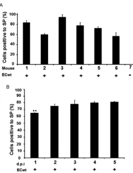
Rotavirus infection of mice - The rotavirus inoculum consisting of cesium chloride-purified ECwt triple-lay- triple-lay-ered particles (TLPs) was adjusted to produce the first diarrhoeal event at 24 h post-inoculation (h.p.i.). Differ-ent volumes (0.1, 1, 2 and 5 µL) from a stock preparation [7.8 x 10⁷ focus forming units (FFU)/mL)] were diluted with PBS to reach 100 µL and then directly applied into the 10-12 day old mouse pharynx. The lowest stock vol-ume (0.1 µL) was routinely used to produce diarrhoea at 24 h.p.i. To determine the proportion of intestinal epithe-lial cells (IEC) infected in each mouse following three days post-inoculation (d.p.i.), six mice were inoculated with ECwt TLPs whereas one mouse was left uninocu-lated as a control. The percentage of infected IEC was determined from the small intestinal villi isolated from virus-infected mice and uninfected control using the im-munochemistry assay described below. The time-course of the percentage of infected IEC was determined by inoculating 10 mice with ECwt TLPs and slaughtering them at one-five d.p.i. The percentage of infected IEC was visualised on isolated intestinal villi by immuno-chemistry. Comparisons were made against cells from uninoculated control mice.
Immunochemistry assay - Small intestinal villi from infected mice were prepared for immunochemistry ana- lysis as previously described (Guerrero et al. 2010). Cells were fixed with ice-cold methanol acetic acid (3:1) for 20 min and then place onto coverslips. After drying, en-dogenous peroxidase was inactivated by treatment with 3% H2O2 in 50% methanol. Fixed cells were reacted with rabbit polyclonal Abs (produced in our animal facilities) against rotavirus SPs or non-structural proteins NSP4 or NSP5. A horseradish peroxidase (HRP)-conjugated goat anti-rabbit Ab (Santa Cruz Biotechnology Inc) was used as secondary Ab. Peroxidase activity was revealed with aminoethylcarbazole (AEC) substrate (Sigma, St. Louis, MO, USA). Small intestinal villi from infected and drug-treated mice were submitted to the same pro-cedure as well as those from non-infected control mice. Ten representative fields were photographed at 40X magnification and the proportion of positive cells to in-fection was expressed as mean percentage relative to the total recorded cells.
ELISA - Capture ELISA was used for determining the accumulation of viral antigen in intestinal villi from infected mice which were either treated with drugs or left untreated. Procedures were essentially as those pre-viously described (Guerrero et al. 2010). Briefly, villi preparations isolated from the entire small intestine were gently agitated immediately before taking sam-ples for assay to ensure homogeneity in villi composi-tion. Isolated villi samples containing about 10 mg/mL were lysed in modified radio immunoprecipitation assay (RIPA) buffer (50 mM Tris-HCl, pH 7.5, 150 mM NaCl, 0.1% Tritonin X-100, 1% NP-40, 0.5% deoxycholate) and subjected to centrifugation. The 2,500 g supernatant was applied to ELISA plate wells previously coated with pol-yclonal guinea pig Abs (1:1000) against rotavirus SP. Af-ter PBS washing, polyclonal rabbit anti-rotavirus SP Abs (1:3000) were added and after washing away unbounded
Abs with PBS, HRP-conjugated donkey anti-rabbit IgG secondary Abs (0.08 µg/mL Santa Cruz Biotechnology Inc) (SC-2313) were added. The reaction was revealed using o-phenylenediamine dihydrochloride in stable peroxidase buffer (Pierce, Rockford, IL, USA). Optical density (OD)492 nm was determined using an ELISA plate reader. Background OD values from uninfected villus lysates were subtracted from all infected samples. The cut-off value was calculated as the mean plus two stand-ard deviations of the negative control wells. ECwt TLPs were used as an internal positive control. Expression of Hcs70 and PDI in intestinal villi isolated from ECwt in-fected mice which had or had not been treated with NAC (18 mg/kg/day) were also analysed by ELISA.
Intestinal villus isolation from infected mice - A previously described procedure for the small intestinal villus isolation was used (Guerrero et al. 2010). Briefly, entire small intestines were flushed with ice-cold mini-mum essential medium (MEM) containing 1.5 mM eth-ylenediamine tetraacetic acid (EDTA) and antibiotic/ antimycotic solution before being opened longitudinally and cut into 0.5-cm-long segments. Following incuba-tion and agitaincuba-tion in the same medium at 37ºC for 15 min, the cell/tissue preparation was further dispersed by gentle pipetting and immediately passed through a ster-ile metal net (1 mm2). After the dispersed preparation was filtered twice, the filtered product was collected by low centrifugation to be subsequently resuspended and washed in MEM containing antibiotic/antimycotic solu-tion. The intestinal villus-enriched preparation was used for recording the percentage of ECwt infected cells and the in vitro titration of virus infectivity.
Virus purification - Small intestines isolated from ECwt infected mice were homogenised with MEM con-taining antibiotic/antimycotic solution and submitted to a freeze-thaw cycle before 1,1,2-trichlorotrifluoroethane (Sigma, St. Louis, MO, USA) extraction and sucrose/ce-sium chloride gradient centrifugation as previously de-scribed (Gualtero et al. 2007). Visible virus bands were collected by aspiration with a syringe, diluted with tris buffered saline (TBS) (10 mM Tris–HCl, pH 7.4, 150 mM NaCl, 1 mM MgCl2, 5 mM CaCl2) and collected by high speed centrifugation (150,000 g for 1.5 h at ºC) prior to resuspension in TBS. Virus infectivity was ti-trated in vitro on isolated intestinal villus (Guerrero et al. 2010) using an immunochemistry assay as previ-ously described.
uninfected mice administered with PBS and ECwt in-fected mice treated with PBS were used as controls. The specificity of NAC inhibitory effect on ECwt infection was determined by infecting comparable ICR mice with purified human reovirus type 1 (Sharpe et al. 1978) using two oral inoculations (100 µL; 8 x 10⁴ FFU/mL) separated by 12 h. Following the first diarrhoeal episode (approximately 48 h.p.i), reovirus-infected mice were orally administered NAC (18 mg/kg/day) three times a day for three days. Six hours after the last dose, the mice were killed and their small intestinal villi isolated for immunochemistry analysis of the percentage of virus in-fected cells. Anti-rotaviral effect of NAC and NTZ were compared by treating ECwt infected mice with NAC (18 mg/kg/day) or NTZ (7.5 mg/kg/day) as described above. Antiviral effect was determined by immunocytochemis-try analysis in terms of the mean percentage of virus in-fected villus cells relative to villus cells from uninin-fected and drug-untreated control mice.
Expression of COX, ERp57, Hsc70, NF-κB, Hsp70, PDI and PPARγ in villus cells from infected mice - Fol-lowing three d.p.i., expression of COX, ERp57, Hsc70, NF-κB, Hsp70, PDI and PPARγ was assessed by indi -rect immunofluorescence in cross-sections of mouse small intestines or flow cytometry analysis of IEC. Small intestine cross-sections (1 cm) were fixed in 10% paraformaldehyde in PBS and embedded into paraffin wax. Paraffin-embedded sections (3-5 µm) were placed onto coverslips, deparaffinised in xylene and rehydrat-ed in a gradrehydrat-ed ethanol series. Endogenous peroxidase activity was quenched by treating sections with 3% H2O2 in 50% methanol for 15 min and then cells were permeabilised with 1% sodium dodecyl sulfate (SDS) for 5 min. After washing with PBS, non-specific bind-ing was blocked with 1% bovine serum albumin (BSA) for 15 min. ECwt infection was assessed by applying rabbit polyclonal anti-rotavirus SP Ab to the sections followed by PBS washing and addition of HRP-conju-gated goat anti-rabbit secondary Ab. Immunostaining was visualised using AEC chromogen. The same cov-erslips were washed three times with PBS and then in-cubated with 50 mM NH4Cl for 30 min. Sections were incubated with the first goat Ab against COX-2, ERp57, Hsc70, Hsp70, PDI, PPARγ (0.2 µg/mL; SC-1747, SC18619, SC1059, SC1060, SC17222, SC6285, Santa Cruz Biotechnology Inc) or rabbit Ab against NF-κB (0. 25 µg/mL; 51-3500, Zymed) in PBS containing 50 mM NH4Cl for 1 h at 37ºC. After three washes with PBS, fluorescein isothiocyanate (FITC)-conjugated donkey anti-goat Ab (0.08 mg/mL; SC-2024, Santa Cruz Bio-technology Inc) or FITC-conjugated donkey anti-rabbit Ab (0.08 mg/mL; SC-2090, Santa Cruz Biotechnology Inc) diluted in PBS containing 1% BSA were added. Following PBS washing, sections were mounted in 70% glycerol in PBS and examined with a fluorescence microscope (VanGuard).
Flow cytometry analysis of COX-2, Hsc70, NF-κB, Hsp70, PDI or PPARγ expression was performed by fix -ing PBS/EDTA (5 mM)-isolated IEC from ECwt infected mice (3 d.p.i.) in methanol/acetic acid (3:1 v/v) for 1-4ºC
before incubation with 50 mM NH4Cl. Goat primary Abs against the above mentioned proteins and rabbit primary Abs against rotavirus SP were mixed together diluted in PBS containing 50 mM NH4Cl and then added to IEC. Following three washes with PBS, FITC-conjugated mouse anti-goat IgG secondary Abs (0.88 µg/mL; SC-2356, Santa Cruz Biotechnology Inc) and phycoerythrin-conjugated anti-rabbit IgG Abs (0.88 µg/mL; SC-3753, Santa Cruz Biotechnology Inc) diluted in PBS supple-mented with 50 mM NH4Cl and 1% BSA were added. Immunofluorescence analysis was performed on a Cyan (Dako) flow cytometer using FlowJo software (Tree Star, Ashland, OR, USA). Flow cytometry analysis was also used for assessing Hsc70 and PDI expression in IEC iso-lated from ECwt infected mice (3 d.p.i.) which had or had not been treated with NAC (18 mg/kg/day).
For confocal microscopy analysis of Hsc70 and PDI expression, deparaffinised and rehydrated sections from intestines isolated from ECwt-infected mice that had or had not been treated with NAC (18 mg/kg/day), IBF (20 mg/kg/day), DCF (1 mg/kg/day) or PGZ (30 mg/kg/day) were incubated with primary goat anti-Hsc70 or anti-PDI Abs mixed together with rabbit anti-rotavirus SP Abs. After three washes with PBS, FITC-conjugated donkey anti-rabbit IgG Abs (0.88 µg/mL; SC-2090, Santa Cruz Biotechnology Inc) and Alexa Flour 568-conjugated donkey anti-goat IgG Abs (4.4 µg/mL; A-11057) (Inv-itrogen) diluted in PBS containing 1% BSA were added together to the sections. Subsequently, they were washed with PBS and the nuclei were counterstained with 4’-6-diamidino-2-phenylindole (0.5 µg/mL). Coverslips were mounted in 70% glycerol and images examined using a confocal microscope (Nikon C-1). Pearson’s co-local-isation analysis and image processing software Image J were used to assess the co-localisation of Hsc70, PDI and rotavirus proteins.
sulfate polyacrylamide gel electrophoresis (SDS-PAGE) (10%) and transfer to PVDF membranes. The target pro-teins were probed with primary goat Abs against Hsc70 (0.2 µg/mL; SC-1059, Santa Cruz Biotechnology Inc) or PDI (0.2 µg/mL; 20132, Santa Cruz Biotechnology Inc). HRP-conjugated donkey anti-goat IgG Ab (0.4 µg/mL; SC-2020, Santa Cruz Biotechnology Inc) was used as a secondary Ab and the reaction was developed with intensified luminescence (Pierce) or AEC. Commercial Abs against COX-2, ERp57, Hsc70, Hsp70, PDI, PPARγ or NF-κB were monitored by WB for the absence of Abs against rotavirus proteins. Purified rhesus rotavirus (RRV) triple layered particles (TLPs) were subjected to SDS-PAGE/WB analysis using commercial preparations as primary Abs and the respective HRP-conjugates as secondary Abs and AEC substrate.
Statistical analysis - The difference in mean and ho-mogeneity of variance of infectivity or antigen accumu-lation in cells was tested by one-way analysis of variance and the Student’s t test. Results were considered statisti-cally significant if the p value is under 0.01.
RESULTS
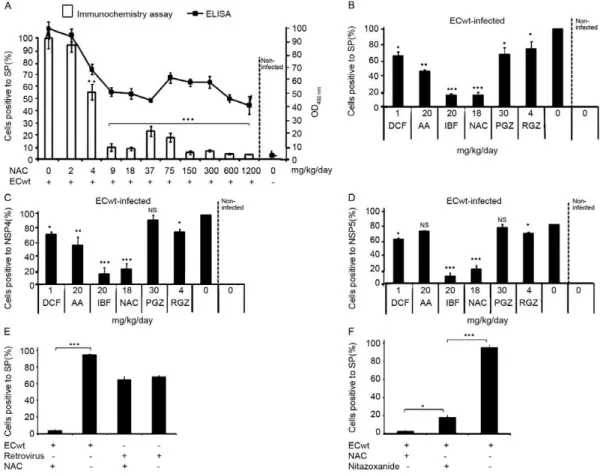
NSAIDs, PPARγ agonists and NAC treatment reduce ECwt infectivity in ICR mice - The cell viability of IEC of small intestinal villi isolated from uninfected mice was estimated to be about 80% by the Trypan blue ex-clusion test. The inoculation of six mice with ECwt re-sulted in the infection of IEC as determined by immuno-chemistry analysis of small intestinal villi isolated from the entire small intestine. After three d.p.i., the propor-tion of IEC resulting positive for virus infecpropor-tion ranged from 57-95%, whereas the mean infectivity was about 75% (Fig. 1A). In all cases, this mean percentage was taken as 100% infectivity for control villi isolated from drug-untreated mice. The absence of differential ECwt infection for small intestine sections has been previously reported for isolated villi (Guerrero et al. 2010). To ana- lyse the time-course of ECwt infectivity, two mice were killed every 24 h following virus inoculation. The re-sults indicated that no significant differences were found in infectivity through four d.p.i. when using an ECwt in-oculum consisting of 7.8 x 10⁴ FFU/mL per mouse (Fig. 1B). The relatively low dispersion values observed for virus infectivity through the period examined suggested that three d.p.i. could be a suitable post-inoculation time for testing drug effects on infectivity. To test for NAC effect on ECwt infectivity, mice at 24 h.p.i. were admin-istered 2 mg NAC/kg/day. After the three-day treatment, infectivity remained similar to that of untreated infected mice (Fig. 2A). Administration of NAC 4 mg/kg/day reduced infectivity to 55%, whilst NAC doses ranging from 9-1,200 mg/kg/day were able to reduce infectiv-ity to 10-4% (Fig. 2A). This NAC dose range thus pro-duced an infectivity reduction that fluctuated between 78.7-94.7%. In searching for a correlation between the percentage of infected cells and the total rotavirus struc-tural antigen accumulated in IEC from isolated villi, an ELISA analysis was conducted. OD492 nm from ELISA plate wells suggested a dose-dependent inhibitory effect
at low NAC concentrations similar to that observed from immunochemistry assay. However, this inhibitory pro-file from ELISA values was not entirely maintained at higher NAC concentrations (Fig. 2A). The effect of other drugs such as NSAIDs (DCF, IBF), PPARγ agonists (PGZ, RGZ), AA and diphenoxylate-HCl on ECwt in-fectivity was determined by detecting the percentage of IEC resulting positive for rotavirus antigen. When using immunocytochemistry detection of SP, NSP4 or NSP5 as rotavirus antigen in isolated villi, the respective in-hibitory effect was as follows: DCF, 35%, 27% and 23%, IBF, 90%, 90% and 93%, PGZ, 32%, 5% and 5%, RGZ, 25%, 20% and 20%, AA, 55%, 50% and 20% and NAC, 90%, 80% and 80% (Fig. 2B-D). The differential rotavi-rus antigen detection led to conclude that regardless of the virus antigen used for determining the percentage of infected cells, the inhibitory trend was similar for each drug tested. However, the highest inhibitory effects were obtained with administration of IBF and NAC.
The specificity of NAC inhibition on rotavirus infec-tion was tested by inoculating mice with human reovi-rus type 1 and then treating them with NAC. This drug treatment did not have any detectable inhibitory effect on reovirus infection as the percentage of infected vil-lus cells remained about 66% which was quite similar to that (67%) found in reovirus infected mice without NAC treatment (Fig. 2E). In contrast, villus cells from ECwt infected mice without NAC treatment showed an
infection of 95% which was reduced to 3% after NAC treatment. To compare the NAC inhibitory effect with that of NTZ (a drug used for treating rotaviral diarrhoea in children over 1 year of age), ECwt infected mice were treated with either drug following the first diarrhoeal episode. Villus cells from NAC-treated mice (18 mg/ kg/day) showed a mean percentage infection of 3% af-ter the three-day treatment as compared with villus cells from NTZ-treated mice (7.5 mg/kg/day) which showed a
mean percentage infection of 17%. Infection percentage for villus cells from control virus-infected mice without drug treatment was 95% (Fig. 2F).
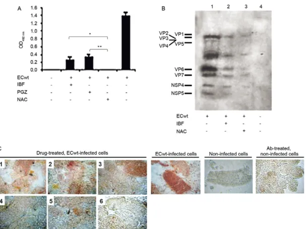
The inhibitory effect of IBF, PGZ or NAC on ECwt infection was further investigated by assessing the viral antigen accumulation in villus cells from ECwt-infected mice after a three-day treatment following the first diar-rhoeal event. ELISA results showed that the total ECwt antigen accumulated was reduced by about 82%, 76.4% and 100% when mice were treated with IBF, PGZ and NAC, respectively. ELISA results were expressed as mean OD492 nm values after subtracting mean background values of villus cell lysates from non-infected mice (Fig. 3A). Similar results were obtained after analysing pro-teins of villus cell lysates from virus-infected mice that had been treated with IBF or PGZ. Western analysis of
lysate proteins led to conclude that both drugs drasti-cally reduced the accumulation of both SP and NSP in cells from drug-treated mice as compared with samples from untreated control mice (Fig. 3B). The inhibitory effect of the above drugs, together with that from AA, DCF, RGZ and diphenoxylate was further confirmed by immunocytochemistry analysis. As shown in the repre-sentative images in Fig. 3C, drug treatment resulted in considerable reduction of cells positive for Abs against rotavirus SP when mice were treated with AA, IBF or NAC. An inhibitory effect, although lesser in degree, was observed in cells from virus-infected mice that were treated with RGZ, PGZ or DCF (Fig. 3C).
The effectiveness of the NAC inhibition of ECwt in-fectivity depends of the frequency of NAC application and the post-infection time of treatment - The
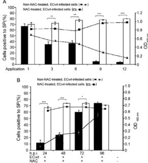
diated inhibition of ECwt infectivity shown above was further investigated in terms of its dependence on the frequency of dose application. When NAC was adminis-tered at only one dose of 6 mg/kg/day following the first diarrhoeal episode at about 24 h, the percentage of ECwt infected villus cells was 68% after 4 h from the appli-cation, whereas the percentage for virus infected and NAC-untreated control cells was 69%. Administration of 6 mg NAC/kg every 8 h for one day (18 mg/kg/day) or for two days, which corresponded to three-six total applications, respectively, led to a reduced proportion of infected cells (34%) in comparison to the percentage ob-served in NAC-untreated control cells (Fig. 4A). Main-taining the same dose but during three (9 applications) or four (12 applications) days resulted in a considerable decrease of the percentage of virus infected villus cells (4%) when comparison was made with that of cells from NAC-untreated control mice (79%). Monitoring the dose frequency effect on NAC inhibition using ELISA for the accumulation of viral antigen in villus cells led to an inhibition profile that was similar to that found us-ing the immunochemistry assay (Fig. 4A). These results suggested that NAC ability to inhibit ECwt infectivity in villus cells increased with increasing the frequency of dose applications.
To determine the post-inoculation time at which NAC was able to exert its inhibitory effect, mice were treated with NAC (18 mg/kg/day distributed in three dai-ly applications) after 24, 48, 72 and 96 h.p.i. This means that each mouse received 12, 9, 6 and three applications, respectively. Six hours after the last application, the pro-portion of villus cells from mice treated with NAC after 24 and 48 h.p.i. resulting positive to ECwt infection was 15% and 23%, respectively, whereas the percentage for cells from NAC-untreated control mice was 74% (Fig. 4B). Starting NAC treatment after 72 h.p.i. resulted in 60% of villus cells resulting positive to viral infection, whilst NAC application after 96 h.p.i. led an infection percentage that was statistically similar to that of cells from untreated control mice (Fig. 4B). The inhibition profiles of NAC application at different post-inoculation times were essentially similar irrespective of whether immunochemistry assay or ELISA were used for mea-suring virus infection (Fig. 4B).
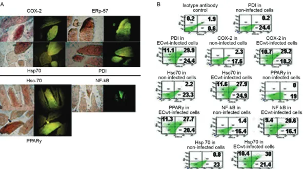
ECwt infection of ICR mice increased expression of COX-2, ERp57, Hsc70, NF-κB, Hsp70, PDI and PPARγ in intestinal villus cells - Some of the molecular mech-anisms involved in the action of the drugs used above have previously been studied (Furuya et al. 2008, Rao & Knaus 2008, Gupta et al. 2010). In addition, there is some previous knowledge about the implication of cer-tain cellular molecules in the rotavirus infection process (Lopez & Arias 2006, Calderon et al. 2012). To get in-sight into the mechanisms supporting the antiviral action of AA, IBF, DCF and NAC, their impact on the expres-sion of COX-2, ERp57, Hsc70, NF-κB, Hsp70, PDI or PPARγ was evaluated using immunochemistry, indirect immunofluorescence, ELISA, WB and confocal micros-copy. It has been reasonably assumed that sera raised in animals susceptible to rotavirus infection may contain
Abs against rotavirus. WB analysis for determining Abs directed against rotavirus proteins in commercially pre-pared sera showed the lack of rotavirus reactivity in the commercial sera used for detecting the above mentioned proteins (data not shown). Immunochemistry and im-munofluorescence analyses of intestinal cross-sections from ECwt infected mice indicated that cells showing the strongest positive signals for viral antigen coincided with those resulting positive for above mentioned pro-teins (Fig. 5A). To further characterise the correlation of virus infection and expression of the above proteins, a flow cytometry analysis was conducted. As shown in Fig. 5B, a simultaneous increase for viral antigen and
proteins COX-2, ERp57, Hsc70, NF-κB, Hsp70, PDI and PPARγ was observed in ECwt infected cells as compared with uninfected control cells.
To examine the effects of NAC treatment on the expression of Hsc70 and PDI in ECwt-infected villus cells, IEC isolated from mice that had been treated with NAC (18 mg/kg/day) during three d.p.i. were analysed by flow cytometry. The results showed that ECwt infec-tion caused the inducinfec-tion of Hsc70 and PDI expression in comparison to control cells, whereas NAC treatment resulted in Hsc70 and PDI expression returning to lev-els similar to those observed in uninfected control cells (Fig. 6A). NAC effect on Hsc70 and PDI expression in ECwt infected mice was also analysed by ELISA. The analysis showed that HSc70 and PDI expression levels were increased in lysates of IEC from ECwt-infected mice, whereas NAC treatment returned these expres-sion levels to values similar to those found in cell lysates from uninfected control cells (Fig. 6B). Induction of Hsc70 and PDI expression by ECwt infection was con-firmed by SDS-PAGE/WB analysis of IEC lysates from infected and uninfected mice. This analysis showed that increased signals corresponding to Hsc70 and PDI pro-tein bands were associated with ECwt infection as com-pared with IEC protein samples from uninfected control
mice (Fig. 6C). Treatment of ECwt infected mice with NAC or IBF returned Hsc70 and PDI protein band in-tensity to a level similar to those observed in IEC pro-tein samples of uninfected control mice (Fig. 6C). RGZ treatment seemed to induce a modest decrease of PDI from ECwt-infected IEC while apparently induced fur-ther increase of the Hsc70 expression level in IEC from infected mice. This latter effect was similarly observed in IEC samples from ECwt-infected mice that had been treated with PGZ (Fig. 6C). In addition, PGZ treatment of infected mice returned the PDI expression level to that observed for IEC samples from uninfected control mice (Fig. 6C). PDI and Hsc70 expression levels detected for IEC protein samples from ECwt-infected mice treated with DCF or AA were similar to those found in IEC pro-tein samples from uninfected control mice (Fig. 6C). Ex-cept for the effect of PGZ and RGZ treatment on Hsc70 expression levels found in IEC samples from ECwt-in-fected mice, all drug treatments appeared to return PDI and Hsc70 expression levels to those observed for IEC protein samples from uninfected control mice.
Confocal microscopy analysis of cross-sections of small intestines from ECwt infected mice indicated that Hsc70 and PDI signals were increased in intestinal cross-sections from virus infected mice that had not been
treat-Fig. 5: increased expression of cyclooxygenase (COX)-2, ERp57, Hsc70, nuclear factor kappa B (NF-κB), Hsp70, protein disulphide isomerase
(PDI) and peroxisome proliferator-activated receptor gamma (PPARγ) in villus cells infected by ECwt. Mice were infected with ECwt and at
thre days post-inoculation (d.p.i.) their small intestinal villi were isolated. Villi were analysed by immunocytochemistry (A) or flow cytometry (B) (n = 2 mice for each analysis). A: the immunocytochemistry analysis of villus cross-sections was performed by using rabbit polyclonal anti-rotavirus structural proteins (SP) antibody (Ab), horseradish peroxidase-conjugated goat anti-rabbit secondary Ab and aminoethylcarbazole chromogen. Cross-sections used in immunocytochemistry were treated with 50 mM NH4Cl and then incubated with goat Ab against COX,
ed with drugs, whilst NAC treatment led to decreased signals of Hsc70 and PDI, which were comparable to those observed in samples from uninfected mice (Sup-plementary data). After quantification of co-localisation using the Pearson’s co-localisation coefficient, it was also found that viral antigen in the villus cells did not co-localised with Hsc70 or PDI (Supplementary data). PGZ also decreased Hsc70 and PDI signals to levels similar to those observed with the NAC treatment (Supplementary data), while IBF treatment caused decreased signals for these proteins, but to a lesser extent than that found with NAC treatment (Supplementary data).
DISCUSSION
The two recently introduced live-attenuated rotavi-rus vaccines have been licensed in various countries for widespread use in massive vaccination programs (Nel-son & Glass 2010, Santosham 2010). However, specific antiviral therapy for rotavirus disease is absent. Oral re-hydration therapy has been useful for treatment of this disease and reduction of mortality, but without having a significant effect on the course of diarrhoea. Alternative, low cost and accessible developments for prophylactic
and therapeutic treatment of the rotavirus-associated di-arrhoeal disease are desirable and needed. In the present study, the inhibitory capacity of NAC, DCF, IBF, PGZ and RGZ on ECwt infection of ICR mice has been dem-onstrated. NAC treatment of infected mice immediately after the first diarrhoeal episode has occurred showed that rotavirus infection process can be interfered with once it has been established. On the other hand, NAC three-day treatment using a dose of 18 mg/kg/day was able to reduce virus infectivity by 80-97% as determined by immunocytochemistry analysis of isolated intestinal villi. However, when infection was determined in terms of rotavirus structural antigen accumulation in intestinal villi, ELISA analysis of lysates from villi showed that NAC treatment reduced viral antigen by 56-100%. The apparent discrepancy found between the immunochem-istry and ELISA calculation of the proportion for the NAC inhibitory effect could be reflecting not only the different number of mice used in each experiment, but also the outbred condition of ICR mice. However, what should be highlighted from our study is that the NAC inhibitory effect was significantly produced on ECwt in-fection as compared with untreated virus-infected
con-Fig. 6: effect of drug treatment on Hsc70 and protein disulphide isomerase (PDI) expression in small intestinal villi isolated from ECwt-infected mice. A: flow cytometric analysis was performed on intestinal epithelial cells (IEC) isolated from villi from ECwt-infected mice (n = 2 mice for each experimental group) that had been treated with ibuprofen (IBF) (20 mg/kg/day), diclofenac (DCF) (1 mg/kg/day), pioglitazone (PGZ) (30 mg/kg/day), rosiglitazone (RGZ) (4 mg/kg/day), N-acetylcysteine (NAC) (18 mg/kg/day), ascorbic acid (AA) (20 mg/kg/day) or difenoxilate sodium (7.5 mg/kg/day) during three days post-inoculation (d.p.i.). Goat primary antibodies (Abs) against Hsc70 or PDI and fluorescein iso-thiocyanate (FITC)-conjugated mouse anti-goat IgG secondary Abs were used. Immunofluorescence analysis was performed on a Cyan (Dako) flow cytometer using a FlowJo software; B: radio immunoprecipitation assay lysates from villi isolated from ECwt-infected mice (n = 3 mice for each experimental group) that had been treated with NAC (18 mg/kg/day) during three days after 24 h post-inoculation. were added to ELISA plates coated with rabbit polyclonal Abs against Hsc70 or PDI. Goat Abs against Hsc70 or PDI were used as detection Abs and the reaction was revealed using horseradish peroxidase (HRP)-conjugated donkey anti-goat Abs and phenylenediamine dihydrochloride substrate before reading at optical density (OD)492 nm. Lysates from uninfected villi were used as a control. Data are expressed as mean OD492 nm Hsc70 or PDI antigen. Graph shows significant increase of Hsc70 and PDI expression (p < 0.01) and their significant reduction by NAC treatment (p < 0.01); C: cell lysates (75 µg protein/well) from villi isolated from ECwt-infected mice that had been treated with drugs as indicated in A were analysed by sodium dodecyl sulfate polyacrylamide gel electrophoresis/Western blotting. Membranes were probed with goat anti-Hsc70 or PDI Abs and
then with HRP-conjugated donkey anti-goat IgG Ab. Β-actin was used as protein loading control. The reaction was developed with intensified
trol mice regardless of the method used for determining virus infection. It is interesting to note that in the ab-sence of NAC treatment, ECwt infection was maintained in about 75% of villus cells at least through the six d.p.i. tested. ECwt appears to be unable to infect 100% of vil-lus cells during this post-infection period. These may be because mature enterocytes from the villus apex seem to be more susceptible to rotavirus infection than those located in the villus base (Pearson et al. 1978, Pearson & McNulty 1979). Although an estimate of the number of diarrheal episodes would have been a very useful vari-able in our work, we were unvari-able to obtain a relivari-able and quantitative recording of the number diarrheal episodes of suckling mice even upon gentle abdominal palpation. This fact prompted us to use the villi isolated from small intestine from ECwt-infected mice as an approach to quantify rotavirus infection.
In searching for an explanation for the inhibitory ac-tivity exhibited by NAC, it should be noted that NAC has been shown to be a potent inhibitor of NF-κB activation in vascular endothelial cells (Schubert et al. 2002). NAC and other antioxidants have been reported to inhibit hy-drogen peroxide-induced NF-κB activation (Gupta et al. 2010). The central role played by NF-κB signal pathway in physiological and pathological conditions has made it a potential target for pharmacological intervention (Magné et al. 2006, Gupta et al. 2010). On the other hand, some rotavirus strains have been shown to activate NF-κB (Rollo et al. 1999, Halasz et al. 2008, Holloway et al. 2009, Bagchi et al. 2010). Inhibition of rotavirus infectivity in MA104 cells has recently been observed in the presence of NAC (Guerrero et al. 2012). Despite these previous findings, further experimental work re-mains to be done in order to assess the implication of NF-κB activation during rotavirus infection of intestinal villus cells and the NAC contribution in interfering with this particular signal pathway.
The administration of IBF showed to have a high in-hibitory capacity on virus infection being comparable to that of NAC, whereas DCF administration resulted in a much lower inhibitory activity, but slightly higher than that observed for RGZ. It is worth noting the differen-tial inhibitory effect exhibited by both NSAIDs tested as IBF caused virus infectivity reduction by about 90% compared with only 35% for DCF. This might suggest that these NSAIDs use different mechanisms for inter-fering with rotavirus infection process in villus cells. Indomethacin, a NSAID, has proven to be effective in inhibiting rotavirus infection in Caco-2 cells where PKA-mediated ERK1/2, p38 MAPK and NF-κB path -ways have been found to be involved (Rossen et al. 2004). IBF and indomethacin have been found to have PPARγ agonistic properties whereas IBF is known for its inhibi-tory effects on NF-κB activation which are absent in in -domethacin (Poligone & Baldwin 2001, Youssef & Mo-stafa 2004, Little et al. 2007). The two PPARγ agonists assayed, PGZ and RGZ, showed relatively low inhibi-tory effect which was about 32% and 25%, respectively. These results might lead to the assumption that PPARγ activation is less involved in the mechanisms used by the rotavirus infection process in villus cells. Moreover,
the inhibitory activity caused by AA might be related with the implication of redox reactions during the ECwt infection process. Several studies have demonstrated the implication of redox imbalance in the establishing of viral infections and the progression of virus-induced diseases (Beck et al. 2000).
To unravel whether NAC inhibitory action has some specificity for rotavirus infection, mice were infected with human reovirus type 1, also a member of the family Reoviridae, but differing in several respects from rota-virus (Roy 2006). As a result, NAC treatment did not bring about any inhibitory effect on reovirus infection. This finding suggested that the rotavirus inhibiting ef-fect of NAC has some specificity at least for the rotavi-rus strain used to infect ICR mice. In order to figure out whether NAC has inhibitor capacity similar to that of NTZ, ECwt-infected mice were treated with either in-hibitor. As indicated in Results section, NAC treatment had higher inhibitory activity than that seen for NTZ treatment. Although the NTZ dose used was equivalent to that used in children for treating rotavirus infection (Bailey & Erramouspe 2004, Rossignol et al. 2006), this result should be taken with caution since a more systematic study should be performed in which several NTZ concentrations are used in order to confirm this result. Taking into account that the highest frequency of rotavirus infection is found in children between six-24 months of age and infants under six months of age show the next highest frequency (Brandt et al. 1979, Crawley et al. 1993, Carraro et al. 2008), NAC could have an ad-ditional advantage over NTZ as the former can be used even in pre-term new-born infants (Ahola et al. 1999), whilst the latter may only be administered to children older than one year of age (Bailey & Erramouspe 2004, Rossignol et al. 2006).
suggest that viral antigen is on average reduced by drug treatment when the antigen is considered in relation to all villus cells, which, when taken together with the percent-age of infected cells, leaves open the question about the possible differential inhibitory impact of drugs on either virus entry or virus-directed protein synthesis or both.
The NAC inhibitory effect was frequency-dependent as 18 mg/kg/day distributed in three applications dur-ing three-day treatment was more effective than less frequent treatment schedules. In addition, NAC admin-istration as early as the occurrence of the first diarrhoeal episode may allow a more successful therapeutic result. However, experiments aimed at determining the spe-cific stage of viral life cycle affected by NAC were not conducted. It remains to be determined whether NAC had any inhibitory effect by directly contacting the en-terocyte luminal surface following oral drug delivery or whether its inhibitory effect was more related with the amount of drug absorbed into the enterocytes from the lumen. Determining the half maximal effective con-centration for NAC requires knowledge about its return kinetics from the systemic circulation back to the small intestinal cells either as NAC itself or cysteine. Nev-ertheless, NAC effects on rotavirus infection might be discussed within the context of signalling pathways af-fected by ROS, NSAIDs and PPARγ agonists.
In some cases, viral infections can cause inhibition of host protein synthesis whilst in other cases a virus-induced differential expression of host proteins has been found. ECwt infection of villus cells induced the expres-sion of host-encoded proteins COX-2, ERp57, Hsc70, NF-κB, Hsp70, PDI and PPARγ as detected in cross-sections from intestinal villi. It deserves to be mentioned that Hsc70 and PDI have been identified as rotavirus-interact-ing proteins durrotavirus-interact-ing the infection of MA104 cells (Guer-rero et al. 2002, Calderon et al. 2012), whereas COX-2 has been found to be required and induced by rotavirus infection of Caco-2 cells (Rossen et al. 2004). On the oth-er hand, the influenza virus-induced ER stress has been associated with increased levels of ER chaperone ERp57 (Roberson et al. 2012), whilst recruitment of Hsp70 chap-erones has been found to be an important component of viral survival strategies (Mayer 2005). Some viruses ben-efit from the anti-apoptotic properties of NF-κB whose activation allows enhancement of viral replication, host cell survival and evasion of host immune responses (His-cott et al. 2001). However, in the case of NF-κB, it should be specified in our study that since its detection was per-formed using an Ab directed against one of the NF-κB subunits (p50), its level does not reflect the activity of the factor. More detailed analyses are needed to determine whether the protein changes induced by ECwt infection of villus associated cells are truly components of a spe-cific cellular response to rotavirus infection or are reflect-ing a general stress response. It is worth notreflect-ing that NAC treatment of ECwt-infected mice returned the Hsc70 and PDI expression levels to values similar to those found in uninfected villus-associated cells. However, evidence is not provided that the effective NAC-inhibitory effect is specifically mediated by the NAC-induced decrease of
Hsc70 and PDI expression. Therefore, further study is needed to unravel the complete role of stress-inducible proteins in rotavirus infection.
Taking together the present results, it appears to be that rotavirus infection benefits from inducing both oxidative stress and activation of proinflammatory sig-nalling pathways from host epithelial cells lining the villi since the treatment of rotavirus-infected mice with NAC, NSAIDs or PPARγ agonist led to significantly reduced infection of villus cells. The results described here suggest the possibility of designing preventive and therapeutic strategies aimed at interfering with rotavirus infection of children or young zootechnic species.
REFERENCES
Ahola T, Fellman V, Laaksonen R, Laitila J, Lapatto R, Neuvonen PJ, Raivio KO 1999. Pharmacokinetics of intravenous N-acetyl-cysteine in pre-term new-born infants. Eur J Clin Pharmacol55: 645-650.
Atkuri KR, Mantovani JJ, Herzenberg LA, Herzenberg LA 2007. N-acetylcysteine - a safe antidote for cysteine/glutathione defi-ciency. Curr Opin Pharmacol7: 355-359.
Bagchi P, Dutta D, Chattopadhyay S, Mukherjee A, Halder UC, Sarkar S, Kobayashi N, Komoto S, Taniguchi K, Chawla-Sarkar M 2010. Rotavirus nonstructural protein 1 suppresses virus-in-duced cellular apoptosis to facilitate viral growth by activating the cell survival pathways during early stages of infection. J Virol 84: 6834-6845.
Bailey JM, Erramouspe J 2004. Nitazoxanide treatment for giardiasis and cryptosporidiosis in children. Ann Pharmacother38: 634-640.
Bartlett S, Sawdy R, Mann G 1999. Induction of cyclooxygenase-2 expression in human myometrial smooth muscle cells by interleu-kin-1-beta: involvement of p38 mitogen-activated protein kinase. J Physiol520: 399-406.
Bassaganya-Riera J, Song R, Roberts PC, Hontecillas R 2010. PPAR-gamma activation as an anti-inflammatory therapy for respira-tory virus infections. Viral Immunol23: 343-352.
Beck M, Handy J, Levander OA 2000. The role of oxidative stress in viral infections. Ann NY Acad Sci917: 906-912.
Brandt C, Kim HW, Yolken RH, Kapikian AZ, Arrobio JO, Rodri-guez WJ, Wyatt RG, Chanock RM, Parrott RH 1979. Compara-tive epidemiology of two rotavirus serotypes and other viral agents associated with pediatric gastroenteritis. Am J Epidemiol 110: 243-254.
Cai J, Chen Y, Seth S, Furukawa S, Compans RW, Jones DP 2003. Inhibition of influenza infection by glutathione. Free Radic Biol Med34: 928-936.
Calderon M, Guerrero CA, Acosta O, Lopez S, Arias CF 2012. In-hibiting rotavirus infection by membrane-impermeant thiol/dis-ulfide exchange blockers and antibodies against protein disthiol/dis-ulfide isomerase. Intervirology55: 451-464.
Carraro E, Sitta PAH, Siqueira I, Pasternak J, Vale MMD 2008. Ro-tavirus infection in children and adult patients attending in a ter-tiary hospital of São Paulo, Brazil. Braz J Infect Dis12: 44-46.
Casola A, Garofalo RP, Crawford SE, Estes MK, Mercurio F, Crowe SE, Brasier AR 2002. Interleukin-8 gene regulation in intesti- Brasier AR 2002. Interleukin-8 gene regulation in intesti-nal epithelial cells infected with rotavirus: role of viral-induced Ikappa-B kinase activation. Virology298: 8-19.
nu-clear factor-kappa B and I kappa B kinase-alpha in human color-ectal cancer epithelial cells. Br J Cancer 88: 1598-1604.
Circu M, Aw TY 2012. Intestinal redox biology and oxidative stress. Semin Cell Dev Biol23: 729-737.
Clark A, Sanderson C 2009. Timing of children’s vaccinations in 45 low-income and middle-income countries: an analysis of survey data. Lancet 373: 1543-1549.
Clément S, Pascarella S, Negro F 2009. Hepatitis C virus infection: molecular pathways to steatosis, insulin resistance and oxidative stress. Viruses1: 126-143.
Crawley J, Bishop R, Barnes GL 1993. Rotavirus gastroenteritis in infants aged 0-6 months in Melbourne, Australia: implications for vaccination. J Paediatr Child Health29: 219-221.
Cui H, Kong Y, Zhang H 2012. Oxidative stress, mitochondrial dys-function and aging. J Signal Transduct2012: ID 646354.
Danchin MH, Bines JE 2009. Defeating rotavirus? The global recom-mendation for rotavirus vaccination. N Engl J Med361: 1919-1921.
de Flora S, Grassi C, Carati L 1997. Attenuation of influenza-like symptomatology and improvement of cell-mediated immuni-ty with long-term N-aceimmuni-tylcysteine treatment. Eur Respir J10: 1535-1541.
Esposito D, Holman RC, Haberling DL, Tate JE, Podewils LJ, Glass RI, Parashar U 2011. Baseline estimates of diarrhea-associated mortality among United States children before rotavirus vaccine introduction. Pediatr Infect Dis J30: 942-947.
Estes MK, Kapikian AZ 2007. Rotaviruses. In DM Knipe, PM How-ley, Fields Virology, Lippincott Williams & Wilkins, Philadel-phia, p. 1917-1974.
Fraternale A, Paoletti MF, Casabianca A, Orlandi C, Schiavano GF, Chiarantini L, Clayette P, Oiry J, Vogel JU, Cinatl Jr J, Magnani M 2008. Inhibition of murine AIDS by pro-glutathione (GSH) molecules. Antiviral Res 77: 120-127.
Fujisawa K, Nishikawa T, Kukidome D, Imoto K, Yamashiro T, Mo-toshima H, Matsumura T, Araki E 2009. TZDs reduce mitochon-drial ROS production and enhance mitochonmitochon-drial biogenesis. Biochem Biophys Res Commun 379: 43-48.
Furuya A, Uozaki M, Yamasaki H, Arakawa T, Arita M, Koyama AH 2008. Antiviral effects of ascorbic and dehydroascorbic acids in vitro. Int J Mol Med22: 541-345.
Garland M, Fawzi WW 1999. Antioxidants and progression of human immunodeficiency virus (HIV) disease. Nutr Res19: 1259-1276.
Garozzo A, Tempera G, Ungheri D, Timpanaro R, Castro A 2007. N-acetylcysteine synergises with oseltamivir in protecting mice from lethal influenza infection. Int J Immunopathol Pharmacol 20: 349-354.
Ghezzi P, Ungheri D 2004. Synergistic combination of N-acetylcysteine and ribavirin to protect from lethal influenza viral infection in a mouse model. Int J Immunopathol Pharmacol17: 99-102.
Go Y, Jones DP 2008. Redox compartmentalization in eukaryotic cells. Biochim Biophys Acta1780: 1273-1290.
Gualtero D, Guzman F, Acosta O, Guerrero CA 2007. Amino acid domains 280-297 of VP6 and 531-554 of VP4 are implicated in heat shock cognate protein Hsc70-mediated rotavirus infection. Arch Virol152: 2183-2196.
Guerrero C, Bouyssounade D, Zarate S, Isa P, Lopez T, Espinosa R, Romero P, Méndez E, Lopez S, Arias CF 2002. Heat shock cognate protein 70 is involved in rotavirus cell entry. J Virol 76: 4096-4102.
Guerrero C, Murillo A, Acosta O 2012. Inhibition of rotavirus
infec-tion in cultured cells by N-acetyl-cysteine, PPARγ agonists and
NSAIDs. Antiviral Res 96: 1-12.
Guerrero C, Santana AY, Acosta O 2010. Mouse intestinal villi as a model system for studies of rotavirus infection. J Virol Methods 168: 22-30.
Gupta S, Sundaram C, Reuter S, Aggarwal BB 2010. Inhibiting
NF-κB activation by small molecules as a therapeutic strategy. Bio
-chim Biophys Acta1799: 775-787.
Halasz P, Holloway G, Turner SJ, Coulson BS 2008. Rotavirus replica-tion in intestinal cells differentially regulates integrin expression by a phosphatidylinositol 3-kinase-dependent pathway, resulting in increased cell adhesion and virus yield. J Virol 82: 148-160.
Hiscott J, Kwon H, Génin P 2001. Hostile takeovers: viral appropria-tion of the NF-κB pathway. J Clin Invest107: 143-151.
Ho W, Dougla SD 1992. Glutathione and N-acetylcysteine suppres-sion of human immunodeficiency virus replication in human monocyte/macrophages in vitro. AIDS Res Hum Retroviruses 8: 1249-1253.
Holloway G, Truong TT, Coulson BS 2009. Rotavirus antagonizes cellular antiviral responses by inhibiting the nuclear accumula-tion of STAT1, STAT2 and NF-κB. J Virol83: 4942-4951.
Jackson A, Kammouni W, Zherebitskaya E, Fernyhough P 2010. Role of oxidative stress in rabies virus infection of adult mouse dorsal root ganglion neurons. J Virol84: 4697-4705.
Jiang C, Ting AT, Seed B 1998. PPAR-gamma agonists inhibit produc-tion of monocyte inflammatory cytokines. Nature391: 82-86.
Jones DP, Go YM 2010. Redox compartmentalization and cellular stress. Diabetes Obes Metab12: 116-125.
Jones DP 2006. Redefining oxidative stress. Antioxid Redox Signal 8: 1865-1879.
Kavouras J, Prandovszky E, Valyi-Nagy K, Kovacs SK, Tiwari V, Ko-vacs M, Shukla D, Valyi-Nagy T 2007. Herpes simplex virus type 1 infection induces oxidative stress and the release of bioactive li-pid peroxidation by-products in mouse P19N neural cell cultures. J Neurovirol13: 416-425.
Kregel KC, Zhang HJ 2007. An integrated view of oxidative stress in aging: basic mechanisms, functional effects and pathological considerations. Am J Physiol Regul Integr Comp Physiol292: R18-R36.
Lai K, Ng WY, Osburga, Chan PK, Wong KF, Cheng F 2010. High-dose N-acetylcysteine therapy for novel H1N1 influenza pneu-monia. Ann Intern Med152: 687-688.
LaMonica R, Nazarova J, Kocer S, Dowling W, Geimonen E, Shaw RD, Mackow ER 2001. VP4 differentially regulates TRAF2 signaling, disengaging JNK activation while directing NF-κB to effect rotavirus specific cellular responses. J Biol Chem276: 19889-19896.
Lee K, Kim SR, Park SJ, Park HS, Min KH, Jin SM, Lee MK, Kim UH, Lee YC 2006. Peroxisome proliferator activated receptor-g modulates reactive oxygen species generation and activation of
nuclear factor-kB and hypoxia-inducible factor 1α in allergic air -way disease of mice. J Allergy Clin Immunol 118: 120-127.
Lim G, Lee JH 2012. N-acetylcysteine in children with dengue-asso-ciated liver failure: a case report. J Trop Pediatr58: 409-413.
Little D, Jones SL, Blikslager AT 2007. Cyclooxygenase (COX) in-hibitors and the intestine. J Vet Intern Med21: 367-377.
Lopez S, Arias CF 2006. Early steps in rotavirus cell entry. Curr Top Microbiol Immunol309: 39-66.
Maruri-Avidal L, Lopez S, Arias CF 2008. Endoplasmic reticulum chaperones are involved in the morphogenesis of rotavirus infec-tious particles. J Virol82: 5368-5380.
Mayer M 2005. Recruitment of Hsp70 chaperones: a crucial part of viral survival strategies. Rev Physiol Biochem Pharmacol153: 1-46.
Mirazimi A, Svensson L 1998. Carbohydrates facilitate correct dis-ulphide bond formation and folding of rotavirus VP7. J Virol75: 3887-3892.
Nelson EA, Glass RI 2010. Rotavirus: realising the potential of a promising vaccine. Lancet 376: 568-570.
Nencioni L, Iuvara A, Aquilano K, Ciriolo MR, Cozzolino F, Rotilio G, Garaci E, Palamara AT 2003. Influenza A virus replication is dependent on an antioxidant pathway that involves GSH and Bcl-2. FASEB J17: 758-760.
Newton R, Kuitert LM, Bergmann M, Adcock IM, Barnes PJ 1997. Evidence for involvement of NF-kappaB in the transcriptional control of COX-2 gene expression by IL-1beta. Biochem Biophys Res Commun237: 28-32.
Parashar U, Bresee J, Glass R 2006. Rotavirus and severe childhood diarrhea. Emerg Infect Dis12: 304-306.
Pearson GR, McNulty MS 1979. Ultrastructural changes in small in-testinal epithelium of neonatal pigs infected with pig rotavirus. Arch Virol59: 127-136.
Pearson GR, McNulty MS, Logan EF 1978. Pathological changes in the small intestine of neonatal calves naturally infected with reo-like virus (rotavirus). Vet Rec 102: 454-458.
Poligone B, Baldwin AS 2001. Positive and negative regulation of
NF-κB by COX-2. Roles of different prostaglandins. J Biol Chem 276: 38658-38664.
Ramig RF 2004. Pathogenesis of intestinal and systemic rotavirus in-fection. J Virol78: 10213-10220.
Rao P, Knaus EE 2008. Evolution of nonsteroidal anti-inflammatory drugs (NSAIDs): cyclooxygenase (COX) inhibition and beyond. J Pharm Pharm Sci 11 (Suppl.): 81S-110S.
Roberson E, Tully JE, Guala AS, Reiss JN, Godburn KE, Pociask DA, Alcorn JF, Riches DW, Dienz O, Janssen-Heininger YM, Anathy V 2012. Influenza induces endoplasmic reticulum stress, caspase-12-dependent apoptosis and c-Jun N-terminal kinase-mediated transforming growth factor-B release in lung epithelial cells. Am J Respir Cell Mol Biol46: 573-581.
Rollo E, Kumar KP, Reich NC, Cohen J, Angel J, Greenberg HB, Sheth R, Anderson J, Oh B, Hempson SJ, Mackow ER, Shaw RD 1999. The epithelial cell response to rotavirus infection. J Im-munol163: 4442-4452.
Rossen J, Bouma J, Raatgeep RH, Buller HA, Einerhand AW 2004. Inhibition of cyclooxygenase activity reduces rotavirus infection at a post-binding step. J Virol78: 9721-9730.
Rossignol J, Abu-Zekry M, Hussein A, Santoro MG 2006. Effect of ni-tazoxanide for treatment of severe rotavirus diarrhoea: randomised double-blind placebo-controlled trial. Lancet368: 124-129.
Roulston A, Marcellus RC, Branton PE 1999. Viruses and apoptosis. Annu Rev Microbiol53: 577-628.
Roy P 2006. Reoviruses: entry, assembly and morphogenesis, Spring-er, New York, 279 pp.
Santosham M 2010. Rotavirus vaccine - a powerful tool to combat deaths from diarrhoea. N Engl J Med362: 358-360.
Schubert S, Neeman I, Resnick N 2002. A novel mechanism for the inhibition of NF-kB activation in vascular endothelial cells by natural antioxidants. FASEB J16: 1931-1933.
Severi T, Ying C, Vermeesch JR, Cassiman D, Cnops L, Verslype C, Fevery J, Arckens L, Neyts J, van Pelt JF 2006. Hepatitis B virus replication causes oxidative stress in HepAD38 liver cells. Mol
Cell Biochem290: 79-85.
Sharpe A, Ramig RF, Mustoe TA, Fields BN 1978. A genetic map of reovirus I. Correlation of genome RNAs between serotypes 1, 2 and 3. Virology84: 63-74.
Sheth R, Anderson J, Sato T, Oh B, Hempson SJ, Rollo E, Mackow ER, Shaw RD 1996. Rotavirus stimulates IL-8 secretion from cultured epithelial cells. Virology221: 251-259.
Straus D, Pascual G, Li M, Welch JS, Ricote M, Hsiang CH, Sen-gchanthalangsy LL, Ghosh G, Glass CK 2000. 15-deoxy-D12,14-prostaglandin J2 inhibits multiple steps in the NF-κB signalling pathway. Proc Natl Acad Sci USA 97: 4844-4849.
Subbaramaiah K, Hart JC, Norton L, Dannenberg AJ 2000. Microtu-bule-interfering agents stimulate the transcription of cyclooxyge-nase-2. Evidence for involvement of ERK1/2 and p38 mitogen-ac-tivated protein kinase pathways. J Biol Chem275: 14838-14845.
Subbaramaiah K, Lin DT, Hart JC, Dannenberg AJ 2001. Peroxisome proliferator-activated receptor gamma ligands suppress the tran-scriptional activation of cyclooxygenase-2. Evidence for involve-ment of activator protein-1 and CREB-binding protein/p300. J Biol Chem276: 12440-12448.
Svensson L, Dormitzer PR, von Bonsdorff CH, Maunula L, Greenberg HB 1994. Intracellular manipulation of disulphide bond formation in rotavirus proteins during assembly. J Virol68: 5204-5215.
Tian Y, Jiang W, Gao N, Zhang J, Chen W, Fan D, Zhou D, An J 2010. Inhibitory effects of glutathione on dengue virus production. Bio
-chem Biophys Res Commun397: 420-424.