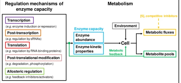
virulence in the human pathogen
Streptococcus pneumoniae
Oeiras,
Sandra M. Carvalho
Dissertation presented to obtain the Ph.D. degree in Biochemistry
Instituto de Tecnologia Química e Biológica | Universidade Nova de Lisboa
Glucose
G6P
Pyruvate
SpxB
Ac-PAcetate
Lactate
Ac-CoA
Formate
Ethanol
PFL
CcpA
α-G1P α-Gal1P GalGalactose
UDP-sugars
UTP Uracil
Sandra M. Carvalho
Dissertation presented to obtain the Ph.D. degree in Biochemistry
Instituto de Tecnologia Química e Biológica | Universidade Nova de Lisboa
Oeiras, September, 2012
central metabolism and virulence in the
human pathogen
Streptococcus pneumoniae
Supervisor: Dr. Ana Rute Neves
Apoio financeiro da Fundação para a Ciência e Tecnologia e do FSE no âmbito do Quadro Comunitário de apoio, Bolsa de Doutoramento com a referência SFRH/BD/35947/2007
Cover page figure: Schematic illustration of the pathways for glucose and galactose
metabolism in a Streptococcus pneumoniae cell. Uracil, an important constituent of
the capsule building blocks, and the non-classical virulence proteins (CcpA, SpxB and PFL) studied in this thesis are highlighted in different colours.
Background of the cover page figure: Transmission electron micrograph of S.
In 2006, at the Lactic Acid Bacteria & in vivo NMR laboratory, I was introduced to
scientific research and really enjoyed the experience. The excitement led me to
accept the opportunity to integrate a PhD project to study central metabolism in S.
pneumoniae, offered by my supervisor, Dr. Ana Rute Neves. During the course of
the thesis I experienced a lot of emotions: the excitement and joy of new ideas
and results, the disappointments of bad or confusing results, the persistence of
not giving up and now the relief and happiness of achieving the final product. The
present thesis would have never been possible without the help and
encouragement of many people.
I thank my supervisor, Dr. Ana Rute Neves, for the opportunity to join her group
and for introducing me to research and to the field of microbial physiology and
biochemistry. I am grateful for her intellectual and scientific support, good ideas,
advices, discussions and critical reading and writing of this thesis. I also thank her
guidance during this PhD project.
I thank my co-supervisor, Prof. Dr. Oscar Kuipers, for the stimulating and pleasant
collaboration with Dr. Ana Rute Neves, and the fruitful discussions and ideas. I
also thank him for accepting me in the Molecular Genetics (MolGen) group, at the
University of Groningen (The Netherlands), where I have been for 6 months,
learning several techniques. I thank him all the support provided during this stay,
which contributed not only to my research experience but also to my personal
Neves during my first year as a research student and for all the help provided
during the course of this thesis. I acknowledge Prof. Dr. Helena Santos for the
valuable discussions in lab meeting presentations and for the critical reading of
the abstract of this thesis.
I thank Prof. Dr. Jan Martinussen for supervising me during a short stay at the
Technical University of Denmark (DTU), Lyngby, and for the determination of
pyrimidine nucleotides. I also thank him and his family for the warm reception and
generous hospitality.
I thank Prof. Dr. Peter Andrew and Dr. Hasan Yesilkaya for the collaboration that
led to the “PFL publication”.
I thank Dr. Jetta Bijlsma (UMCG, Groningen) for the good collaboration in the
work involving pyruvate oxidase (SpxB) and for all the helpful discussions.
I thank Dr. Susana Vinga for the good collaboration in the development of
descriptive and predictive models of S. pneumoniae metabolism. I thank José
Caldas for the fast assistance in microarray analysis.
When I started my PhD, in 2007, I was the only student working with S.
pneumoniae in the LAB & in vivo NMR group, but I was never alone, I found good
company among the people at the Cell Physiology and NMR group. In 2008/9,
during 6 months, I got settled in Groningen, and in the MolGen group I got support
from many people, especially the “Strep” group. When I got back, the LAB & in
partners. Having more people working with S. pneumoniae in the lab and sharing
many questions, “Strep” related, was an additional excitement. I am truly thankful
for the help and friendship of many of you.
I thank Patrícia Almeida, Ana Manso and Cristiana Faria for being helpful
colleagues and good friends. Thank you for listening to me and motivating me
when I needed it.
I thank Mafalda Cavaleiro, Joana Oliveira, Anabela Vieira, Irene Gonzalez and
João Jorge for the stimulating and fun environment inside and outside the lab.
Thank you all for the support and help provided.
I thank Nuno Borges, Rute Castro and Ana Lúcia Carvalho for their invaluable
help in the lab and for being always available to teach. I thank Luís Fonseca for
the useful discussions and for always being available to help.
All my other colleagues in the LAB & in vivo NMR and Cell Physiology and NMR
groups and short-stay lab visits: Ana Esteves, Dusica Rados, Nelson, Claudia
Sanchez, Marta Rodrigues, Teresa Maio, Paula Gaspar, Ricardo Sequeira, Ana
Isabel Mingote, Ana Laura Paixão, Luís Gafeira, Tiago Pais, Tiago Faria, Pedro
Lamosa, Marta Conchinha, Carla Jorge, Melinda Noronha, Tony Collins, Pedro
Quintas, Teresa Ferreira, Margarida Santos, Catarina Silva, Sónia Neto, Márcia,
Andreia, Alexandre, Eva Lourenço, Francesca Spissu, Ana Belém, Alessandro
there. In particular, I would like to acknowledge the “Strep” group for making
Groningen a nice place to remember. I thank Tomas Kloosterman for the
supervision of my work at the MolGen, for all the help provided during this thesis
and for the nice and friendly talks. I thank Sulman Shafeeq for being a great
colleague and friend, for helping me finding things in the lab and for the useful
discussions. I also thank him and Munir for the delicious and spicy Pakistani food.
I thank Asia, Rutger, Agata and Michael for all the help and company.
During this thesis, I have crossed with many other great people. I thank Nicolas
Bernier, Manuel Marques Pita, Daniela, Yuki Miyagaki and Chantal Fernandes for
the pleasant talks and excursion activities.
I thank the invaluable technical support of many people, Teresa Batista, Luís
Gonçalves, Miguel Loureiro, people from the academic services, D. Fátima and
Ana Maria Portocarrero, and from the washing room of the 5th floor and D. Alice.
I thank Fundação para a Ciência e Tecnologia (FCT) for the financial support that
made this doctoral work possible (SFRH/BD/35947/2007).
Agradeço aos meus pais e ao meu irmão o amor incondicional, a compreensão
nos meus momentos de ausência involuntários, e o apoio em todas as
circunstâncias. A toda a minha família e amigos, obrigada por tudo.
Finalmente gostaria de agradecer ao Nuno, com quem partilho de todo o coração
a minha vida. Obrigada pela tua paciência, incentivo, conselhos, inspiração e
Streptococcus pneumoniae is a normal inhabitant of the human
nasopharynx, but it is better known for its role in a plethora of human diseases.
Growing emergence of antibiotic-resistant streptococci and non-type vaccine
strains increases the urgency of finding new targets for the development of novel
therapeutic and preventive drugs. As a major concern for global public health,
S. pneumoniae has always attracted great attention from the scientific community,
which has translated into knowledge on pathogenesis and virulence and the
development of a considerable “toolbox” for genetic manipulation and genomic
analysis, as well as a large number of deciphered genome sequences.
Interestingly, genome-wide studies have consistently pinpointed genes involved in
carbohydrate uptake and metabolism as essential for the virulence of
S. pneumoniae. These global studies offered the opportunity to investigate in
greater depth the potential connections between basic physiology, and in
particular central metabolism, and pneumococcal virulence and pathogenesis.
The general goal of this thesis is to achieve a deeper understanding of the
molecular mechanisms underlying sugar metabolism and their relation to
virulence factors in S. pneumoniae, with a special focus on capsule production. In
the present work, glucose (Glc) and galactose (Gal) were used as carbon sources
for the study of pneumococcal sugar metabolism. This choice was made for two
reasons: Firstly, Glc is a common preferred sugar and is also found as a major
carbon source in niches potentially occupied by S. pneumoniae during host
inflammation or hyperglycaemia. Secondly, Gal, generally a slowly metabolized
non-preferred sugar, is a major carbohydrate in the human nasopharynx, the
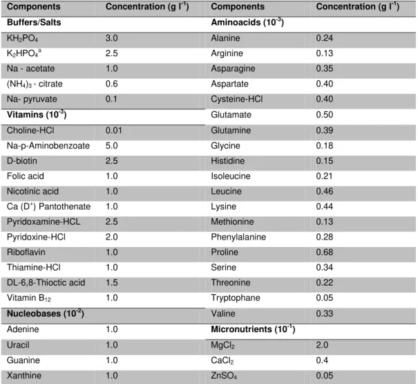
sugar metabolism is largely facilitated by the use of well-defined conditions. We
developed a chemically defined medium and growth conditions for high yield
streptococcal growth, applicable to in vivo NMR studies. Time series data on
metabolite pools have been obtained in vivo by 13C- and 31P- NMR, during Glc
metabolism of S. pneumoniae. The major end-product formed from the
metabolism of [1-13C]Glc by resting cells of S. pneumoniae acapsular strain R6
was lactate (about 35 mM). Fructose 1,6-bisphosphate (FBP) was the only
glycolytic intermediate detected. The pool of FBP increased at the expense of
Glc, reached a steady concentration and decreased to undetectable levels before
Glc exhaustion. In 31P-NMR experiments, upon Glc addition, the levels of
inorganic phosphate (Pi) decreased, accompanying the consumption of Glc, and
the levels of NTP reached very small values (circa 2 mM). Moreover, during the
optimization of growth conditions for S. pneumoniae, an extensive comparative
metabolic characterization between strains D39 and its non-encapsulated
derivative R6 was obtained under different environmental and nutritional
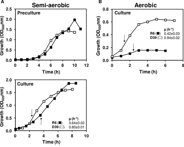
conditions. The effect of oxygen, Glc and nucleobases on the physiology of
growth of these strains was studied under controlled conditions of pH,
temperature and gas atmosphere (anaerobic, semi-aerobic or aerobic) in 2-l
bioreactors. Independently of the growth conditions tested, both strains displayed
a typical homolactic behavior. However, the growth rates of D39 and R6 were
stimulated under semi-aerobic and strictly anaerobic conditions, respectively. This
difference was mainly attributed to lower activity of pyruvate oxidase in strain D39,
in the presence of oxygen. It was notable that the maximal biomass reached by
strain D39 was substantially enhanced by supplementation of the culture medium
with extra uracil, whereas in strain R6 no effect was observed, suggesting a link
UTP and dTTP. UTP is formed from UMP in the salvage pathway of pyrimidine
biosynthesis. UMP may also be synthesized de novo from amino acids and CO2
or via the salvage pathway using external pyrimidine nucleobases (e.g. uracil) or
nucleosides. A remarkable adaptation of S. pneumoniae to fluctuating
environmental conditions in the human host consists in the switch between
opaque and transparent phenotypes, distinguished by different capsule amounts.
In this work, the role of uracil on capsule expression and production was
assessed by studying D39 and a spontaneous non-reversible D39 mutant
displaying an underproducing capsule phenotype. A comparative transcriptome
analysis between these strains in medium with and without uracil suggested a
connection between pyrimidine metabolism and capsule production. In medium
lacking uracil, strain D39 exhibited a long lag phase, decreased growth rate and
higher biomass than in the presence of the nucleobase. In accordance, capsule
promoter activity and capsule amounts were decreased under uracil-deprived
conditions in strain D39. In contrast, uracil showed no effect on the mutant strain.
Growth and CFU analysis of co-cultures of both strains in medium without uracil
clearly showed a prevalence of the mutant strain at mid-exponential phase of
growth, independently of the initial cell concentrations used for inoculation. Thus,
we propose that sensing uracil is a potential mechanism by which S. pneumoniae
alters capsule production.
Other than uracil, oxygen variability in host niches may also trigger the
capsular phenotype switch in S. pneumoniae. Pyruvate oxidase (SpxB) is an
oxygen-consuming enzyme involved in the aerobic metabolism of S. pneumoniae.
SpxB converts pyruvate, the end-product of glycolysis, O2 and Pi into CO2, H2O2
and acetyl-phosphate. We show that inactivation of spxB in D39 leads to
addition, the spxB mutation increased the expression of two putative operons
involved in carbohydrate uptake and processing, leading to altered sugar
utilization capabilities of D39, as shown by a combination of metabolite profiling
analysis and growth experiments. In environments with oxygen and Glc as major
carbon source, SpxB activity presumably minimizes the use of alternative sugar
utilization pathways and the amount of capsule, hence improving the fitness of
S. pneumoniae.
Besides SpxB, pyruvate formate-lyase (PFL) is also a competing activity
for the glycolytic end-product, pyruvate. This oxygen-sensitive enzyme is involved
in mixed-acid fermentation and requires post-translational activation by the
pyruvate formate-lyase activating enzyme (PFL-AE). PFL activity is associated
with formation of formate and acetyl-CoA. The sequenced genome of D39
possesses two copies of pfl and pfl-ae. Mutant strains disrupted in these genes
were created, and their Glc and Gal metabolism was analyzed. In the presence of
Glc, all strains, including D39, exhibited homolactic fermentation. However, in Gal,
strain D39 produced formate, acetate and ethanol in a 2:1:1 ratio, denoting
activity of PFL, but no formate production was observed by mutants defective in
spd_0420 and spd_1774, indicating that spd_0420 codes for PFL and spd_1774
for PFL-AE. The spd_0420 and spd_1774 mutants showed impaired colonization
and infection in mouse studies, indicating a direct link between pneumococcal
fermentative metabolism and virulence.
S. pneumoniae has to adapt to changing carbon sources in the fluctuating
environments of the human host. Pathways metabolizing sugars other than the
preferred one are silenced through mechanisms of catabolite repression. A global
regulator involved in this process in several Gram-positive bacteria is CcpA. In
affecting the expression of up to 19% of the genome, covering multiple cellular
functions. Remarkably, CcpA was not only a major repressor in response to Glc,
but also to Gal. Additionally, we show that Leloir and tagatose 6-phosphate
pathways for Gal metabolism are functionally active, despite repression of the
Leloir genes by CcpA. Of surprise was the shift from mixed-acid towards a more
homolactic fermentation in the ccpA mutant during growth on Gal, as genes
involved in mixed-acid fermentation were mostly under CcpA repression. This
result indicates regulation at other cellular layers, which is further supported by
dissimilar accumulation in the ccpA and parent strain of intracellular metabolites
potentially involved in metabolic regulation. Interestingly, capsule amounts were
higher in response to Gal than to Glc, regardless of CcpA. Overall, the
whole-genome transcriptome analyses provided solid evidence that S. pneumoniae
optimizes basic metabolic processes in a CcpA-mediated manner. This view is
further corroborated by the metabolic profiles obtained under the conditions
studied. Moreover, integration of transcriptional and metabolic data revealed a link
between CcpA and the association of surface molecules (capsule and
phosphorylcholine) recognized as virulence factors, to the cell wall. This finding
indicates an important role of CcpA in modulating the interaction of
S. pneumoniae with its host.
Overall, the work presented in this thesis represents an important step
towards the understanding of basic metabolic processes and, in particular, sugar
metabolism, in S. pneumoniae. We believe that the insights gained from this study
will help to better comprehend the role of metabolism in the interaction between
S. pneumoniae and its host. In this light, the work here described makes a unique
bridge between the often disconnected fields of fundamental physiology, and
A bactéria Streptococcus pneumoniae é uma habitante normal da
nasofaringe humana, conhecida principalmente por ser a causa de várias
doenças humanas. Em ambientes hospitalares bem como na comunidade, o
aparecimento galopante de estreptococos resistentes a antibióticos bem como de
estirpes não integradas no programa de vacinação incentiva novos estudos com
o objectivo de identificar alvos para o desenvolvimento de novas drogas
terapêuticas ou preventivas. Dado o seu impacto ao nível de saúde pública
mundial, S. pneumoniae tem atraído desde sempre a atenção da comunidade
científica. Este grande interesse permitiu grandes avanços no conhecimento da
patogenicidade e virulência deste organismo, e levou ao desenvolvimento de
muitas ferramentas de manipulação genética e análise genómica e à
descodificação de um número elevado de sequências genómicas. Em estudos de
análise pangenómica, genes envolvidos no transporte e metabolismo de hidratos
de carbono foram consistentemente identificados como sendo essenciais para a
virulência de S. pneumoniae. Estes estudos globais ofereceram a oportunidade
de investigar em profundidade uma ligação potencial de processos metabólicos
básicos, em particular envolvidos no metabolismo central, com a virulência e
patogenicidade de pneumococos. Assim sendo, este trabalho de tese tem por
objectivo geral elucidar fundamentos necessários para a compreensão de
mecanismos moleculares relacionados com o metabolismo de açúcares e sua
ligação a factores de virulência de S. pneumoniae, em especial a cápsula. As
hexoses glucose (Glc) e galactose (Gal) foram seleccionadas como fontes de
carbono para o estudo do metabolismo de açúcares. Esta escolha baseou-se no
seguinte: i), a Glc é o açúcar preferido por excelência e é a principal fonte de
açúcar não preferido e de metabolismo lento, é um dos hidratos de carbono em
maior quantidade na nasofaringe humana, o nicho de colonização de S.
pneumoniae.
O estudo de mecanismos de regulação complexos, como os envolvidos
em metabolismo de açúcares, é grandemente facilitado pelo uso de condições
bem definidas. Neste trabalho, foram desenvolvidos um meio quimicamente
definido e condições de crescimento para produção de biomassa elevada de
estreptococos, aplicável também a estudos de NMR in vivo. Pela primeira vez, as
concentrações de metabolitos formados e consumidos ao longo do tempo,
durante o metabolismo de Glc em S. pneumoniae, foram obtidas in vivo
recorrendo a NMR de 13C e 31P. O produto final maioritário do metabolismo de
[1-13C]Glc pelas células R6 em repouso foi o lactato (cerca de 35 mM). A frutose
1,6-bisfosfato (FBP) foi o único intermediário glicolítico detectado. Os níveis de
FBP aumentaram à custa do consumo de Glc, atingiram uma concentração
estacionária e decresceram para níveis não detectáveis antes da exaustão de
Glc. Nas experiências de 31P-NMR, após a adição de Glc, os níveis de fosfato
inorgânico (Pi) decresceram, acompanhando o consumo de Glc, e os níveis de
NTP atingiram valores muito baixos (cerca de 2 mM). Além disso, durante a
optimização das condições para crescimento de S. pneumoniae, uma
caracterização metabólica comparativa entre as estirpes D39 e a sua derivada
não encapsulada R6 foi obtida em diferentes ambientes manipulados. O efeito do
oxigénio, Glc e nucleobases na fisiologia do crescimento destas estirpes foi
estudado em condições controladas de pH, temperatura e atmosfera gasosa
(anaeróbica, semi-aeróbica e aeróbica), em reactores de 2-l. Independentemente
das condições de crescimento testadas, ambas as estirpes exibiram um
estritamente anaeróbicas, respectivamente. Esta diferença foi atribuída
essencialmente à baixa actividade de piruvato oxidase na primeira estirpe, na
presença de oxigénio. Curiosamente, a biomassa máxima atingida pela estirpe
D39 foi substancialmente melhorada suplementando o meio de cultura com
uracilo adicional, enquanto na estirpe R6 nenhum efeito foi observado, indicando
uma ligação entre a quantidade de uracilo e produção de cápsula.
A síntese dos açúcares ligados quimicamente a NDP, precursores da
cápsula da estirpe D39, requer UTP e dTTP. O UTP é sintetizado a partir de UMP
na via de salvamento de síntese de pirimidinas. O UMP pode ser sintetizado de
novo a partir de aminoácidos e CO2 ou na via de salvamento das nucleobases
(e.g. uracilo) ou nucleósidos de pirimidinas exógenas. Um mecanismo notável de
adaptação do S. pneumoniae a condições ambientais inconstantes no hospedeiro
humano consiste na permuta entre fenótipos opaco e transparente, que se
distinguem pela quantidade diferente de cápsula exibida. Neste trabalho, o papel
do uracilo na expressão e produção de cápsula foi demonstrado usando D39 e
um mutante não reversível da D39 exibindo características associadas à baixa
produção de cápsula. Uma análise de transcriptómica comparativa entre estas
estirpes em meio com e sem uracilo sugeriu uma ligação entre o metabolismo de
pirimidinas e produção de cápsula. Em meio sem uracilo, a estirpe D39 exibiu
uma fase de adaptação (lag) longa, taxa de crescimento reduzida e biomassa
mais elevada do que em meio contendo a nucleobase. Em conformidade, a
actividade do promotor do operão que codifica para a cápsula bem como a
quantidade de cápsula decresceram em meio sem uracilo na estirpe D39. Em
contraste, o uracilo não teve qualquer efeito na estirpe mutante. O crescimento e
análise de CFUs de co-culturas de ambas as estirpes mostrou claramente a
para inoculação das co-culturas. Posto isto, propomos que a detecção de
quantidades variáveis de uracilo no ambiente constitui um mecanismo pelo qual o
S. pneumoniae poderá alterar a produção de cápsula.
Para além do uracilo, o oxigénio poderá também desencadear a permuta
dos fenótipos relacionados com diferentes quantidades de cápsula em
S. pneumoniae. A piruvato oxidase (SpxB) é uma enzima consumidora de
oxigénio envolvida no metabolismo aeróbio de S. pneumoniae. A SpxB converte
piruvato, o produto final da glicólise, O2 e Pi em CO2, H2O2 e acetil-fosfato. Neste
trabalho, mostramos que a inactivação de spxB em D39 leva a um aumento na
quantidade de cápsula produzida em meio com Glc. Este aumento foi
parcialmente mediado pela indução de cps2A, o primeiro gene do locus capsular.
Adicionalmente, a mutação da spxB aumentou a expressão de dois operões
putativamente envolvidos no transporte e processamento de hidratos de carbono,
levando a uma alteração da capacidade de utilização de açúcares na estirpe
D39, como indicado pela combinação de análise de perfis de metabolitos e
experiências de crescimento. Em ambientes com oxigénio e Glc como principal
fonte de carbono, a actividade de SpxB minimiza presumivelmente o uso de vias
alternativas de utilização de açúcares e a quantidade de cápsula, melhorando
assim a aptidão fisiológica de S. pneumoniae.
Para além da SpxB, a piruvato formato-liase (PFL) também compete por
piruvato. Esta enzima sensível ao oxigénio, está envolvida na fermentação a
ácidos mistos e sofre activação pós-traducional pela enzima activadora da
piruvato formato-liase (PFL-AE). A actividade de PFL está associada à formação
de formato e acetil-coenzima A. O genoma de D39 apresenta duas cópias de pfl
e pfl-ae. Neste trabalho, estirpes mutantes inactivadas nestes genes foram
D39 produziu formato, acetato e etanol na razão 2:1:1, indicando actividade de
PFL, mas não se observou formação de formato nos mutantes com inactivação
dos genes spd_0420 e spd_1774, indicando que spd_0420 codifica para a PFL e
spd_1774 para a PFL-AE. Os mutantes spd_0420 e spd_1774 mostraram
deficiências na colonização e infecção de ratos, indicando uma ligação directa
entre o metabolismo fermentativo de pneumococos e patogenicidade.
Nos ambientes inconstantes do hospedeiro humano S. pneumoniae é
forçado a adaptar-se a variações na acessibilidade de fontes de carbono. As vias
envolvidas no metabolismo de açúcares, que não o preferido, são silenciadas
através de mecanismos de repressão catabólica. Um regulador global envolvido
neste processo em várias bactérias é a CcpA. Em S. pneumoniae, uma avaliação
sistémica do papel da CcpA na fisiologia desta bactéria não existia. Neste
trabalho, mostrou-se claramente que a CcpA é um regulador global que afecta a
expressão de cerca de 19% do genoma, abrangendo inúmeros processos
celulares. Curiosamente, o papel repressor da CcpA não foi observado apenas
em Glc, mas também em Gal. Adicionalmente, mostrou-se que as vias Leloir e
tagatose 6-fosfato envolvidas no metabolismo de Gal estão funcionalmente
activas, apesar da CcpA reprimir os genes da via de Leloir. Considerando que os
genes envolvidos na fermentação a ácidos mistos estão sob repressão pela
CcpA, o desvio do metabolismo de Gal de ácidos mistos para fermentação
homoláctica no mutante da ccpA foi claramente surpreendente. Esta observação
indica regulação ao nível de outras camadas celulares, e é fortemente
corroborada pelos diferentes conteúdos intracelulares no mutante e na estirpe
parental potencialmente envolvidos em regulação metabólica. Notavelmente, os
níveis de cápsula foram maiores em Gal do que em Glc, independentemente da
metabólicos básicos através da CcpA. Esta observação é corroborada pelos
perfis metabólicos obtidos nas condições estudadas. Mais ainda, a integração
dos dados de transcriptómica e de metabolismo revelaram uma ligação entre a
CcpA e a associação de moléculas da superfície celular (cápsula e fosforilcolina),
reconhecidas como factores de virulência, com a parede celular. Desta forma, a
CcpA pode ter um papel preponderante nas interações hospedeiro-micróbio.
Em suma, o trabalho apresentado nesta tese representa um passo
importante para a compreensão de processos metabólicos básicos e, em
particular, metabolismo de açúcares, em S. pneumoniae. Estamos convictos que
os conhecimentos adquiridos neste estudo contribuirão para um conhecimento
mais profundo do papel do metabolismo na interacção entre S. pneumoniae e o
hospedeiro. O trabalho aqui descrito estabelece uma ponte única entre duas
áreas, muitas vezes desconectadas, ou sejam, a fisiologia fundamental e
Thesis Outline
Abbreviations
xxiii
xxv
Chapter 1 General introduction 1
Chapter 2 Environmental and nutritional factors that affect growth and metabolism of the pneumococcal serotype 2 strain D39 and its non-encapsulated derivative strain R6
47
Chapter 3 Interplay between capsule expression and uracil
metabolism in Streptococcus pneumoniae
103
Chapter 4 Pyruvate oxidase influences the sugar utilization pattern
and capsule production in Streptococcus pneumoniae
141
Chapter 5 The functional pyruvate formate-lyase (PFL) and
PFL-activating enzymes of Streptococcus pneumoniae
185
Chapter 6 CcpA ensures optimal metabolic fitness of Streptococcus pneumoniae
207
Chapter 7 Overview and concluding remarks 261
Diseases caused by Streptococcus pneumoniae constitute a major global
health problem. The ever-increasing emergence of antibiotic resistant strains
exacerbates the need to find new targets for the development of effective
therapeutic and prophylactic drugs. The work in this thesis aimed at gaining
further knowledge on central metabolism and its connections to virulence in
S. pneumoniae.
Chapter 1 starts with an introduction to the pneumococcus and its
virulence factors. Next, an overview of carbohydrate metabolism, including
capsule synthesis in the pneumococcus, is presented. Finally, a detailed
description of the current knowledge concerning regulation of sugar metabolism in
Streptococcaceae and links between basic physiology in S. pneumoniae and
virulence is provided.
Chapter 2 describes the development of a culture medium and growth
conditions for high yield pneumococcal growth, that enable the application of in
vivo NMR techniques to study metabolism in S. pneumoniae. During this
optimization a comparative metabolic characterization between the model strain
D39 and its unencapsulated derivative R6 was performed.
In Chapter 3, the effect of uracil in the physiology of growth and capsule
expression/production of D39 and D39SM, a spontaneous mutant displaying
characteristics of underproducing capsule phenotype, was analyzed. Additionally,
a genome wide transcriptional response of both strains to uracil and uracil
limitation was studied by using DNA microarrays.
In Chapter 4, the transcriptional response of S. pneumoniae to spxB,
encoding pyruvate oxidase, deletion was examined. The effect of this mutation in
quantification, respectively. The effect of the spxB knock-out on the utilization of a
number of sugars was also studied.
The identification of S. pneumoniae pyruvate formate-lyase (PFL) and the
PFL activating enzyme (PFL-AE) is described in Chapter 5. Deletion of putative
genes coding for these proteins was performed and functional assignment was
determined by measuring the fermentation products of the mutant strains during
metabolism of Glc and Gal.
Chapter 6 describes the carbon catabolite protein A (CcpA) regulon on Glc
and Gal and the impact of this transcriptional regulator on the expression of
virulence factors. In this work CcpA is demonstrated to act as a global regulator in
S. pneumoniae. Overall, our results support the hypothesis that S. pneumoniae
optimizes basic metabolic processes, likely enhancing in vivo fitness, in a
CcpA-mediated manner.
A general discussion of the findings resulting from this work is presented
ABC ATP-binding cassette
ACKA Acetate kinase A
Ac-P Acetyl-phosphate
ADH Alcohol dehydrogenase
BgaA β-galactosidase A
CBP Choline-binding protein
CcpA Carbon catabolite protein A
CCR Carbon catabolite repression
CDM Chemically defined medium
CDP-Cho CDP-Choline
CFU Colony forming unit
COG Clusters of Orthologous Groups
Cre Catabolite responsive element
DHAP Dihydroxyacetone phosphate
D39SM D39 spontaneous mutant
dTDP-Rha dTDP-rhamnose
EMP Embden-Meyerhof-Parnas
Eno Enolase
FAD Flavine adenine dinucleotide
FBP Fructose 1,6-bisphosphate
Gal Galactose
Gal1P Galactose 1-phosphate
GAP Glyceraldehyde 3-phosphate
GAPDH Glyceraldehyde 3-phosphate dehydrogenase
GlcNAc N-acetylglucosamine
GlcUA Glucuronic acid
G1P Glucose 1-phosphate
G6P Glucose 6-phosphate
H2O2 Hydrogen peroxide
HPLC High Performance Liquid Chromatography
HPr Histidine-containing phosphocarrier protein
HPr(His15)~P HPr phosphorylated at histidine 15
HPrK/P HPr kinase/phosphorylase
HPr(Ser46)~P HPr phosphorylated at serine 46
LAB Lactic acid bacteria
LDH Lactate dehydrogenase
LOX Lactate oxidase
MIC Minimal inhibitory concentration
NAD+ Nicotinamide adenine dinucleotide
NADH Dihydronicotinamide adenine dinucleotide
NMR Nuclear magnetic resonance
PCho Phosphorylcholine
PDHc Pyruvate dehydrogenase complex
PEP Phosphoenolpyruvate
PFK 6-Phosphofructokinase
PFL Pyruvate formate-lyase
PFL-AE Pyruvate formate-lyase activating enzyme
3-PGA 3-Phosphoglycerate
6-PGD 6-Phosphogluconate dehydrogenase
PK Pyruvate kinase
PRDs PTS-regulatory domains
PTA Phosphotransacetylase
PTS Phosphoenolpryruvate:carbohydrate phosphotransferase system
PTSMan Mannose-PTS (domains IIAMan, IIBMan, IICMan and IIDMan)
RT-PCR Real time PCR
SpxB Pyruvate oxidase
TBP Tagatose 1,6-bisphosphate
TEM Transmission electron microscopy
UDP-Gal UDP-galactose
UDP-Glc UDP-glucose
UDP-GlcUA UDP-glucuronic acid
Und-P Undecaprenyl-phosphate
Chapter 1
Chapter 1
–
Contents
Streptococcus pneumoniae ... 3
General characteristics of S. pneumoniae ... 3
Virulence factors ... 6
Carbohydrate metabolism in S. pneumoniae ... 13
Transport of glucose and galactose ... 15
Metabolism of glucose and galactose... 18
Pyruvate metabolism in S. pneumoniae ... 22
Synthesis of capsule ... 25
Regulation of carbohydrate metabolism ... 31
Control of glucose and galactose metabolism in Streptococcaceae ... 35
Carbon catabolite repression (CCR) and CcpA ... 38
Streptococcus pneumoniae
Streptococcus pneumoniae, also called the pneumococcus, was first
isolated and identified independently by George M. Sternberg and Louis Pasteur,
in 1880 (Gray and Musher, 2008). During the following decade the pneumococcus
would be recognized as the major cause of human lobar pneumonia (Gray and
Musher, 2008). In the beginning of the 20th century the pneumococcus was
among the leading causes of human death and a major concern in medicine
(Gray and Musher, 2008). For this reason, S. pneumoniae was the subject of
intensive research in a number of groundbreaking scientific discoveries, including
the discovery of DNA as the carrier of genetic inheritance, and the therapeutic
efficacy of the antibiotic penicillin (Avery et al., 1944; Watson et al., 1993). In the
present time, an increase in antibiotic-resistant pneumococcal strains due to the
widespread and uncontrolled antibiotic usage encourages the scientific
community to find and develop novel preventive and prophylactic drugs (López,
2006).
General characteristics of
S. pneumoniae
S. pneumoniae is a Gram-positive, low-GC microorganism. The
pneumococcus may appear singly, as two joined cells (diplococcus) or in short or
long chains (Fig. 1.1). The individual cells are ovoid, spherical or lancet-shaped
with a size of 0.5 – 1.25 µm, and display a capsule surrounding the cell wall (Fig.
1.1). S. pneumoniae is motile and spore forming. This organism is a
non-respiring, catalase-negative, aerotolerant fermentative anaerobe that requires
several nutrients for growth, normally supplied by its natural habitats (mucosal
secretions or blood); it produces mainly lactic acid from fermentation of
pneumococcus is 37ºC and 6.5 – 7.5, respectively. In batch cultures, this
microorganism undergoes autolysis at stationary phase (Severin et al., 1997).
Fig. 1.1.Transmission electron micrograph of S. pneumoniae cells grown in liquid medium. The red arrow points to the polysaccharide capsule. This image was obtained at the University of Groningen during the course of this work (Chapter 4).
Due to its appearance as pairs of cocci under the microscope the
pneumococcus was generally known as Diplococcus pneumoniae. This
denomination remained official until 1974, when the bacterium was reclassified as
Streptococcus pneumoniae based on its growth as chains in liquid media and the
proximity with other streptococcal species (López, 2006; Gray and Musher, 2008).
The genus Streptococcus includes other highly relevant pathogenic bacteria (e.g.
S. agalactiae, S. pyogenes, S. mutans, S. mitis, S. gordonii, S. sanguis, S. bovis,
and S. suis), infecting humans and/or animals, and a dairy bacterium, S.
thermophilus (reviewed in Facklam, 2002). Some of these microorganisms
colonize niches (oral cavity) in close proximity to that of S. pneumoniae
(nasopharynx). Differentiation of nasopharyngeal swab isolated pneumococci
of inulin, sensitivity to optochin, agglutination with anti-polysaccharide capsule
antibodies (Quellung reaction) and alpha-haemolytic activity in blood agar plates,
namely the formation of a green halo surrounding pneumococcal colonies
(Dowson, 2004). The genus Streptococcus belongs to the family
Streptococcaceae, which also includes the genus Lactococcus (Schleifer et al.,
1985).
S. pneumoniae is a frequent colonizer of the human nasopharynx of
healthy individuals, especially children (Obaro and Adegbola, 2002; Bogaert et al.,
2004; Cardozo et al., 2006). Prevalence of carriage as high as 90% has been
reported for children in some African regions, whereas in western societies the
values can be as high as 70% (Bogaert et al., 2004; Cardozo et al., 2006). Hence,
children are believed to be an important vehicle for the horizontal spread of S.
pneumoniae within the communities (Obaro and Adegbola, 2002; Bogaert et al.,
2004; Cardozo et al., 2006). The transmission of the pneumococcus occurs
through direct droplet contact with colonized individuals. S. pneumoniae is also an
opportunistic bacterium, therefore, when the host immune system is immature
(e.g. in children) or debilitated (e.g. in elderly and immuno-compromised people),
it can migrate from the nasopharynx to normal sterile parts of the human body,
such as middle ear, paranasal sinuses, lungs, blood or meninges, causing otitis
media and sinusitis, or other less prevalent, but more severe conditions such as
pneumonia, septicaemia and meningitis (Obaro and Adegbola, 2002; Mitchell,
2003; Cardozo et al., 2006). The mortality caused by pneumococcal infections is
very high and is often age-associated. For instance, in the developed world
serious pneumococcal infections occur in children below two years of age and in
the elderly (> 65 years); in developing countries (e.g. African and South Asian) S.
pneumoniae kills more than 1 million children under the age of 5 years anually,
Cardozo et al., 2006). In these countries, pneumonia is the leading cause of children death, earning the name of “The Forgotten Killer of Children” (UNICEF/WHO, Pneumonia: The Forgotten Killer of Children, 2006
(http://www.unicef.org/publications/files/Pneumonia_The_Forgotten_Killer_of_Chil
dren.pdf). The progression from carriage to invasive disease is often associated
with changes in the expression of pneumococcal virulence factors (reviewed in
Mitchell, 2003). Virulence factors enable the microorganism to overcome host
defence mechanisms and survive in different host niches. The properties
presented by some of these factors make them potential candidates for vaccine
design.
Virulence factors
In classical terms, a virulence factor is any molecule of a pathogen that
damages the host (Casadevall and Pirofski, 1999). In S. pneumoniae most of
these factors are surface exposed proteins that interact directly with the host,
soluble toxins and structures protecting the pneumococcus from the host immune
system (reviewed in Jedrzejas, 2001). The role of many virulence factors in the
pathogenicity of S. pneumoniae has been reported (reviewed in Jedrzejas, 2001;
Kadioglu et al., 2008; Mitchell and Mitchell, 2010). Major pneumococcal virulence
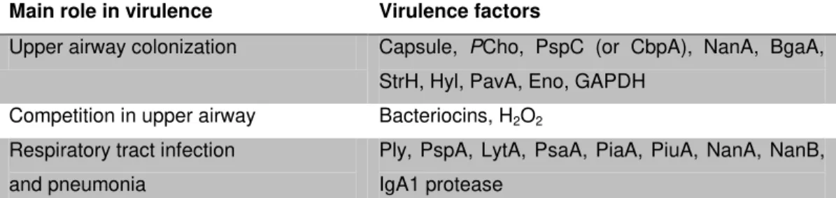
Table 1.1. Pneumococcal virulence factors and their main role in virulence (adapted from Kadioglu et al., 2008)
Main role in virulence Virulence factors
Upper airway colonization Capsule, PCho, PspC (or CbpA), NanA, BgaA, StrH, Hyl, PavA, Eno, GAPDH
Competition in upper airway Bacteriocins, H2O2
Respiratory tract infection and pneumonia
Ply, PspA, LytA, PsaA, PiaA, PiuA, NanA, NanB, IgA1 protease
PCho, phosphorylcholine; PspC, pneumococcal surface protein C (or CbpA, choline-binding protein
A); NanA, neuraminidase A; BgaA, β-galactosidase A; StrH, β-N-acetylglucosaminidase; Hyl, hyaluronidase; PavA, pneumococcal adhesion and virulence A; Eno, enolase; GAPDH, glyceraldehyde 3-phosphate dehydrogenase; H2O2, hydrogen peroxide; Ply, pneumolysin; PspA,
pneumococcal surface protein A; LytA, autolysin A; PsaA, pneumococcal surface antigen A; PiaA, pneumococcal iron acquisition A; PiuA, pneumococcal iron uptake A; NanB, neuraminidase B; IgA1, immunoglobulin A1.
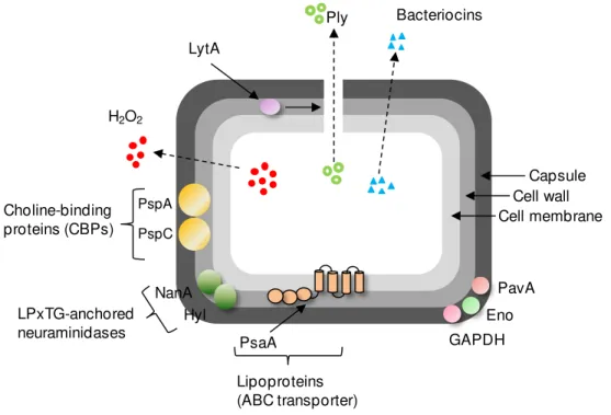
Of all classical virulence factors, capsule is the only one meeting the
criteria of condition sine qua non of virulence (Iannelli et al., 1999; Ogunniyi et al.,
2002). Capsule is a hydrophilic polysaccharide structure surrounding the cell wall
(see the red arrow in Fig. 1.1, Fig. 1.2). The pneumococcal serotypes are defined
by the composition of the capsular polysaccharide. When this work started 90
serotypes were documented, but currently at least 3 more have been described
(Song et al., 2012), denoting the high genetic plasticity of this microorganism. The
intrinsic properties of each capsule type, i.e., carbohydrate chemical nature,
degree of electronegative charge and thickness, confer specific host
anti-phagocytic and neutrophil responses and therefore, different ability to cause
invasive disease (Lee et al., 1991; AlonsoDeVelasco et al., 1995; Wartha et al.,
2007; Kadioglu et al., 2008; Mitchell and Mitchell, 2010). Capsule interferes with
the binding of antibodies (e.g. Fc of IgG) and complement system (e.g. iC3b
specific receptors in phagocytic cells (AlonsoDeVelasco et al., 1995; Abeyta et al.,
2003; Kadioglu et al., 2008; Mitchell and Mitchell, 2010). Capsule has also been
shown to reduce transformation (Weiser and Kapoor, 1999) and spontaneous or
antibiotic-induced lysis, contributing to tolerance to antibiotics (Fernebro et al.,
2004; Kadioglu et al., 2008). Moreover, capsule is essential for colonization since
it prevents mechanical exclusion of the pneumococcal cells by the mucus in
mucosal surfaces (Nelson et al., 2007; Kadioglu et al., 2008; Mitchell and Mitchell,
2010). Intra-strain and serotype-dependent variation of the amount of capsule is
also an important feature in colonization and disease: a very thick capsule is
detrimental for adhesion of S. pneumoniae surface proteins to host cell receptors.
On the other hand, in blood a thick capsule contributes to persistence of the
pneumococcus in this planktonic environment (Weiser et al., 1994; Cundell et al.,
H2O2
Bacteriocins Ply
LytA
PsaA Lipoproteins (ABC transporter)
Capsule Cell wall Cell membrane
PspA
PspC
Choline-binding proteins (CBPs)
LPxTG-anchored neuraminidases
NanA Hyl
PavA Eno GAPDH
Fig. 1.2. Schematic representation depicting pneumococcal virulence factors and their localization in the cell. Abbreviations: LytA, autolysin A; Ply, pneumolysin; PspA and PspC, pneumococcal surface proteins A and C; NanA, neuraminidase A; Hyl, hyaluronidase; PsaA, pneumococcal surface antigen A; PavA, pneumococcal adhesion and virulence A; Eno, enolase; GAPDH, glyceraldehyde 3-phosphate dehydrogenase; H2O2, hydrogen peroxide.
The nasopharynx is a densely populated niche. In assymptomatic
individuals, and particularly in children, various S. pneumoniae serotypes may
cohabit with other species, including Haemophilus influenzae, Moraxella
catarrhalis, Neisseria meningitidis and even Staphylococcus aureus (Pericone et
al., 2000; García-Rodríguez and Martínez, 2002; Pettigrew et al., 2008; Brugger
competition phenomena may occur. Among the streptococcal virulence factors,
the bactericidal bacteriocins (e.g. the bacterial pneumocins BlpM and BlpN) and
hydrogen peroxide (H2O2) are known for their roles targeting others bacteria in the
upper airway (Fig. 1.2) (Pericone et al., 2000; Dawid et al., 2007; Pettigrew et al.,
2008). The H2O2 produced by S. pneumoniae may reach extracellular
concentrations in the mM range, higher than those generated by many other
species, and sufficient to kill the competitors (Pericone et al., 2000). Interestingly,
H2O2 was also shown to have cytotoxic effects on epithelial cells (Duane et al.,
1993; Hirst et al., 2000; Braun et al., 2002). Another important virulence factor is
pneumolysin (Ply). Ply is a pore-forming cytotoxin belonging to the family of
cholesterol-dependent cytolysins (CDCs). Pneumococcal autolysis triggers the
release of this cytoplasmic soluble protein (Fig. 1.2), which displays multiple
functions important for infection. In short, Ply oligomers polymerize in
cholesterol-containing host cell membranes forming a transmembrane pore inducing cell
lysis; Ply activates and binds the complement of the host immune system, e.g.
C3b protein, diverting it from the surface of pneumococcal cells; it affects host cell
activities at sub-lytic concentrations (reviewed in Kadioglu et al., 2008; Mitchell
and Mitchell, 2010). The major autolysin inducing pneumococcal lysis, and thus,
release of intracellular content (e.g. pneumolysin and DNA), is autolysin A (LytA)
(Fig. 1.2) (reviewed in Kadioglu et al., 2008; Mitchell and Mitchell, 2010). LytA is
also a virulence factor and belongs to the group of choline-binding proteins
(CBPs). CBPs are surface exposed proteins that anchor non-covalently to the cell
wall teichoic acid and membrane-bound lipoteichoic acids through residues of
phosphorylcholine (PCho). PCho is an atypical bacterial cell wall component,
present in S. pneumoniae, that stimulates adherence of bacterial cells to the
platelet-activating factor receptor (rPAF) in epithelial surfaces of the nasopharynx
an N-acetylmuramoyl-L-alanine-amidase that digests peptidoglycan; its major role
in virulence is presumably mediated by the release of pneumolysin and cell wall
(Fig. 1.2) (reviewed in Kadioglu et al., 2008; Mitchell and Mitchell, 2010). Cell wall
has a pivotal function in triggering inflammatory processes (reviewed in Kadioglu
et al., 2008; Mitchell and Mitchell, 2010). Other two known virulent CBPs are the
pneumococcal surface proteins C (PspC, also known as choline-binding protein
A, CbpA) and A (PspA) (Fig. 1.2), with function in colonization and infection,
respectively. During transcytosis across the surface epithelium, PspC binds the
immunoglobulin A (IgA) receptor and the complement factor H-binding protein,
inhibiting complement activation (reviewed in Kadioglu et al., 2008; Mitchell and
Mitchell, 2010). PspA binds the bactericidal protein lactoferrin and the C3 protein
of the complement system preventing its deposition on the surface of
pneumococcal cells (reviewed in Kadioglu et al., 2008; Mitchell and Mitchell,
2010). Among the surface proteins, several LPxTG-proteins, which are anchored
by an amino acid LPxTG motif to peptidoglycan, are well described virulence
factors (Fig. 1.2) (reviewed in Bergmann and Hammerschmidt, 2006; Kadioglu et
al., 2008). Of these, neuraminidase A (NanA), also called sialidase A, is a good
example. NanA cleaves N-acetylneuraminic acid or sialic acid from glycosylated
proteins or lipids and oligosaccharides in the mucosal cell surfaces. By cleaving
these residues, NanA unmasks potential receptors in the host surface epithelium,
promoting colonization, and modifies the function of sialidated proteins of the host
involved in clearance of bacteria, stimulating invasion (reviewed in Kadioglu et al.,
2008; Mitchell and Mitchell, 2010). The pneumococcal surface antigen A (PsaA),
a lipoprotein that is part of the Psa(ABC) transporter (Fig. 1.2), is involved in the
translocation of manganese ions into the cell. The contribution of this protein in
the resistance to oxidative stress and in increased adhesion has been shown
proteins involved in adherence to host cells and/or invasion are the pneumococcal
adherence and virulence A (PavA), enolase (Eno), glyceraldehyde 3-phosphate
dehydrogenase (GAPDH), 6-phosphogluconate dehydrogenase (6-PGD), β
-galactosidase A (BgaA), β-N-acetylglucosaminidase (StrH) and hyaluronate lyase
A (HylA) (Fig. 1.2) (Berry and Paton, 2000; Bergmann et al., 2001; Holmes et al.,
2001; Bergmann et al., 2003; Pracht et al., 2005; King et al., 2006; Daniely et al.,
2006; Terra et al., 2010; Dalia et al., 2010; Limoli et al., 2011). BgaA, StrH and
HylA are involved in the hydrolysis of surface glycoproteins and unmasking of
host receptors, which ultimately facilitates pneumococcal adherence to epithelial
cells. Remarkably, BgaA and Hyl were also found to be important for growth in
certain media (van Opijnen et al., 2009; Marion et al., 2012). The glycolytic
enzymes Eno and GAPDH bind the host plasminogen, which prevents activation
of plasmin and cleavage of fibrin clots (Henderson and Martin, 2011). The
moonlighting facet of these proteins suggests a more relevant role of sugar
metabolic enzymes than previously imagined (Henderson and Martin, 2011). In
accordance, a number of studies showed that deletion of genes involved in sugar
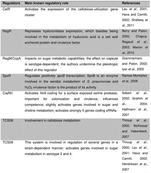
metabolism and regulation (e.g. pyruvate oxidase (SpxB)) and the global regulator
CcpA leads to virulence attenuation (Giammarinaro and Paton, 2002; Iyer et al.,
2005; Ramos-Montañez et al., 2008). Although not classical virulence factors,
these proteins, also common to non-pathogenic bacteria, are very important for
the fitness of S. pneumoniae in host niches. The study of sugar metabolism may
therefore disclose unforeseen metabolic factors as potential targets for the
Carbohydrate metabolism in
S. pneumoniae
Over the past few decades, most investigators centered their attention in
trying to understand the mechanisms by which S. pneumoniae classical virulence
factors interact with the host (reviewed in Kadioglu et al., 2008). However, factors
involved in basic metabolic physiology, allowing for fitness and adaptation of this
strictly fermentative bacterium in the microenvironments of the host, have been
largely ignored. In the nasopharynx, free sugars, and in particular the often
preferred hexose glucose ([Glc]nasopharynx < 1 mM), are scarce (Shelburne et al.,
2008a). In this environment, S. pneumoniae most likely relies on wide range of
glycosylated proteins (mucins) present in the mucus lining the epithelial cell
surfaces of airway structures (e.g. nasopharynx, tracheae, bronqui) (Rose and
Voynow, 2006; Yesilkaya et al., 2008). The most abundant mucin present in
mucus contains a chain of different sugars (acetylgalactosamine,
N-acetylglucosamine, fucose, sialic acid, galactose and sulfated sugars) in varied
proportions that is O-glycosydically linked to the polypeptide (apomucin)
(Yesilkaya et al., 2008; Terra et al., 2010). The determination of the relative
proportion of each sugar in these chains showed that Gal is one of the sugars
present at higher concentrations (Holmén et al., 2004; Yesilkaya et al., 2008;
Terra et al., 2010). Release of Gal from deglycosylation of human mucins by
exoglycosidases expressed by S. pneumoniae has been reported (King et al.,
2006). Contrastingly, in other host niches, such as blood, S. pneumoniae
encounters Glc as major carbon source, [Glc]blood ~ 4-6 mM (Shelburne et al.,
2008a). Glc and Gal are thus relevant sugars for the study of sugar metabolism in
S. pneumoniae.
Complete genome sequences of S. pneumoniae are available since 2001
genomes brought to light the importance of sugars to the lifestyle of
S. pneumoniae and provided the framework for the development of a number of
genome-wide analysis (e.g. microarray analysis and signature-tagged
mutagenesis) (Hava and Camilli, 2002; Orihuela et al., 2004a; Shelburne et al.,
2008a). Interestingly, studies designed to identify genes essential for virulence at
the genome level consistently revealed genes involved in sugar catabolism.
Capitalizing on these genome-wide studies, several research efforts have shown
that non-classical virulence factors such as carbohydrate uptake systems,
metabolic enzymes and a global regulator of carbon metabolism (CcpA) directly
contribute to S. pneumoniae colonization and disease (Giammarinaro and Paton,
2002; King et al., 2004; Iyer et al., 2005; King et al., 2006; Iyer and Camilli, 2007;
Burnaugh et al., 2008; Ramos-Montañez et al., 2008; Yesilkaya et al., 2009;
Trappetti et al., 2009; Marion et al., 2011; Marion et al., 2012).
Although the value of sugar metabolic proteins for the infectivity of
S. pneumoniae is starting to be recognized, the need for in depth studies enabling
integration of basic metabolic processes, regulation and connections to virulence
in S. pneumoniae is evident. In the following subsections a genome and literature
based overview of the putative sugar transport and metabolic capabilities,
particularly for the sugars Glc and Gal, of S. pneumoniae will be presented.
Subsequently, the cross talk between the synthesis of the polysaccharide capsule
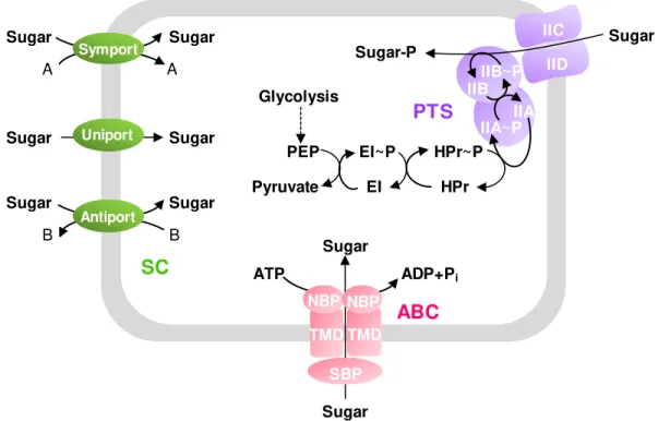
Transport of glucose and galactose
The transport of any exogenous carbohydrate across the
sugar-impermeable lipidic membrane is the first step committed to its metabolism. In the
Streptococacceae family this translocation may occur through three different
types of carbohydrate uptake systems: secondary carriers (uniporters, symporters
and antiporters), ABC-transporters and phosphoenolpyruvate: carbohydrate
phosphotransferase systems (PTS-systems) (Fig. 1.3) (Lorca et al., 2007). The
driving force impelling the transport of carbohydrates through secondary carriers
is an electrochemical gradient of other solutes across the cell membrane
(Poolman and Konings, 1993). In ABC transporters, also called ATP-binding
cassettes, this driving force is the intracellular hydrolysis of ATP (reviewed in
Rees et al., 2009). The PTS systems are multiprotein complexes comprising both
cytoplasmatic and transmembranar proteins (reviewed in Postma et al., 1993).
Although more complex, this system is more efficient in energetic terms, since
rather than ATP it utilizes phosphoenolpyruvate (PEP) to phosphorylate the
incoming sugar (Fig. 1.3) (reviewed in Postma et al., 1993). In S. pneumoniae, a
genome survey revealed the occurrence of twenty-one PTS systems, seven
ABC-transporters, one symporter and a permease (uniporter) for carbohydrate uptake
(Bidossi et al., 2012). The number of sugar transporters in the pneumococcus is
higher than for any other prokaryote relative to genome size (Tettelin et al., 2001;
Bidossi et al., 2012) and is variable between serotypes (Bidossi et al., 2012). The
ensemble of transporters have specificity for several sugars, such as Glc, Gal,
N-acetylglucosamine, mannose, fructose, sialic acid, cellobiose, fructose and
β-glucosides (Bidossi et al., 2012). However, firm functional assignments are only
Sugar B B A A Sugar-P IIB~P IIB IIA IIA~P HPr HPr~P EI~P EI Pyruvate PEP
PTS
SC
Uniport Symport AntiportTMD TMD
SBP NBP NBP
Sugar Sugar
ATP ADP+Pi
ABC
Glycolysis Sugar Sugar Sugar Sugar Sugar Sugar IID IICFig. 1.3. Schematic figure showing the three classes of carbohydrate uptake systems in Streptococcaceae. SC (in green), secondary carriers; ABC (in pink), ABC-transporters; PTS (in purple), phosphoenolpyruvate: carbohydrate phosphotransferase system. Abbreviations: NBP, nucleotide-binding protein; TMD, transmembrane domain; SPB, sugar/solute-binding protein; PEP, phosphoenolpyruvate; EI, HPr are the cytoplasmatic proteins of the PTS-system; IIAB and IICD are the cytoplasmatic and transmembranar proteins of the carbohydrate-specific PTS, respectively. The PTS-system depicted here represents a PTS-system from the PTS mannose-fructose-sorbose family (PTSMan).
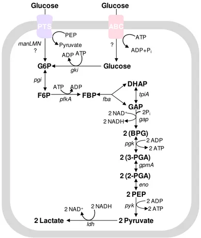
As for the hexose Glc, its transport is not fully elucidated in
S. pneumoniae, but a PTS system (spd_0264-2 or manLMN) of the PTS
mannose-fructose-sorbose (Man) family, referred as PTSMan, is presumably the
was, however, not abolished by a manLMN mutation, a result consistent with the
presence of other uptake systems (Bidossi et al., 2012). Furthermore, the ability
of a ptsI mutant, disrupted for the general PTS protein EI, to grow on Glc supports
the occurrence of non-PTS systems for Glc transport (Bidossi et al., 2012).
Current work in our laboratory is aimed at identifying additional Glc uptake routes
in S. pneumoniae.
Transport of Gal in S. pneumoniae is even more obscure; however, the
PTS system of the family PTS-galactitol (Gat) encoded by spd_0559-61 aka
gatABC was pointed out in two independent studies as playing a role on Gal
uptake (Kaufman and Yother, 2007; Bidossi et al., 2012). Confirmation has been
hampered by the weak phenotype of the null mutant. Which other transporters are
involved in Gal uptake remain to be elucidated, but Bidossi et al. reported a
decreased ability of a manLMN mutant to grow on Gal (Bidossi et al., 2012).
Hitherto, firm identification of Glc and Gal transporters and their ensuing
biochemical characterization in S. pneumoniae is still missing. Interestingly, PTS
sugar transporters are only present in prokaryotes. Considering their location in
bacterial cells (superficial), these transporters could be good targets for the
development of drugs.
Metabolism of glucose and galactose
S. pneumoniae is a strictly fermentative bacterium, relying on sugar
substrates for growth. Since it lacks a respiratory chain, this organism depends on
substrate-level phosphorylation to form ATP (Tettelin et al., 2001; Hoskins et al.,
2001; Lanie et al., 2007). In this process, ATP is originated from the direct transfer
of a phosphoryl group from phosphorylated reactive intermediates to ADP (Nelson
and Cox, 2000).
S. pneumoniae is a homofermentative bacterium that converts hexoses to
pyruvate via the Embden-Meyerhof-Parnas (EMP) pathway; recycling of reducing
equivalents is achieved mainly through reduction of pyruvate to lactate (Fig. 1.4).
Glc can be transported and concomitantly phosphorylated by PTS
systems or enter the cell via non-PTS transporters and be subsequently
phosphorylated to G6P by intracellular glucokinases (Fig. 1.4). The ensuing
Glucose PEP Pyruvate PTS G6P ATP ADP gki 2 NADH 2 NAD+
FBP DHAP GAP fba 2 (BPG) 2 (3-PGA) 2 (2-PGA) 2 PEP pyk eno 2Pi 2 ATP tpiA gap pgk gpmA F6P pfkA pgi ADP ATP 2 ADP Glucose ATP ADP+Pi ABC Glucose ? 2 ATP 2 ADP 2 Pyruvate 2 NADH
2 NAD+
ldh
2 Lactate
manLMN
?
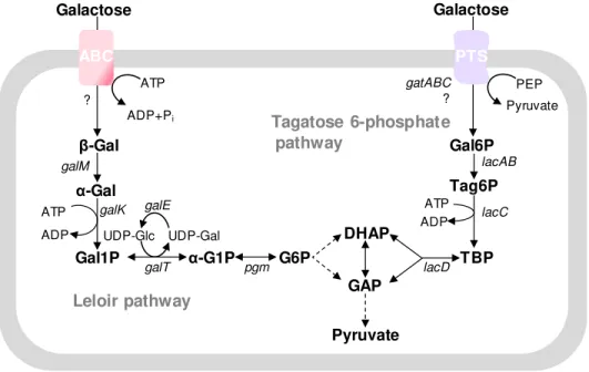
Gal is generally recognized as a slowly metabolized carbohydrate
(reviewed in Deutscher et al., 2006). The metabolism of Gal is well documented
for a number of Streptococcaceae, including S. mutans, S. pyogenes, L. lactis,
and S. salivarius) (Steele et al., 1954; Pierce, 1957; Thomas et al., 1980;
Abranches et al., 2004; Neves et al., 2010). In these organisms, Gal can be
metabolized by the Leloir and/or tagatose 6-phosphate pathways (Fig. 1.5). In
S. mutans, Gal can only be efficiently metabolized when both pathways are
operating simultaneously (Abranches et al., 2004). In L. lactis, the use of the
Leloir and/or the tagatose 6-phosphate pathway for Gal utilization is currently
viewed as strain-dependent, but the relative efficacy in the degradation of the
sugar has not been established (Thomas et al., 1980; Neves et al., 2010). In
S. salivarius, due to absence of Gal-PTS transporters only the Leloir pathway is
functional (Chen et al., 2002). In S. pneumoniae, the functionality of the two
pathways is yet to be proven, but the enzymatic steps of both pathways can be
inferred from the genome (Fig. 1.5) (Tettelin et al., 2001; Hoskins et al., 2001;