VARIATIONS IN THE SENSITIVITY OF DIFFERENT PRIMERS FOR DETECTING WOLBACHIA IN
ANASTREPHA (DIPTERA: TEPHRITIDAE)
Helena Sanches Marcon1*, Virgínia Elias Coscrato1, Denise Selivon2, André Luiz Paranhos Perondini2, Celso Luis Marino1
1
Departamento de Genética, Instituto de Biociências, Universidade Estadual Paulista Júlio de Mesquita Filho, Botucatu, SP,
Brasil; 2Departamento de Genética e Biologia Evolutiva, Instituto de Biociências, Universidade de São Paulo, São Paulo, SP, Brasil.
Submitted: May 02, 2010; Returned to authors for corrections: August 23, 2010; Approved: January 13, 2011.
ABSTRACT
Wolbachia are endosymbiont bacteria of the family Rickettsiacea that are widespread in invertebrates and
occur between 20% and 60% of Neotropical insects. These bacteria are responsible for reproductive
phenomena such as cytoplasmic incompatibility, male killing, feminization and parthenogenesis.
Supergroups A and B of Wolbachia are common in insects and can be identified using primers for 16S
rDNA, ftsZ and wsp; these primers vary in their ability to detect Wolbachia. The ftsZ primer was the first
primer used to detect Wolbachia in Anastrepha fruit flies. The primers for 16S rDNA, ftsZ and wsp and the
corresponding PCR conditions have been optimized to study the distribution of Wolbachia and their effect
on the biology of Anastrepha in Brazil. In this work, we examined the ability of these primers to detect
Wolbachia in Anastrepha populations from three regions in the State of São Paulo, southeastern Brazil. All
of the samples were positive for Wolbachia supergroup A when screened with primers for 16S A rDNA and
wsp A; the wsp B primer also gave a positive result, indicating cross-reactivity. The ftsZ primer showed a
poor ability to detect Wolbachia in Anastrepha and generated false negatives in 44.9% of the samples. These
findings indicate that reliable PCR detection of Wolbachia requires the use of primers for 16S rDNA and
wsp to avoid cross-reactions and false negatives, and that the ftsZ primer needs to be redesigned to improve
its selectivity.
Key words:Anastrepha, cross reaction, ftsZ, wsp
INTRODUCTION
Wolbachia are intracellular obligatory bacteria of the
family Rickettsiacea that occur in a wide range of arthropods
and nematodes. These bacteria are maternally inherited by
horizontal transmission (26, 29) and enhance their propagation
by altering the reproductive system of their host in various
ways, e.g., by cytoplasmic incompatibility, male killing,
feminization and parthenogenesis (19, 24).
Molecular markers have been extensively used to detect
Wolbachia. For example, primers for 16S rDNA (a ribosomal
gene), ftsZ (a regulatory gene of the bacterial cell cycle) and
*Corresponding Author. Mailing address: Departamento de Genética, Instituto de Biociências, Universidade Estadual Paulista Júlio de Mesquita Filho,
wsp (a gene for an cell membrane protein) have been used to
study the phylogenetics of Wolbachia (2, 6, 18) and have led to
the division of Wolbachia into eight taxonomic supergroups (A
to H) (12, 13, 27). The initial studies in this field were done
with 16S rDNA primers and identified two groups, A and B,
that diverged about 50 million years ago and are widely
distributed in insects (14, 27).
The ftsZ gene was extensively used by Werren and
colleagues (27, 28) to detect Wolbachia, but several studies
have demonstrated its low sensitivity (3, 9, 30, 31). This low
sensitivity can lead to false negatives in bacterial detection
when this primer is used alone, i.e., without another marker or
technique to detect Wolbachia. The 16S and wsp primers have
greater sensitivity for detecting Wolbachia (30, 31), although
the wsp primer can generate false positives in discriminating
between supergroups A and B (8). This lack of absolute
specificity means that there is a need to use specific primers in
the characterization of these bacteria.
Between 20% and 60%of Neotropical insects are infected
with Wolbachia (28). Studies in different regions of Brazil
have detected Wolbachia in fruit flies of the genus Anastrepha
(3, 4, 17, 23). Anastrepha fruit flies are a major pest insect of
Brazilian fruit crops because of the losses they cause to
commercial fruit growers (5, 15). The presence of Wolbachia
in Anastrepha is therefore of considerable interest since these
bacteria may be exploited as biological controls of pest insects,
as suggested by Bourtzis (1).
Coscrato (4) and Mascarenhas (17) showed that 16S and
wsp primers varied in their ability to detect Wolbachia in
different Anastrepha species; studies with other fruit fly
genera, such as Bactrocera (8, 11, 25), Rhagoletiscerasi (20)
and Ceratitis capitata (21), have reported similar findings. In
this work, we examined the ability of 16S, ftsZ and wsp
primers to detect Wolbachia in populations of Anastrepha from
different regions of the State of São Paulo in southeastern
Brazil.
MATERIALS AND METHODS
Sixty-six fruit fly larvae collected from guava (Psidium
guajava) and chapeu-do-sol (Terminalia catappa) fruits in
three regions of the State of São Paulo, southeastern Brazil
(Table 1), were stored in boxes with vermiculite and
transported to the laboratory, where the larvae were allowed to
develop to pupal stage. The pupae were subsequently removed
and allowed to grow to the adult stage for species
identification, after which they were stored in 70% ethanol at
-20oC.
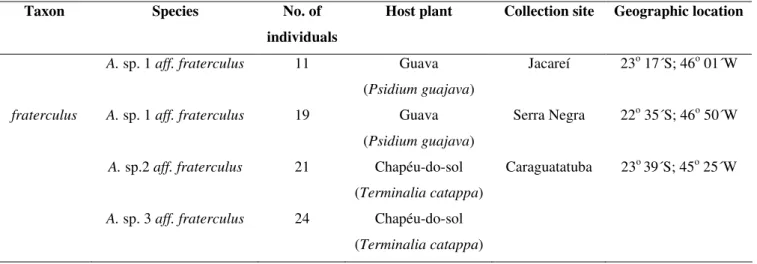
Table 1. Number of individuals, host plants, collection sites and geographic location of the fruit fly populations examined in this
study. All collection sites were in the State of São Paulo.
Taxon Species No. of
individuals
Host plant Collection site Geographic location
A. sp. 1 aff. fraterculus 11 Guava
(Psidium guajava)
Jacareí 23o 17´S; 46o 01´W
fraterculus A. sp. 1 aff. fraterculus 19 Guava
(Psidium guajava)
Serra Negra 22o 35´S; 46o 50´W
A. sp.2 aff. fraterculus 21 Chapéu-do-sol
(Terminalia catappa)
Caraguatatuba 23o 39´S; 45o 25´W
A. sp. 3 aff. fraterculus 24 Chapéu-do-sol
DNA was extracted from each fly abdomen using the
protocol described by Jowett (10), with some modifications.
Amplification reactions were done as described below.
The reaction mixture for the wsp A and B primers
consisted of 50 ng of DNA, 10 X buffer (Invitrogen), 1.0 µl of
50 mM MgCl2, 0.4 µl of dNTPs (10 µM each), 0.5 µl of
forward (F) primer (20 µM), 0.5 µl of reverse(R) primer (20
µM), 0.5 µl of Taq DNA polymerase (5 U/µl) (Invitrogen) and
water in a final volume of 20 µl. The amplification reaction
consisted of one cycle of 1 min at 94oC, 1 min at 58oC and 2 min at 72oC, followed by 35 cycles of 15 s at 94oC, 1 min at 58oC and 2 min at 72oC, and one cycle of 15 s at 94oC, 1 min at 58oC and 7 min at 72oC. These conditions yielded PCR products of ~600 base pairs (bp) (2, 32).
The reaction mixture for the ftsZ A and B primers (6)
consisted of 50 ng of DNA, 10 X buffer (Invitrogen), 0.6 µl of
50 mM MgCl2, 0.4 µl of dNTPs (10 µM each), 0.5 µl of F
primer (10 µM), 0.5 µl of R primer (10 µM), 0.5 µl of Taq
DNA polymerase (5 U/µl) (Invitrogen) and water in a final
volume of 20 µl. The amplification reaction consisted of an
initial 4 min incubation at 94ºC, followed by one cycle of 1
min at 58ºC and 2 min at 72ºC, 38 cycles of 15 s at 94ºC, 1 min
of 58ºC and 2 min at 72ºC, one cycle of 15 s at 94ºC and 1 min
at 58ºC, with a final extension of 7 min at 72ºC. These
conditions yielded fragments of 1043-1055 bp.
The reaction mixture for the 16S A and B rDNA primers
(18) consisted of 50 ng of DNA, 10X buffer (Invitrogen), 0.75
µl of 50 mM MgCl2, 0.5 µl of dNTPs (10 µM each), 0.35 µl of
Fprimer(20 µM), 0.35 µl of Rprimer(20 µM),0.25 µl of Taq
DNA polymerase (5 U/µl) (Invitrogen) and water in a final
volume of 25 µl. The amplification reaction consisted initially
of 2 min at 95ºC, 35 cycles of 30 s at 95ºC, 1 min at 55ºC and 1
min at 72ºC, with a final extension of 3 min at 72ºC. These
conditions yielded fragments of ~259 bp.
In all cases, the PCR products were analyzed by
electrophoresis in 1% agarose gels in Tris-borate EDTA buffer
(TBE 1X) containing 1% ethidium bromide. After
electrophoresis, the gels were examined in ultraviolet light and
documented with an Eagle Eye II photodocumentation system
(Stratagene).
For sequencing, the PCR products were purified with
GFXTM PCR DNA and gel band purification kits (Amersham Pharmacia Biotech), after which DNA (100 ng/µl) was mixed
with 1.0 µl of buffer (1 M Tris-HCl, pH 9.0, containing 50 mM
MgCl2), 2.0 µl of Big Dye, 1.0 µl of primer (5 pmol/ul) and
water in a final volume of 10 µl. Sequencing was done in a
Genetic Analyzer 3100 (Applied Biosystems) sequencer. The
sequences generated from these samples and the corresponding
consensus sequences were assembled with phredPhrap/consed
v. 14.0 and then used to search the National Center for
Biotechnology Information (NCBI) database for homology
with Wolbachia sequences; the searches were done using
BLASTN (http://www.ncbi.nlm.nih.gov).
RESULTS AND DISCUSSION
Wolbachia was detected in all of the DNA fragments
generated by the 16S and wsp primers in Anastrepha species 1,
2 and 3. The sequence of the DNA fragment amplified with the
wsp primer showed 98% and 96% similarity with Wolbachia
from Anastrepha sp.2 aff. fraterculus (EU116316.1) and
Wolbachia from Brugia pahangi (AY527208.1), respectively.
The results with the 16S rDNA and wsp primers allowed us to
classify the bacteria as belonging to Wolbachia supergroup A.
These results were similar to studies in other Anastrepha
species (4, 17).
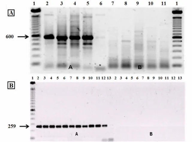
In some samples, the wsp B primer generated fragments
that suggested the presence of supergroup B (figure 1A).
However, this result was considered to be a false positive since
no fragments were generated by the primer for 16S B rDNA
(figure 1B). Kittayapong (11) and Ruang-Areerate (22) have
previously shown that the wsp B primer yields false positive
results for this Wolbachia supergroup because of
cross-reactions; similar findings have been described by Coscrato (3)
conclusive identification of Wolbachia supergroup A in Anastrepha requires the use of wsp and 16S rDNA primers.
Figure 1. Electrophoresis of Anastrepha sp.1 samples in 1% agarose gels containing 1% ethidium bromide, after amplification. In
(A), the primers used were wsp A in lanes 2A-6A and wsp B in lanes 7B-11B. In (B), the primers used were for 16S A rDNA in
lanes 2A-13A and 16S B rDNA in lanes 2B-13B.
We also compared the results obtained for the ftsZprimer
with those for the 16S and wsp primers, particularly since data
generated by the former primer have sometimes led to the
misidentification of Wolbachia. No Wolbachia were detected
in the 66 samples incubated with the ftsZ primer, a finding in
agreement with previous studies that have also used this primer
to screen for these bacteria in other insects (7, 9, 30).
The ftsZ gene is of particular importance because of the
potential usefulness of its product for detecting Wolbachia,
identifying supergroups and performing phylogenetic analyses.
We therefore sought to optimize the PCR protocol for this
primer by altering the concentrations of DNA, MgCl2, dNTPs,
primersand Taq DNA polymerase and quality of the DNA in
order to detect Wolbachia in Anastrepha (Table 2). When the
volume of DNA in the reaction was decreased to 3 µl (50
ng/ul), Walbachia was detected in 27% of Anastrepha sp.1
(Jacareí) samples (Table 3). Based on these results, this volume
Table 2. Modifications in the reagent concentrations and volumes of the PCR reactions (ftsZ I-VIII) used to detect Wolbachia in Anastrepha with the ftsZprimer.
*Alteration in PCR conditions (reagent concentration and/or volume).
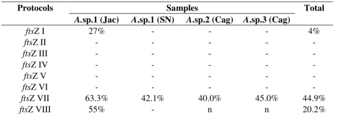
Table 3. Efficiency of the ftsZ primer in detecting Wolbachia in Anastrepha samples using the altered protocols (ftsZ I-VIII) described in Table 2.
Samples Protocols
A.sp.1 (Jac) A.sp.1 (SN) A.sp.2 (Cag) A.sp.3 (Cag)
Total
ftsZ I 27% - - - 4%
ftsZ II - - - - -
ftsZ III - - - - -
ftsZ IV - - - - -
ftsZ V - - - - -
ftsZ VI - - - - -
ftsZ VII 63.3% 42.1% 40.0% 45.0% 44.9%
ftsZ VIII 55% - n n 20.2%
Collection sites: Jac – Jacareí; SN – Serra Negra; Cag – Caraguatatuba. (-): absence of fragment.
(n): not tested with the protocol indicated.
PCR reaction Reagents Concentration Volume (µµµµl)
ftsZ I DNA 50 ng/µl 3.00*
ftsZ II MgCl2 50 mM 0.50*
DNA 50 ng/µl 3.00
ftsZ III Primer F 8 µM* 0.50
Primer R 8 µM* 0.50
DNA 50 ng/µl 3.00*
ftsZ IV MgCl2 50 mM 0.50*
Primer F 8 µM* 0.50
Primer R 8 µM* 0.50
DNA 50 ng/µl 3.00
ftsZ V DNTP 10 mM 0.50*
MgCl2 50 mM 0.50*
DNA 50 ng/µl 3.00*
ftsZ VI DNTP 10 mM 0.50*
MgCl2 50 mM 0.50*
Primer F 8 µM* 0.50
Primer R 8 µM* 0.50
DNA 50 ng/µl 3.00*
ftsZ VII Taq DNA polymerase 1 unit 0.25*
DNA 50 ng/µl 3.00
No Wolbachia were detected with protocols ftsZ II, III,
IV, V and VI (Table 2), indicating that the alterations
incorporated in these reactions did not improve the efficiency
of detection. In reaction ftsZ VII in which the volume of Taq
DNA polymerase was changed and the number of cycles was
increased from 35 to 40 (in order to enhance the number of
DNA fragments) (Table 2), Wolbachia was detected in 44.9%
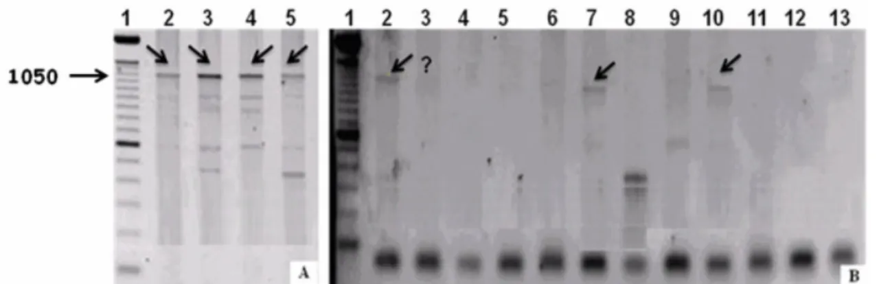
of the samples (Table 3). However, this protocol showed poor
reproducibility for the same sample analyzed at different times
(Figure 2A, B). Similar findings were reported by Jeyaprakash
and Hoy (9) for other arthropod species and these authors
proposed that DNA present in the reaction could interfere with
Taq DNA polymerase activity to generate false negatives.
Werren and Windsor (30) observed that the quality of
DNA was a determinant factor in the successful detection of
Wolbachia with the ftsZ primer and recommended that only
newly extracted DNA be used for the PCR, i.e., one should
avoid using DNA stored at -20oC. To examine the influence of DNA quality on the detection of Wolbachia with the ftsZ
primer we extracted DNA from Anastrepha sp.1 (Jacareí and
Serra Negra) and used it in protocol VIII, along with the wsp
and 16S rDNA primers. In these conditions, Walbachia was
detected in 20.2% of the samples screened with the ftsZ primer
(Table 3), whereas all of the samples tested with the wsp and
16S rDNA primers were positive for the bacteria. These
findings confirm the low sensitivity of the ftsZ primer in
detecting Wolbachia in Anastrepha, despite the alterations in
the extraction and amplification protocols.
Figure 2. Electrophoresis of Anastrepha sp. 1 (Jacareí) samples in 1% agarose gels containing 1% ethidium bromide, after amplification with the primers ftsZ (A) and ftsZ (B). The Anastrepha sp.1 samples are in lanes 2-5 in (A) and lanes 2-13 in (B).
Lane 1 - 100 bp ladder. Arrows indicate the presence of fragments. ? = presence of fragment in column B3 uncertain.
Together, the results of this study indicate that the most
efficient way of detecting Wolbachia in Anastrepha, and of
identifying the relevant supergroup and making phylogenetic
inferences, is through the combined use of 16S rDNA and wsp
primers. The 16S rDNA primer can be used by itself to detect
Wolbachia and identify supergroups. However, since this
primer is for preserved gene it is inappropriate for phylogenetic
and population analyses. This limitation can be overcome by
concomitant use of the wsp primer, which by itself is
inappropriate for identifying Wolbachia supergroups A and B.
The variability of wsp makes primers of this gene particularly
useful for phylogenetic and population analyses.
ACKNOWLEDGMENTS
This work was supported by Fundação de Amparo à
Coordenadoria de Aperfeiçoamento de Pessoal de Nível
Superior (CAPES).
REFERENCES
1. Bourtzis, K. (2008). Wolbachia-based technologies for insect pest population control. Adv. Exp. Med. Biol. 627, 104-113.
2. Braig, H.R.; Zhou, W.; Dobson, S.; O’Neil, S.L. (1998). Cloning and characterization of the gene encoding the major surface protein of the bacterial endosymbiont Wolbachia. J. Bacteriol. 180, 2373-2378. 3. Coscrato, V.E. (2006). Detecção e filogenia da bactéria endossimbionte
Wolbachia em espécies de moscas-das-frutas do gênero Anastrepha e
Ceratitis (Diptera: Tephritidae). Botucatu – S.P., Brazil. 144 p. (PhD Dissertation. Instituto de Biociências. UNESP).
4. Coscrato, V.E.; Braz, A.S.; Perondini, A.L.P.; Selivon, D.; Marino, C.L. (2009). Wolbachia in Anastrepha fruit flies (Diptera: Tephritidae). Curr. Microbiol. 59, 295-301.
5. Duarte, A.L. and Malavasi, A. (2000). Tratamentos quarentenários. In: Malavasi, A. and Zucchi, R.A. (eds). Moscas-das-frutas de importância econômica no Brasil (conhecimento básico e aplicado). FAPESP-Holos, Ribeirão Preto, Brazil, p.187-192.
6. Holden, P.R.; Brookfield, J.F.Y.; Jones, P. (1993). Cloning and characterization of an ftsZ homologue from a bacterial symbiont of
Drosophila melanogaster. Mol. Gen. Genet. 240, 213-220.
7. Hong, X.Y.; Gotoh, T.; Noda, H. (2002). Sensitivity comparison of PCR
primers for detecting Wolbachia in spider mites. Appl. Entomol. Zool. 37, 379-383.
8. Jamnongluk, W.; Kittayapong, P.; Baimai, V.; O'Neill. S.L. (2002).
Wolbachia infections of tephritid fruit flies: molecular evidence for five distinct strains in a single host species. Curr. Microbiol. 45, 255-260. 9. Jeyaprakash, A. and Hoy, M.A. (2000). Long PCR improves Wolbachia
DNA amplification: wsp sequences found in 76% of sixty-three arthropod species. Insect Mol. Biol. 9, 393-405.
10. Jowett, T. (1986). Preparation of nucleic acids. In: Roberts, D.B. (ed).
Drosophila: a practical approach. Oxford University Press, Oxford. p. 275-286.
11. Kittayapong, P.; Milne, J.R.; Tigvattananont, S.; Baimai, V. (2000). Distribution of the reproduction-modifying bacteria, Wolbachia, in natural populations of tephritid fruit flies in Thailand. Sci. Asia.26, 93-103.
12. Lo, N.; Casiraghi, M.; Salati, E.; Bazzocchi, C.; Bandi, C. (2002). How many Wolbachia supergroups exist? Mol. Biol. Evol. 19, 341-346. 13. Lo, N.; Paraskevopoulos, C.; Bourtzis, K.; O'Neill, S.L.; Werren, J.H.;
Bordenstein, S.R.; Bandi, C. (2007). Taxonomic status of the intracellular bacterium Wolbachiapipientis. Int. J. Syst. Evol. Microbiol. 57, 654-657.
14. Lukenhaus, J.F. (1990). Regulation of cell division in E. coli. Trends Genet. 6, 22-25.
15. Malavasi, A.; Zucchi, R.A.; Sugayama, R.L. (2000). Biogeografia, In: Malavasi, A. and Zucchi, R.A. (eds). Moscas-das-frutas de importância econômica no Brasil (conhecimento básico e aplicado). FAPESP-Holos, Ribeirão Preto, Brazil, p. 93-98.
16. Marcon, H.S. (2009). Identificação da bactéria endossimbionte
Wolbachia em populações de moscas-das-frutas do complexo
Anastrepha fraterculus (Diptera: Tephritidae). Botucatu – S.P., Brazil. 110 p. (MSc Dissertation. Instituto de Biociências. UNESP).
17. Mascarenhas, R.O. (2007). Endossimbionte Wolbachia em moscas-das-frustas do gênero Anastrepha (Thephritidae) e em vespas parasitóides (Braconidae) associadas. São Paulo – S.P., Brazil. 80 p. (MSc Dissertation. Instituto de Biociências. USP).
18. O’Neil, S.L.; Giordano, R.; Colbert, A.M.; Karr, T.L.; Robertson, H.M. (1992). 16S rRNA phylogenetic analysis of the bacterial endosymbionts associated with cytoplasmic incompatibility in insects. Proc. Natl. Acad. Sci. USA 89, 2699-2702.
19. O’Neil, S.L.; Hoffmann, A.A.; Werren, J.H. (1997). Influential passengers: inherited microorganisms and arthropod reproduction. Oxford University Press, New York, 214 p.
20. Riegler, M. and Stauffer, C. 2002. Wolbachia infections and superinfections in cytoplasmically incompatible populations of the European cherry fruit fly Rhagoletis cerasi (Diptera, Tephritidae). Mol. Ecol. 11, 2425-2434.
21. Rocha, L.S.; Mascarenhas, R.O.; Perondini, A.L.P.; Selivon, D. (2005). Occurrence of Wolbachia in Brazilian samples of Ceratitis capitata
(Wiedmann) (Diptera: Tephritidae). Neotrop. Entomol. 34, 1013-1015. 22. Ruang-Areerate, T.; Kittayapong, P.; Baimai, V.; O’Neill, S.L. (2003).
Molecular phylogeny of Wolbachia endosymbionts in Southeast Asian mosquitoes (Diptera: Culicidae) based on wsp gene sequences. J. Med. Entomol. 40, 1-5.
23. Selivon, D.; Perondini, A.L.P.; Ribeiro, A.F.; Marino, C.L.; Lima, M.M.A.; Coscrato, V.E. (2002). Wolbachia endosymbiont in a species of
Anastrepha fraterculus complex (Diptera: Tephritidae). Invertebr. Reprod. Dev. 42, 121-127.
24. Stouthamer, R.; Breewer, J.A.J.; Hurst, G.D.D. (1999). Wolbachia pipientis: microbial manipulator of arthropod reproduction. Annu. Rev. Microbiol. 53, 71-102.
26. Vavre, F.; Fleury, F.; Lepetit, D.; Fouillet, P.; Bouletreau, M. (1999). Phylogenetic evidence for horizontal transmission of Wolbachia in host-parasitoid associations. Mol. Biol. Evol. 16, 1711-1723.
27. Werren, J.H.; Windsor, D.; Guo, L.R. (1995a). Distribution of Wolbachia
among neotropical arthropods. Proc. R. Soc. Lond. Ser. B: Biol. Sci. 262, 197-204.
28. Werren, J.H.; Zhang, W.; Guo, L.R. (1995b). Evolution and phylogeny of Wolbachia: reproductive parasites of arthropods. Proc. R. Soc. Lond. Ser. B: Biol. Sci. 261, 55-63.
29. Werren, J.H. (1997). Biology of Wolbachia. Annu. Rev. Entomol. 42,
587-609.
30. Werren, J.H., and Windsor, D.M. (2000). Wolbachia infection frequencies in insects: evidence of a global equilibrium? Proc. R. Soc. Lond. Ser. B: Biol. Sci. 267, 1277-1285.
31. Xiao-Yue, H.; Gotoh, T.; Noda, H. (2002). Sensitivity comparison of PCR primers for detecting Wolbachia in spider mites. Appl. Entomol. Zool. 37, 379-383.
32. Zhou, W.; Rousset, F.; O’Neill, S.L. (1998). Phylogeny and PCR based classification of Wolbachia strains using wsp gene sequences. Proc. R. Soc. Lond. Ser. B: Biol. Sci. 265, 509-515.