online | memorias.ioc.fiocruz.br
Toxoplasmosis is a worldwide zoonosis caused by
Toxoplasma gondii, an intracellular protozoan of a het-eroxenic life-cycle that is capable of infecting a variety of warm-blooded vertebrates. Up to 30% of the human popu-lation may harbour the parasite (Tenter et al. 2000). In im-munocompetent hosts, toxoplasmosis may be asymptom-atic and induces a long-lasting immune response, which is sufficient to prevent the transplacental transmission of the parasite. However, if primary infection occurs during pregnancy, T. gondii can be transmitted to the foetus and cause inflammatory lesions, resulting in potentially se-vere neurological and ophthalmological damage (Petersen 2007). The early diagnosis of congenital toxoplasmosis (CT) may allow for prompt treatment of these children, possibly leading to more favourable outcomes (Altcheh et al. 2006, Vasconcelos-Santos et al. 2009). Thus, serologi-cal tests for the detection of antibodies may be valuable tools for the early diagnosis of such infections (Rorman et al. 2006). The use of recombinant antigens in serologi-cal tests has advantages over total antigens because they preferentially react with the IgG antibodies of individuals with acute infections (Li et al. 2000, Altcheh et al. 2006).
This study evaluated the use of soluble (STAg) and recombinant (rSAG1 and rMIC3) T. gondii antigens in ELISA-based testing for total IgG antibodies (IgGt) and its subclasses in serum samples of newborns with con-genital toxoplasmosis.
SUBJECTS, MATERIALS AND METHODS
Patients and clinical samples - This study was con-ducted as part of a prospective investigation on the im-pact of CT in the state of Minas Gerais from 1 November 2006-31 May 2007 with children participating in the State Program for Neonatal Screening (Vasconcelos-Santos et al. 2009). In this program, filter paper-dried blood sam-ples from 146,307 newborns were tested for anti-T. gondii
IgM (TOXO IgM Q-preven®, Symbiosis, Leme, Brazil) with 217 presenting positive or inconclusive serological results. These newborns were retested for IgG and IgM one month after birth (ELFA-VIDAS®, BioMérieux AS, Lyon, France) and were classified into two groups: (i) group I (infected children), which comprised 175 infants who had persistent, specific IgG antibodies (ELFA-VI-DAS®) at 12 months of life and (ii) group NI (non-infected children), which comprised 42 infants who tested negative for specific IgG (ELFA-VIDAS®)at the end of the first year of life. The sera used in the ELISA with recombinant
T. gondii antigens were collected from infants at an aver-age aver-age of 55.6 ± 16.6 days of life. The study protocol was approved by the local Ethical Committee (Federal Univer-sity of Minas Gerais, protocol 298/06).
Antigens- STAgof T. gondii was obtained as previ-ously described (Elsaid et al. 2001). Briefly, tachyzoites
Financial support: SES/MG, FAPEMIG (CBB-APQ-00129-09), CNPq (301110/2009-3)
RWAV is a CNPq research fellow.
+ Corresponding author: ricardovitor@icb.ufmg.br Received 22 June 2011
Accepted 1 December 2011
Early diagnosis of congenital toxoplasmosis in newborn infants using
IgG subclasses against two
Toxoplasma gondii
recombinant proteins
Carlos Henryque de Souza e Silva1, Gláucia Queiroz de Andrade2,
José Nélio Januário2, Ana Carolina de Aguiar Vasconcelos Carneiro1, Mariangela Carneiro1,
Daniel Vitor Vasconcelos-Santos2, Ricardo Wagner de Almeida Vitor1/+
1Departamento de Parasitologia, Instituto de Ciências Biológicas 2Faculdade de Medicina,
Universidade Federal de Minas Gerais, Av. Antônio Carlos 6627, 31270-901 Belo Horizonte, MG, Brasil
The aim of this work was to evaluate the utility of ELISA-based testing of total IgG (IgGt) antibodies and its subclasses (IgG1, IgG2, IgG3 and IgG4) against soluble (STAg) and recombinant (rSAG1 and rMIC3) antigens of
Toxoplasma gondii for diagnosing congenital toxoplasmosis. Sera from 217 newborns initially testing positive for specific IgM in filter paper dried blood spots were tested for specific IgM and IgG by ELFA-VIDAS®. Congenital
toxoplasmosis was confirmed in 175 and ruled out in 42 infants. The validity of the ELISA tests was determined using the persistence of IgG antibodies (ELFA-VIDAS® kit) at the end of 12 months, which is considered the reference test
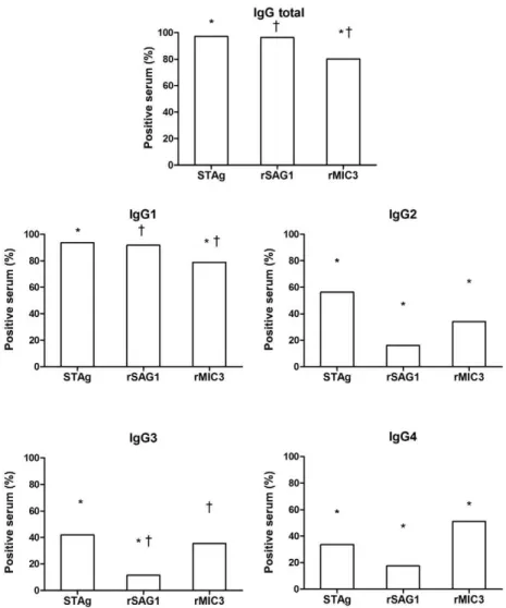
for the diagnosis of congenital toxoplasmosis. The frequency of positivity with IgGt against STAg, rSAG1 and rMIC3 was found in 97.2%, 96.3% and 80.2%, respectively, of the newborns with confirmed congenital toxoplasmosis. IgG1 reacted with all three antigens, while IgG3 and IgG4 reacted preferentially with rMIC3. Higher mean values of re-activity (sample optical density/cut-off) were found for all subclasses when using rMIC3. All of the antigens showed high sensitivity and low specificity in detecting anti-T. gondii IgGt and IgG1 and low sensitivity and high specificity in detecting IgG3 and IgG4. In conclusion, the combined detection of IgG antibody subclasses against recombinant toxoplasmic antigens may be useful for the early diagnosis of congenital toxoplasmosis.
of the RH strain were sonicated using five 30-sec peri-ods at 40 Hz and 1 min intervals. After centrifugation at 4ºC (30 min at 15,000 g), the supernatant was collected and used as STAg. Additionally, two commercially avail-able recombinant T. gondii antigens (SAG1 and MIC3, GenWay Biotech, Inc, San Diego, CA) were used in the ELISA for the diagnosis of CT. Recombinant antigens were expressed in Escherichia coli as fusion proteins with glutathione S-transferase (GST).
ELISA- Maxisorb plates (Nunc, Denmark) were sen-sitised overnight with 100 µL of STAg (10 ug/mL), rSAG1 (2.5 ug/mL) or rMIC3 (2.5 ug/mL) and blocked for 1 h at 37ºC with 2% foetal calf serum in phosphate buffered saline, pH 7.2. Serum samples were diluted to previous-ly determined optimum titres and incubated for 1 h at 37ºC (STAg: IgGt - 1:400, IgG1 - 1:100, IgG2, IgG3 and IgG4 - 1:50; rSAG1: IgGt, IgG1, IgG2 and IgG3 - 1:100, IgG4 - 1:50; rMIC3: IgGt, IgG2, IgG3 and IgG4 - 1:50, IgG1 - 1:100). After washing, the plates were incubated with biotin-conjugated murine monoclonal anti-human antibody (anti-IgGt, -IgG1, -IgG2, -IgG3 or -IgG4) at a previously determined optimum dilution for 1 h at 37ºC (STAg: IgGt - 1:20,000, IgG1 - 1:8,000, IgG2, IgG4 - 1:5,000 and IgG3 - 1:4,000; rSAG1: IgGt - 1:20,000, IgG1, IgG2, IgG4 - 1:5,000 and IgG3 - 1:10,000; rMIC3: IgGt, IgG1, IgG4 - 1:5,000, IgG2 and IgG3 - 1:2,500). A solution of streptavidin-peroxidase (SIGMA), diluted 1:4,000, was then added. After 30 min at 37ºC, the plates were washed and incubated with the chromogen (o-phe-nylenediamine in 0.1 M citric acid, using hydrogen per-oxide as the substrate) for 20 min. The assay was inter-rupted by the addition of 4N H2SO4 and the absorbance was read at 492 nm (reference wavelength at 650 nm) using an ELISA reader (BIO-RAD 3550). In the read-ings obtained with the two recombinant proteins, the absorbance value for each serum was subtracted against readings taken of 2.5 µg/mL GST (SIGMA) which was used to sensitise parallel wells of the same plate. The cut-off value was considered the mean absorbance of eight samples of human sera negative for T. gondii (for each isotype) plus three standard deviations tested on each plate. Each serum sample was assayed in duplicate. The results were expressed as the reactivity index (RI) (average sample optical density/cut-off). The sera with RI values equal to or greater than 1 were considered positive (Altcheh et al. 2006).
Statistical analysis - The validity of the ELISA tests was determined by a comparison with the results of the reference test, IgG ELFA-VIDAS®. The persistence of specific IgG antibodies at 12 months of life confirmed congenital toxoplasmosis. The sensitivity and specific-ity of the tests were determined, as well as the posi-tive and negaposi-tive likelihood ratios (LR) and respecposi-tive 95% confidence intervals. A chi-squared test was used to compare the frequencies between infected and non-infected children (p < 0.05). Differences between serum reactivity for the different antigens were analysed using the McNemar test. The mean RI values for IgGt and sub-classes against the different antigens were compared by Student’s t test (p < 0.05).
RESULTS
Initially, IgGt and its subclasses reacting with STAg, rSAG1 and rMIC3 were assessed in the sera of the 217 children positive for IgM. The frequency of positivity for IgGt was higher with STAg (97.2%) and rSAG1 (96.3%) compared with rMIC3 (80.2%) (Fig. 1). Similar results were observed for IgG1, while for IgG2 and IgG3 the fre-quency of positivity against rMIC3 was higher than that against rSAG1. IgG4 reacted more frequently with rMIC3 (51.2%) than with STAg (33.6%) or rSAG1 (17.5%).
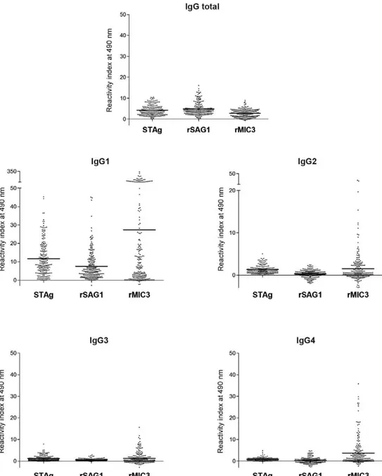
All sera reacted with at least one of the antigens. However, different mean RI values were observed for IgGt and its subclasses (Fig. 2). IgGt was less reactive with rMIC3 than with STAg or rSAG1. For the IgG sub-classes, the mean RI values were higher for rMIC3. IgG antibodies to rSAG1 were least reactive. The reactivity of sera obtained from 175 children with confirmed CT (group I) was compared with that of the 42 non-infected (group NI) control children, whose antibodies had been passively acquired from the mother. The ELISA for IgGt showed reactivity in more than 84% of infected infants, regardless of the antigen used (Table I). IgGt in non-infected infants reacted with less intensity than in in-fected infants with either the rSAG1 or rMIC3 antigen. For IgG1, the proportion of non-infected infants recog-nising the three antigens was significantly lower than that of the infected children. For IgG2, IgG3 and IgG4, reactivity was moderate, with significant differences be-tween the proportion of positive ELISAs for infected and non-infected children, especially when using rMIC3. For IgG4, only the reaction with rMIC3 showed significantly different frequencies between the infected (62.3%) and non-infected (4.8%) newborns (Table I).
Table II shows the sensitivity and specificity values and positive and negative LRs of the ELISA-based testing for IgGt and its subclasses in the sera of the infected and non-infected newborns, considering the persistence of IgG (ELFA-VIDAS®)at 12 months of life as the reference (gold standard). Better sensitivity and negative LR results were found for IgGt and IgG1, particularly against STAg or rSAG1. Better specificity and positive LR results were observed for IgG3 and IgG4, particularly against rMIC3.
DISCUSSION
In this study, we analysed the presence of IgGt and its subclasses through an ELISA of serum samples from 217 newborns with suspected CT. The IgG antibodies from these children reacted with STAg and the recom-binant proteins SAG1 and MIC3. IgGt sensitivity varied between 86-99%. In contrast, the specificity ranged from 7-35%, depending on the antigen employed. A signifi-cant difference was observed when using recombinant antigens when comparing the proportions of the reac-tive samples of the infected and non-infected newborns. Similar results were observed by Buffolano et al. (2005), who found that over 75% of the samples from infected children reacted with the recombinant antigens.
negative LRs for IgGt and IgG1 were low, indicating that the probability is high that a newborn testing negative is not infected. The high IgG levels (mainly IgG1) in these newborns can be explained by the passive transfer of maternal antibodies during pregnancy, which decrease after birth (Correa et al. 2007, Petersen 2007).
For IgG2, low sensitivity and specificity were ob-served using STAg (61% and 64%, respectively), while rMIC3 showed a higher sensitivity (95.9%). This result could explain the significant difference between the pro-portions of positive results for infected and uninfected children using STAg vs. rMIC3. The IgG3 and IgG4 sub-classes revealed higher specificities (despite a low sensi-tivity) and higher positive LRs on the ELISA when com-pared with IgGt. These results show that children with a positive ELISA are probably infected, as IgG2 and IgG3 to T. gondii are synthesised by the newborns and their presence indicates an active response against infection (Cañedo-Solares et al. 2008). As observed for IgG2, a significant difference between the proportion of
posi-tive ELISAs for IgG3 in the infected and non-infected newborns was found using STAg or rMIC3. ELISA for IgG4 using rMIC3 also showed a significant difference between the proportion of positive tests for the infected and non-infected infants. Only 5% of the non-infected newborns reacted with rMIC3, while 62% of the infected children reacted with this antigen.
Higher mean RI values were observed for all IgG subclasses on the ELISA using rMIC3 when compared with using STAg or rSAG1. Our results show that rMIC3 reacts more frequently with IgG antibodies in children with CT, despite those non-infected newborns present-ing passively acquired maternal antibodies. This findpresent-ing suggests that anti-MIC3 antibodies may indicate the oc-currence of active T. gondii replication in infected indi-viduals, as previously described (Beghetto et al. 2005, Buffolano et al. 2005).
In general, all of the sera samples from the children reacted with at least one recombinant antigen, a find-ing also observed by Buffolano et al. (2005), showfind-ing
the immunogenicity of these antigens. Regarding the subclasses, 98% of the samples tested for IgG1 reacted to at least one of the recombinant antigens, followed by 61% for IgG4, 48% for IgG2 and 41% for IgG3 (data not shown). When testing samples of infected adult patients using an ELISA-STAg, Huskinson et al. (1989) observed that infection with T. gondii caused a predominantly IgG1 response. A less intense response was observed for sub-classes IgG2 and IgG3, while IgG4 was not recognised. In our study, similar results were also observed when us-ing STAg, rSAG1 and rMIC3. However, we also detected considerable levels of IgG4 reacting with rMIC3.
Our study has some limitations. Newborns were di-agnosed with CT by postnatal screening and because the sensitivity of the TOXO IgM Q-preven® and ELFA-VIDAS® kits is not very high, the prevalence of infection may have been underestimated. Gilbert et al. (2007) ob-served the highest sensitivity of postnatal screening for T. gondii-specific IgM and IgA when tests were performed between one and two weeks after birth. Specificity var-ied depending on the time of the sampling of newborns and was higher by four weeks after birth. In the present study, newborns were tested one month after birth and this delay may have resulted in false-negative cases that
had been infected in the first trimester of pregnancy and may have ultimately had more severe disease.
In addition to ELISA using recombinant antigens, immunoblot is also being currently evaluated for the postnatal diagnosis of CT. Immunoblot identifies infect-ed newborns with a sensitivity and specificity of 82.4% and 93%, respectively (Gross et al. 2000). These results still leave approximately 18% of congenitally infected newborns without diagnosis confirmation. Difficulties in the standardisation of antigens for immunoblot also
hinder its wider application in the serological diagnosis of postnatal CT. However, diagnostic assays based on re-combinant antigens for measuring Toxoplasma-specific IgG subclasses show promising results in infants with or without CT born to mothers who have acquired toxo-plasmosis during pregnancy (Buffolano et al. 2005).
Our results suggest that the assessment of IgG sub-classes using recombinant antigens is a promising com-plementary tool for CT diagnosis, allowing the identi-fication of a large number of newborns infected with T.
TABLE I
Reactivity of anti-Toxoplasma gondii total IgG (IgGt) and IgG subclasses in 217 serum samples: 175 infected children (group I) and 42 non-infected newborns (group NI)
Group
Reactive samples on ELISA for different antigens n (%) pa
STAg rSAG1 rMIC3
IgGt I 172 (98.3) 174 (99.4) 148 (84.6)
NI 39 (92.9) 0.08 35 (83.3) < 0.05 26 (61.9) < 0.05
IgG1 I 168 (96) 167 (95.4) 158 (90.3)
NI 35 (83.3) < 0.05 32 (76.2) < 0.05 13 (31) < 0.05
IgG2 I 107 (61.1) 24 (13.7) 72 (41.1)
NI 15 (35.7) < 0.05 11 (26.2) 0.06 2 (4.8) < 0.05
IgG3 I 85 (48.6) 22 (12.6) 77 (44)
NI 6 (14.3) < 0.05 3 (7.1) 0.42 0 (0) < 0.05
IgG4 I 64 (36.6) 28 (16) 109 (62.3)
NI 9 (21.4) 0.07 10 (23.8) 0.25 2 (4.8) < 0.05
a: pvalue by χ2 comparing infected and non-infected children; rSAG1 and rMIC3: recombinant antigen; STAg: soluble antigen.
TABLE II
Sensitivity, specificity and likelihood ratio of ELISA testing for anti-Toxoplasma gondii
IgG antibodies and their subclasses using recombinant proteins in 217 newborn infants
Sensibility (95% CI)
Especificity (95% CI)
LR positive
LR negative
STAg IgG total 98.8 (95.9-99.7) 7.1 (2.4-19.0) 1.065 0.160
IgG1 97.1 (93.4-98.7) 16.6 (8.3-30.6) 1.165 0.173
IgG2 61.4 (54.1-68.4) 64.2 (49.2-77.0) 1.722 0.599
IgG3 46.5 (39.3-53.9) 85.7 (72.1-93.3) 3.259 0.623
IgG4 36.4 (29.6-43.81) 78.5 (64.0-88.3) 1.699 0.802
rSAG1 IgG total 99.4 (96.3-99.9) 16.6 (8.3-30.6) 1.193 0.034
IgG1 95.4 (91.2-97.7) 23.8 (13.5-38.5) 1.253 0.192
IgG2 13.7 (9.4-19.6) 73.1 (58.9-84.7) 0.523 1.169
IgG3 12.5 (8.4-18.3) 92.8 (81.0-97.5) 1.760 0.941
IgG4 16.5 (11.8-22.8) 76.1 (61.5-86.5) 0.696 1.095
rMIC3 IgG total 86 (80.0-90.4) 34.8 (22.4-49.8) 1.321 0.400
IgG1 90.2 (85.0-93.8) 69.0 (54.0-80.9) 2.917 0.140
IgG2 95.9 (88.7-98.6) 27.2 (20.6-35.1) 1.319 0.140
IgG3 44.0 (36.8-51.4) 100 (91.6-100) ND 0.560
IgG4 62.2 (54.9-69.1) 95.2 (84.2-98.7) 13.080 0.396
gondii. Reactivity to the rMIC3 antigen is particularly useful to differentiate infected from non-infected in-fants. The possibility of standardisation and automation of ELISA with recombinant antigens of T. gondii is ex-pected to make this technique feasible for widespread use in the serological diagnosis of postnatal CT.
ACKNOWLEDGEMENTS
To Rosalida Estevan Nazar Lopes, for her technical as-sistance.
REFERENCES
Altcheh J, Diaz NS, Pepe CM, Martin V, Nigro M, Freilij H, Angel SO 2006. Kinetic analysis of the humoral immune response against 3
Toxoplasma gondii-recombinant proteins in infants with suspected congenital toxoplasmosis. Diagn Microbiol Infect Dis 56: 161-165. Beghetto E, Nielsen HV, Del Porto P, Buffolano W, Guglietta S, Felici
F, Petersen E, Gargano N 2005. A combination of antigenic re-gions of Toxoplasma gondii microneme proteins induces protec-tive immunity against oral infection with parasite cysts. J Infect Dis191: 637-645.
Buffolano W, Beghetto E, Del Pezzo M, Spadoni A, Di Cristina M, Petersen E, Gargano N 2005. Use of recombinant antigens for early postnatal diagnosis of congenital toxoplasmosis. J Clin Mi-crobiol 43: 5916-5924.
Cañedo-Solares I, Galván-Ramires ML, Luna-Pastén H, Pérez LRR, Ortiz-Alegría LB, Rico-Torres CP, Vela-Amieva M, Pérez-An-drade M, Figueroa-Damián R, Correa D 2008. Congenital toxo-plasmosis specific IgG subclasses in mother/newborn pairs. Pe-diatr Infect Dis J27: 457-475.
Correa D, Cañedo-Solares I, Ortiz-Alegría LB, Caballero-Ortega H, Rico-Torres CP 2007. Congenital and acquired toxoplasmosis: diversity and role of antibodies in different compartments of the host. Parasite Immunol29: 651-660.
Elsaid MMA, Martins MS, Frézard F, Braga EM, Vitor RWA 2001. Vertical toxoplasmosis in a murine model. Protection after im-munization with antigens of Toxoplasma gondii incorporated into liposomes. Mem Inst Oswaldo Cruz96: 99-104.
Gilbert ER, Thalib L, Tan HK, Paul M, Wallon M, Petersen E 2007. Screening for congenital toxoplasmosis: accuracy of immu-noglobulin M and immuimmu-noglobulin A tests after birth. J Med Screen14: 8-13.
Gross U, Lüder CGK, Hendgen V, Heeg C, Sauer I, Weidner A, Krc-zal D, Enders G 2000. Comparative immunoglobulin G antibody profiles between mother and child (CGMC test) for early diagno-sis of congenital toxoplasmodiagno-sis. J Clin Microbiol38: 3619-3622. Huskinson J, Stepick-Biek PN, Araujo FG, Thulliez P, Suzuki Y,
Remington JS 1989. Toxoplasma antigens recognized by im-munoglobulin g subclasses during acute and chronic infection.
J Clin Microbiol27: 2031-2038.
Li S, Galvan G, Araujo FG, Suzuki Y, Remington JS, Parmley S 2000. Serodiagnosis of recently acquired Toxoplasma gondii infection using an enzyme-linked immunosorbent assay with a combination of recombinant antigens. Clin Diagn Lab Immunol 7: 781-787. Petersen E 2007. Toxoplasmosis. Semin Fetal Neonatal Med12:
214-223.
Rorman E, Stein C, Rilkis I, Ben-David H 2006. Congenital toxoplas-mosis - prenatal aspects of Toxoplasma gondii infection. Reprod Toxicol21: 458-472.
Tenter AM, Heckeroth AR, Weiss LM 2000. Toxoplasma gondii: from animals to humans. Int J Parasitol 30: 1217-1258.