online | memorias.ioc.fiocruz.br
Profile of total IgG, IgG1, IgG2, IgG3 and IgG4 levels in sera
of patients with paracoccidioidomycosis: treatment follow-up using
Mexo and rPb27 as antigens in an ELISA
Lílian da Silva Santos1/+, Viviane Cristina Fernandes3, Samuel Gonçalves da Cruz2, Weverton César Siqueira2, Alfredo Miranda Goes3, Ênio Roberto Pietra Pedroso1,2
1Programa de Pós-Graduação em Ciências da Saúde, Infectologia e Medicina Tropical 2Faculdade de Medicina 3Departamento de
Bioquímica e Imunologia, Instituto de Ciências Biológicas, Universidade Federal de Minas Gerais, Belo Horizonte, MG, Brasil
The levels of total of IgG, IgG1, IgG2, IgG3 and IgG4 were evaluated in 54 patients with chronic paracoccid-ioidomycosis (PCM) before, during and after treatment using an enzyme-linked immunosorbent assay with Mexo and recombinant Pb27 (rPb27) as the antigens. Mexo was effective in distinguishing PCM patients from individuals in the negative control group (NC) based on total IgG and rPb27 performed worse than Mexo when these two groups were compared. IgG1, IgG2, IgG3 and IgG4 could not be used to clearly distinguish PCM patients from those in the NC group using either antigen. There was no clear relationship between antibody levels and the period of treatment. The majority of patients presented with decreased antibody levels during treatment, with no statistically significant dif-ferences among the different periods of treatment. Only IgG4 presented a negative correlation between its levels and clinical improvement during treatment. In total, 65% of untreated PCM patients showed reactivity against IgG4 when the Mexo antigen was used and this reactivity decreased over the course of treatment. There was a tendency towards decreasing antibody levels during treatment, but these antibody levels did not necessarily clear after the treatment was stopped. Mexo was useful for PCM diagnosis using total IgG; however, more studies are necessary before this antigen can be used in measuring the levels of total IgG and its subclasses for monitoring patients during treatment.
Key words: treatment follow-up - ELISA - Mexo antigen - rPb27 antigen - paracoccidioidomycosis - IgG subclasses
Paracoccidioidomycosis (PCM) is a systemic disease caused by the thermodimorphic fungus Paracoccid-ioides brasiliensis (Marques 1998). This deep mycosis is endemic in many Latin American countries, with the majority of cases occurring in Brazil, followed by Ven-ezuela, Colombia, Ecuador and Argentina (Shikanai-Yasuda 2006, Ameen et al. 2010). The most affected age group is between 30-50 years old, 90% of whom are men who live in rural areas and work in agriculture. Recently, endemic foci of PCM infection were found in urban ar-eas, which can be related to population migration from rural to urban areas (Blotta et al. 1999). Epidemiological studies have demonstrated that the number of patients clinically diagnosed with PCM may represent only a small proportion of infected individuals (Almeida et al. 2003). In endemic areas, up to 50% of inhabitants have been exposed to the fungus, but only a minority devel-ops the disease (Ameen et al. 2010).
PCM is characterised as an infection when patients are positively diagnosed using serological, microbiologi-cal or molecular techniques, even in the absence of signs
Financial support: CAPES, FAPEMIG, CNPq + Corresponding author: lilianufop@yahoo.com.br Received 16 December 2010
Accepted 25 November 2011
or symptoms of the disease because many patients can be asymptomatic at the time of first evaluation. The evo-lution of PCM infection can resolve spontaneously, prog-ress to disease or remain latent, depending on the immune response of the host (Rivitti & Aoki 1999). PCM disease may manifest with several symptoms ranging from lo-cal and benign to disseminated, severe and progressive, leading to fatal outcomes in the absence of treatment. The clinical manifestations of PCM disease may vary depending on several factors, such as the virulence of the fungus, the host’s established immune response, the affected areas and other intrinsic factors of the host (Be-nard 2008, Mendes-Giannini et al. 2008). There are two clinical forms of PCM disease: the acute, or juvenile form, and the adult, or chronic form (Franco et al. 1987, Ra-mos-e-Silva et al. 2008). The acute form affects children and adolescents of both genders and represents 5-15% of all cases. This clinical form is characterised by a rapid and aggressive progression, mainly affecting the reticu-loendothelial system. Frequent manifestations include skin lesions, digestive symptoms and lymphadenopathy (Nogueira et al. 2006). The chronic form occurs in more than 90% of patients, most of them adult males between 30-50 years old. This form progresses slowly, with pul-monary symptoms present in 90% of the affected adults (Shikanai-Yasuda et al. 2006, Wanke & Aidê 2009).
polyclonal B cell activation, hypergammaglobulinaemia and high levels of specific antibodies, which are general-ly correlated with disease severity (Del Negro et al. 2000, Juvenale et al. 2001, Shikanai-Yasuda et al. 2006).
The assessment of the humoral immune response is an important tool for the diagnosis and follow-up care of PCM patients. Different serological techniques are used to measure the levels of IgG, such as double immunodif-fusion (DI), counterimmuno-electrophoresis (CIE), im-munofluorescence (IFI), enzyme-linked immunosorbent assay (ELISA) and immunoblotting. These techniques may use various antigenic preparations to assess anti-bodies against P. brasiliensis. Some of these techniques use crude antigenic preparations, while others use spe-cific antigenic preparations, such as the 19 kDa, 31 kDa, 43 kDa and 70 kDa glycoproteins and recombinant Pb27 (rPb27) (Ortiz et al. 1998, Baida et al. 1999, Díez et al. 2003, Albuquerque et al. 2005, Correa et al. 2007, Reis et al. 2008, Fernandes et al. 2011, Silveira-Gomes et al. 2011). However, there is no consensus on the best tech-niques for the diagnosis and follow-up care of PCM pa-tients (Campos et al. 1990, Alves 1996, Martins et al. 1997, Del Negro et al. 2000, Camargo 2008).
Different techniques have been employed to measure the levels of IgG and its subclasses IgG1, IgG2, IgG3 and IgG4. Some groups have attempted to associate classes of immunoglobulins with clinical forms of PCM or clini-cal improvement during treatment (Mota & Franco 1979, Barbosa et al. 1981, Biagioni et al. 1984, Baida et al. 1999, Del Negro et al. 2000, Juvenale et al. 2001). However, the relationship between the levels of immunoglobulin sub-classes and clinical improvement remains controversial.
Different classes of drugs can be used for the treatment of PCM, including sulphonamides (sulphamethoxazole-trimethoprim), amphotericin B, imidazole derivatives (ketoconazole, itraconazole and fluconazole) and triazolic derivatives (voriconazole). Drug selection is based on dis-ease severity, but the treatment cost can be an important factor in drug choice (Shikanai-Yasuda et al. 2006).
This study aimed to measure the serum levels of total IgG, IgG1, IgG2, IgG3 and IgG4 in both untreated PCM patients and patients after different treatment durations via an ELISA with two different antigenic preparations (Mexo and rPb27) to verify the suitability of these antigens for use in the diagnosis and follow-up care of PCM patients.
SubjECtS, MAtERIALS And MEthodS
Patients and control sera - Sera were collected from 54 patients with chronic PCM before, during and after treatment at the Training Center and Parasitic Infectious Diseases Reference, Hospital and Clinics (HC) of Fed-, Hospital and Clinics (HC) of Fed-eral University of Minas Gerais (UFMG), Brazil. A to-A to-tal of 92 serum samples were assessed and, of these, 38 were obtained from the same patients at different time points during and after treatment. Sera were aliquoted and stored at -20ºC until use. The diagnosis of PCM was determined by biopsy in all patients and, in some cases, conventional serological tests were used in combination with the biopsy results. Patients were treated with ke-toconazole, itraconazole,
sulphamethoxazole-trimeth-oprim or amphotericin B during hospitalisation. The patients in this study were not treated with immunosup-pressive drugs. The first analysis evaluated one serum sample from each PCM patient before or at the begin-ning of treatment to compare the levels of total IgG and its subclasses with those found in healthy individuals (NC group) to show the suitability of Mexo and rPb27 as antigens for use in PCM diagnosis. This assay was per-formed on the 54 initial serum samples from the PCM patients and 10 serum samples from the NC group. Next, a second analysis was performed to assess the levels of total IgG and its subclasses during PCM treatment. To accomplish this analysis, a total of 92 serum samples were obtained (many patients provided more than one serum sample during treatment) and the results of these samples were compared with those from the NC group. Serum samples were classified according to the duration of time over which the patients were treated, the time elapsed since the end of treatment as follows: not treat-ed (NT) (14), treattreat-ed for one month (T1M) (8), treattreat-ed for two-nine months (T2-9M) (19), treated for one year (T1Y) (13), treated for two years (T2Y) (11), treated for three-four years (T3-4Y) (16), six-nine months from the end of treatment (AT6-9M) (3), one year from the end of treatment (AT1Y) (2), two years from the end of treatment (AT2Y) (2) and three years from the end of treatment (ATY3) (1). Additionally, one group contained patients who had relapsed (Rel) (3). In both experiments, sera from 10 NC from the Institute of Biological Sci-ences (UFMG) were assessed to determine cut-off val-ues of the ELISA. Patients with concomitant diseases, such as toxoplasmosis, histoplasmosis, cryptococcosis, infectious mononucleosis, acquired immune deficiency syndrome, tuberculosis, sarcoidosis or lymphoma, were excluded from the study. This study was approved by the Ethical Committee of the HC of School of Medicine of UFMG and informed consent was obtained from each patient before blood collection.
Antigens - The secreted and surface antigen Mexo was obtained from Pb18, a human source of a virulent strain of P. brasiliensis (Reis et al. 2005). Yeast cells were cultured in YPD agar medium (0.5% yeast extract, 0.5% peptone, 1.5% D-glucose and 1.5% agar, pH 7.0) (Sigma, USA) at 35ºC and harvested on the seventh day of culture. Yeast cells were removed from the culture medium and subjected to agitation by a vortex in 0.05 mol L-1 phosphate buffered saline (PBS), pH 7.4, for 30 s. The solution was centrifuged (14,000 g) for 10 min at 4ºC. The amount of protein in the supernatant was quantified using the Bradford method (Bradford 1976) and this preparation was used as the Mexo antigen.
C-terminal his-tag. The protein rPb27 was expressed in
Escherichia coli which according to the manufacturer’s instructions produces a recombinant protein with a C-terminal his-tag. Purification of the recombinant protein was performed using a HiTrapTM Chelating HP (Amer-sham Biosciences, Uppsala, Sweden).
ELISA of total IgG, IgG1, IgG2, IgG3 and IgG4 lev-els using the Mexo and rPb27 antigens - The ELISAs of anti-P. brasiliensis total IgG, IgG1, IgG2, IgG3 and IgG4 levels were performed in flat-bottomed polystyrene plates (Nunc-ImmunoPlate PolySorp Surface, USA) us-ing the Mexo and rPb27 antigens. Briefly, plates were coated overnight at 4ºC with 100 µL of a 1 µg/100 µL solution of Mexo or rPb27 in a 0.5 mol L-1 carbonate buffer, pH 9.6. The plates were washed five times with washing buffer [0.05 mol L-1 PBS with 0.05% Tween 20 (PBS-Tween)] and blocked with 200 µL of blocking so-lution [1.5 mol L-1 PBS with 1.6% casein (PBS-casein)] for 1 h at 37ºC. After incubation, the plates were washed five times with PBS-Tween and filled with 100 µL of either patient sera or negative control sera (in duplicate) diluted 1:400 in 1.5 mol L-1 PBS with 0.25% casein. The plates were re-incubated for 1 h at 37ºC and then washed 10 times. After washing, 100 µL of a rabbit anti-human total IgG peroxidase-conjugated antibody specific to the gamma chain (DAKO, USA) diluted 1:10.000 in 0.15 mol L-1 PBS was added to the wells. This antibody reacts specifically with the gamma chain, detecting only IgG antibodies. The plates were incubated for 1 h at 37ºC and then washed 10 times. The reaction was developed with 100 µL of TMB Plus (Bio-tecnologia, Brazil) for 10 min at room temperature. Colour development was stopped with 50 µL of 2 mol L-1 H
2SO4. The optical den-sity (OD) at 450 nm was determined using an ELISA reader (Anthos 2010, Cambridge, England). Similar pro-tocols were performed for the IgG subclasses, but be-cause these monoclonal antibodies were not conjugated, a goat anti-mouse peroxidase-conjugated antibody was added as an additional step. The monoclonal anti-human IgG1 antibody (Fc-specific, Sigma, USA) was used at a 1:12.000 dilution and a goat anti-mouse IgG2b
perox-idase-conjugated antibody (γ2b-chain specific, South -ernBiotech, USA) was added at a 1:6.000 dilution. The monoclonal anti-human IgG2, IgG3 and IgG4 antibodies (Sigma, USA) were used at a 1:10.000 dilution and a goat
anti-mouse IgG1 peroxidase-conjugated antibody
(γ1-chain-specific, SouthernBiotech) was added at a 1:6.000 dilution. Cut-off values for the detection of IgG and its subclasses were determined using the mean plus three standard deviations of serum levels from 10 NC.
Statistical analysis - Serological results of the PCM patients vs. the negative control group (NC) were analy-sed using the Mann-Whitney test. The groups of patients treated for different durations of time, NT and the NC groups were compared and analysed using the Kruskall-Wallis non-parametric test. The comparison between all groups was calculated using Dunn’s test. The Spearman rank correlation coefficient was used in correlation stud-ies. All data were considered significant when p < 0.05.
RESuLtS
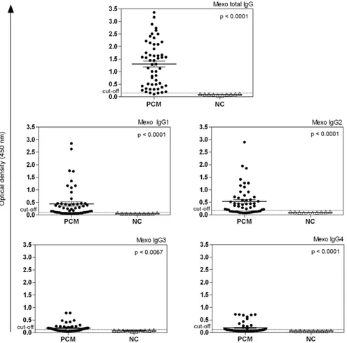
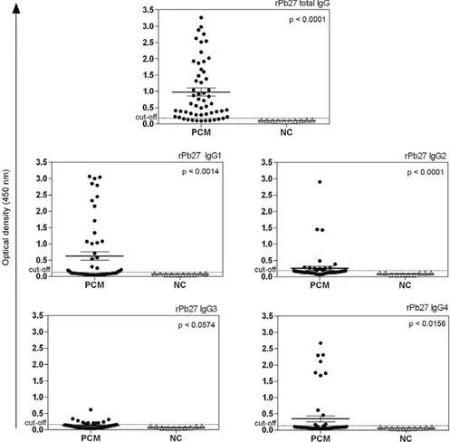
In this study, the sera from 54 patients with chronic PCM were assessed before, during and after treatment. A total of 92 serum samples were analysed using an in-house ELISA with two different antigenic preparations (Mexo and rPb27) to determine total IgG, IgG1, IgG2, IgG3 and IgG4 levels. First, one serum sample from each patient (before treatment or at the beginning of treat-ment) was analysed and compared with NC group. Sig-nificant differences were found between serum samples from the PCM group (54 patients with PCM) and the NC group (sera from 10 NC) using Mexo as the antigen for total IgG and its subclasses. The in-house ELISA using Mexo as the antigen showed higher antibody reactivity for total IgG in all PCM sera analysed. IgG2, IgG1, IgG4 and IgG3 showed decreasing antibody reactivity, respec-tively. Only one PCM patient had an OD value below the cut-off point when total IgG was measured. In contrast, for IgG1, IgG2, IgG3 and IgG4, many PCM patients had OD values below the cut-off point (Fig. 1). When the rPb27 antigen was used, statistically significant differ-ences were found between the PCM patients and the NC group with respect to total IgG, IgG1, IgG2 and IgG4. Using rPb27, total IgG also showed higher antibody re-activity, which was similar to the results observed for the Mexo antigen. However, of the IgG subclasses, IgG1 had the highest antibody reactivity, followed by IgG4, IgG2 and IgG3, respectively. Using rPb27, 10 patients had OD values below the cut-off point for total IgG. For IgG1, IgG2, IgG3 and IgG4, the majority of patients did not have OD values above the cut-off point (Fig. 2).
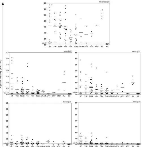
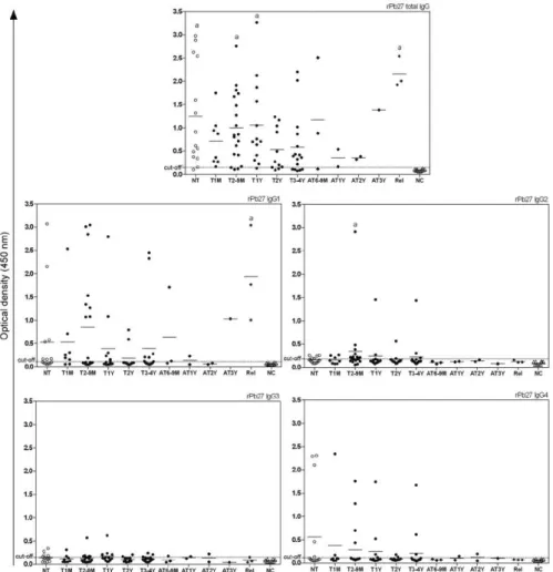
correla-tion between the treatment period and OD of the IgG4 serum. Among sera from NT patients, 65% showed reac-tivity with IgG4 and this reacreac-tivity clearly decreased as the treatment progressed (Fig. 3). When rPb27 was used, no statistically significant differences were found (p = 0.3014) (Fig. 4). It is worth mentioning that all groups were compared with each other, but statistically signifi-cant differences were only found between the NC group and the NT group or between the NC group and other groups with different periods of treatment as explained above. No significant difference was observed among the different treated groups.
Patients were also analysed according to their anti-fun-gal therapy and their clinical disease states. Of 54 patients, only four did not undergo treatment during this study. The remaining 50 patients were divided as follows: 38 were treated with sulphamethoxazole-trimethoprim, seven with itraconazole and five with ketoconazole . The analy-sis of patients treated with sulphamethoxazole-trimethop-rim did not reveal any association between IgG levels and the duration of treatment. Some patients with more than one year of treatment had IgG values similar to patients at the beginning of treatment. For the other subclasses of IgG assayed using Mexo and rPb27, patients had values
similar to the cut-off point. All patients treated with keto-conazole had three years of treatment and two of them had high levels of total IgG, measured using Mexo in one case and rPb27 in the other. Patients treated with itraconazole had between one month and nine months of treatment and of seven patients, five had high levels of total IgG using Mexo and rPb27, regardless of the duration of treatment. No patients were treated exclusively with amphotericin B. This drug was prescribed to a few patients during hospitalisation and it was always combined with itracon-azole, ketoconazole or sulphamethoxazole-trimethoprim. For this reason, it was not possible to verify the effect of amphotericin B on antibody levels. Supplementary data gives the medians and standard deviations of the levels of total IgG and its subclasses in untreated patients and those treated with sulphamethoxazole-trimethoprim, ke-toconazole or itraconazole. Supplementary data shows the similarities in the levels of IgG and its subclasses between the different treatment groups.
classified as having multifocal disease. Similar levels of IgG and its subclasses were detected in patients with uni-focal and multiuni-focal diseases, with no statistically signifi-cant differences for almost all antibodies. Only the analy-sis of IgG2 using rPb27 as the antigen yielded a statistically significant difference between the unifocal and multifocal groups. However, this difference was related to only two patients from the unifocal group that presented with high levels of IgG2, and there is no clear explanation for this re-sult. Supplementary data shows the medians and standard deviations of the levels of total IgG and its subclasses in patients in relation to their clinical disease status.
dISCuSSIon
In this study, 92 serum samples from PCM patients at different points in treatment or after treatment were analysed. All of the patients selected had chronic PCM, varying from unifocal to multifocal disease. The first aim of our study was to assess the utility of Mexo and rPb27 as antigens using an ELISA. This evaluation showed statistically significant differences between PCM serum samples and the NC group for total IgG and all of its subclasses when Mexo was used as the antigen. Using Mexo, total IgG showed the highest level of reactivity
possible explanation for his low levels of IgG is that this patient presented with the unifocal form (he only had a mild oral mucosal lesion) and patients with mild forms of PCM may have a lower humoral immune response. How-ever, many other patients with unifocal PCM had high OD values compared to multifocal PCM patients.
When rPb27 was used as the antigen, total IgG showed the highest levels of reactivity, followed by IgG1, IgG4, IgG2 and IgG3, respectively. Only the analysis of IgG3 showed no statistically significant difference be-tween the PCM patients and the NC group. For total IgG analysis, 10 serum samples of PCM patients were ob-served with OD values below the cut-off point, a worse performance compared with that of the Mexo antigen. The analysis of the IgG subclasses did not allow us to distinguish PCM patients from the NC group. This re-sult is understandable because unlike Mexo, rPb27 is a much more specific molecule, which limits the epitopes recognised by IgG. Reis et al. (2008) immunised mice
with the antigenic rPb27 fraction and measured their humoral immunological response. These mice produced high levels of IgG2b, moderate levels of IgG1 and low levels of IgG2a. The authors also observed high levels of
transforming growth factor beta and interferon (IFN)-γ
tients with different forms of clinical disease. Although the test demonstrated significant sensitivity, with anti-bodies from the majority of PCM patients recognising the antigen used, cross reactivity with aspergillosis and histoplasmosis serum samples was observed (Ortiz et al. 1998). Some patients not producing detectable antibodies against the rPb27 antigen could be related to the specific-ity of this protein because it has a single dominant anti-genic epitope. It would likely be necessary to use a cock-tail of recombinant proteins to achieve higher sensitivity values. Recently, a study performed by our group (Fer-nandes et al. 2011) demonstrated that the combined use of rPb27 and rPb40 in an ELISA provided high sensitivity and specificity for PCM diagnosis, which confirmed that rPb27 was much more efficient as an antigen when paired with another recombinant protein than when isolated.
As shown in other studies, total IgG is generally de-tected at high levels in PCM patients, especially in the acute form or in severe disease, regardless of the antigen
The second part of our study determined the levels of total IgG and its subclasses in chronic PCM patients before, during and after treatment. Some groups have attempted to determine a correlation between antibody levels and disease severity in PCM patients, especially in the follow-up period after treatment (Bueno et al. 1997, Martins et al. 1997, Reis et al. 2005, Anastácio et al. 2007, Bertini et al. 2007, Yoshida et al. 2009), but this association is still controversial. Restrepo et al. (1978) found decreased antibody levels after six months of treatment using DI and CF tests in only three of the 16 patients analysed. Although apparently cured, most patients showed persistent low antibody levels. Ferrei-ra-da-Cruz et al. (1990) used DI to measure antibodies against P. brasiliensis in 66 patients with PCM before and after treatment. Clinical improvement was associ-ated with decreased antibody levels in all patients during treatment. However, after two years of treatment, nine patients continued to show high antibody levels, which showed that the measurement of IgG levels using DI does not indicate active PCM. Reis et al. (2005) dem-onstrated that patients with PCM undergoing treatment for more than one year had a reduced antibody response against the Mexo antigen, suggesting that the presence of antibodies against Mexo could be an indicator of ac-tive disease. In our study, untreated PCM patients had the highest OD values for total IgG when using Mexo or rPb27 as the antigen. However, a clear decrease in anti-body levels associated with clinical improvement during treatment was not observed. Indeed, PCM patients may need several years to clear antibody levels, as suggested by Lopes (1971) and Ferreira-da-Cruz et al. (1990). Del Negro et al. (2000) evaluated antibody responses dur-ing the follow-up care of patients with acute and chronic PCM using ELISA, CIE and CF (complement fixation) techniques. In that study, 43 patients were analysed be-fore treatment and 27 of these patients were followed-up during their treatment for two years. Patients with chronic unifocal disease cleared their antibodies after one year of treatment when CF was used and after two years when ELISA was used, in contrast to the results of our study. It was suggested that patients with chronic unifocal disease needed a shorter course of therapy and patients with acute or multifocal disease needed more than two years of treatment to clear their antibodies. An increase in antibody levels was also associated with the relapse of PCM in five patients. Baida et al. (1999) as-sessed total IgG, IgG1, IgG2, IgG3, IgG4 and IgA in the sera of patients with juvenile and adult PCM using the 43 kDa glycoprotein as the antigen in an ELISA. They found that juvenile PCM patients had higher total IgG levels when compared with the adult form. IgG1 levels were similar between the two groups. However, IgG4 levels were higher in the juvenile form, while IgG2 lev-els were higher in the adult form of PCM. In their study, IgG3 was either absent or detected at low levels in all pa-tients, while IgA was detected mainly in the adult form of PCM. Our study also showed that for both antigens used, high total IgG levels in all patients were observed, independent of the treatment duration. Juvenale et al. (2001) found the same results in sera from acute and
chronic PCM patients before treatment using a crude antigen of P. brasiliensis in an ELISA. Similar to Bai-da et al. (1999) and Juvenale et al. (2001), in our study, IgG3 was detected in few patients and at low levels with both antigens, independent of the treatment duration and clinical disease state. When the Mexo antigen was used, IgG2 was detected at high levels, especially at the base-line of treatment, but showed no differences related to clinical manifestation. Baida et al. (1999) showed higher IgG2 levels in adult PCM patients as a driven response of
IFN-γ. Patients with benign or chronic PCM tend to have
a Th1 immune pattern. However, patients with juvenile PCM have high IgG4 levels, suggesting a Th2 immune pattern. Because our study examined only patients with the chronic form of PCM, low levels of IgG4 in all pa-tients could be anticipated. It is worth mentioning that a negative correlation was found only for IgG4; a decrease in IgG4 levels was associated with clinical improvement during treatment. Antibody titres tend to decrease during therapy, but not all clinically cured patients show nega-tive serology immediately after treatment is stopped. In our study, using Mexo or rPb27 as the antigen, high lev-els of total IgG and its subclasses were detected in some patients after three years of treatment. One explanation for this result could be the severity of PCM presented by these patients. Of 54 patients, 31 presented with multifo-cal disease, with severe lesions in different areas of the body, including the skin, oral mucosa and lungs, simul-taneously. High antibody levels may persist in patients with more severe disease for a longer period of time. In our study, patients with unifocal disease had a tendency to produce lower levels of IgG and its subclasses, but no significant difference in antibody levels was found between patients with mild and severe PCM.
Barbosa SFC, Takeda AK, Chacha J, Cuce LC, Fava Netto C 1981. Anticorpos específicos das classes IgG, IgM e IgA para Para-coccidioides brasiliensis dosados através da reação de imuno- fluorescência indireta no soro de pacientes e sua correlação com o tempo de evolução e forma clínica da doença. Rev Inst Adolfo Lutz41: 121-126.
Benard G 2008. An overview of the immunopathology of human paracoccidioidomycosis. Mycopathol165: 209-221.
Bertini S, Colombo AL, Takahashi HK, Straus AH 2007. Expres-sion of antibodies directed to Paracoccidioides brasiliensis gly-cosphingolipids during the course of paracoccidioidomycosis treatment. Clin Vaccine Immunol14: 150-156.
Biagioni LMJ, Souza MJ, Chamma LG, Mendes RP, Marques SA, Mota NGS, Franco M 1984. Serology of paracoccidioidomyco-sis. II. Correlation between class-specific antibodies and clinical forms of the disease. Trans R Soc Trop Med Hyg78: 617-621.
Blotta MH, Mamoni RL, Oliveira SJ 1999. Endemic regions of paracoccidioidomycosis in Brazil: a clinical and epidemiologic study of 584 cases in the Southeast Region. Am J Trop Med Hyg 61: 390-394.
Bradford RM 1976. A rapid and sensitive method for the quantitation of microgram quantities of protein by the principle of protein-dye binding. Ann Biochem72: 248.
Bueno JP, Mendes-Giannini MJ, Del Negro GM, Assis CM, Takiguti CK, Shikanai-Yasuda MA 1997. IgG, IgM and IgA antibody re-sponse for the diagnosis and follow-up of paracoccidioidomyco-sis: comparison of counterimmunoelectrophoresis and comple-ment fixation. J Med Vet Mycol35: 213-217.
Camargo ZP 2008. Serology of paracoccidioidomycosis. Mycopatho-logia165: 289-302.
Campos EP, Unterkircher C, Camargo ZP 1990. Serological evalu-Serological evalu-ation in follow-up of the paracoccidioidomycosis patients. Rev Microbiol21: 11-17.
Correa MM, Bedoya AM, Guerrero MP, Méndez J, Restrepo A, McE-wen JG 2007. Diagnosis of paracoccidioidomycosis by a dot blot assay using a recombinant Paracoccidioides brasiliensis p27 pro-tein. Mycoses50: 41-47.
Del Negro GM, Garcia NM, Rodríguez EG, Cano MI, de Aguiar MS, Lírio VS, Lacaz CS 1991. The sensitivity, specificity and efficiency values of some serological tests used in the diagnosis of paracoc-cidioidomycosis. Rev Inst Med Trop Sao Paulo33: 277-280.
Del Negro GM, Pereira CN, Andrade HF, Palacios SA, Vidal MM, Charbel CE, Benard G 2000. Evaluation of tests for antibody re-Evaluation of tests for antibody re-sponse in the follow-up of patients with acute and chronic forms of paracoccidioidomycosis. J Med Microbiol49: 37-46.
Díez S, Gómez BL, McEwen JG, Restrepo A, Hay RJ, Hamilton AJ 2003. Combined use of Paracoccidioides brasiliensis recom-binant 27-kilodalton and purified 87-kilodalton antigens in an enzyme-linked immunosorbent assay for serodiagnosis of para-coccidioidomycosis. J Clin Microbiol41: 1536-1542.
Fernandes VC, Coitinho JB, Veloso JM, Araújo SA, Pedroso EP, Góes AM 2011. Combined use of Paracoccidioides brasiliensis recom-binant rPb27 and rPb40 antigens in an enzyme-linked immuno-sorbent assay for immunodiagnosis of paracoccidioidomycosis.
J Immunol Methods31: 78-84.
Ferreira-da-Cruz MF, Francesconi-do-Vale AC, Espinera MC, Wanke B, Galvão-Castro B 1990. Study of antibodies in paracoccidioid-Study of antibodies in paracoccidioid-omycosis: follow-up of patients during and after treatment. J Med Vet Mycol28: 151-157.
Franco M, Montenegro MR, Mendes RP, Marques SA, Dillon ML, Mota NGS 1987. Paracoccidioidomycosis: a recently proposed classifica-tion of its clinical forms. Rev Soc Bras Med Trop20: 129-132. OD value above the cut-off point for total IgG and IgG1.
This result shows the direct relationship between the measurement of antibody levels and the antigenic frac-tions used in the different serological tests. It is worth mentioning that the levels of total IgG were high in Rel patients using either Mexo or rPb27 as the antigen. This is an important finding because the majority of follow-ups of PCM patients are performed for total IgG detec-tion and not for its subclasses (Del Negro et al. 2000, Fer-nandes et al. 2011). However, this result does not allow for a conclusion about the relationship between relapses and increases in the levels of antibodies because only three patients presented relapses and a statistical determination could not be performed with this small group.
Ultimately, no clear association between antibody levels and treatment follow-up was found in our study. Many patients had decreased antibody levels during therapy, but there was no significant difference between the different treatment groups. A significant difference was found only between patients at different times of treatment and the NC group for total IgG and its sub-classes. Only IgG4 presented a negative correlation be-tween its levels and clinical improvement during treat-ment. With this result, it can be inferred that antibody levels tend to decrease during treatment, but these levels do not clear in most patients after treatment is stopped. Because many patients in this study had the multifocal form of PCM, they will likely require a longer time to clear their antibody levels. These results also suggest an increase in total IgG levels in patients with relapses using either Mexo or rPb27 as the antigen. Finally, the Mexo antigen was useful in discriminating between PCM patients and the NC group, especially for total IgG, showing its suitability for use in PCM diagnosis. It is worth noting that serological follow-up of PCM patients during and after treatment is of great impor-tance, but more studies are necessary to establish an ideal serological technique for this purpose.
REFEREnCES
Albuquerque CF, Marques da Silva SH, Camargo ZP 2005. Improve-ment of the specificity of an enzyme-linked immunosorbent as-say for diagnosis of paracoccidioidomycosis. J Clin Microbiol43: 1944-1946.
Almeida OP, Jacks J Jr, Scully C 2003. Paracoccidioidomycosis of the mouth: an emerging deep mycosis. Crit Rev Oral Biol Med 14: 377-383.
Alves JR 1996. Comparação entre três métodos sorológicos no segui-mento de pacientes com paracoccidioidomicose, MSc Thesis, Universidade Federal de São Paulo, São Paulo, 73 pp.
Ameen M, Talhari C, Talhari S 2010. Advances in paracoccidioi- domycosis. Clin Exp Dermatol 35: 576-580.
Anastácio VM, Passeto MPA, Góngora DVN, Soares MMCN, Al-meida MTG 2007. Paracoccidioidomycosis: correlation between clinical and laboratorial findings in São José do Rio Preto region.
Arq Cienc Saude14: 181-185.
Baida H, Biselli PJC, Juvenale M, Del Negro GMB, Mendes-Gian-nini MJS, Duarte AJS, Benard G 1999. Differential antibody isotype expression to the major Paracoccidioides brasiliensis
Juvenale M, Del Negro GM, Duarte AJS, Benard G 2001. Antibody isotypes to Paracoccidioides brasiliensis somatic antigen in sub-acute and chronic form of paracoccidiodomycosis. J Med Micro-biol50: 127-134.
Lopes CF 1971. Evaluación de los resultados obtenidos en el tratami-ento de la blastomicosis sudamericana con sulfamida de adminis- tración semanal. Med Cutanea15: 357-366.
Marques AS 1998. Paracoccidioidomicose. An Bras Dermatol 73: 455-469.
Marquez ADES, Moreira AP, Leonello PC, Nakanishi FA, Itano EN 2009. Serum proteins and fractions, HDL-cholesterol and total IgG and IgE levels in cases of acute and chronic paracoccidioi- domycosis. Rev Soc Bras Med Trop42: 245-249.
Martins R, Marques S, Alves M, Fecchio D, Franco MF 1997. Sero-logical follow-up of patients with paracoccidioidomycosis treated with itraconazole using dot-blot, ELISA and Western blot. Rev Inst Med Trop Sao Paulo39: 187-195.
McEwen JG, Ortiz BL, García AM, Florez AM, Botero S, Restrepo A 1996. Molecular cloning, nucleotide sequencing and charac-terization of a 27-kDa antigenic protein from Paracoccidioides brasiliensis. Fungal Genet Biol 20: 125-131.
Mendes-Giannini MJ, Monteiro da Silva JL, de Fátima da Silva J, Donofrio FC, Miranda ET, Andreotti PF 2008. Interactions of
Paracoccidioides brasiliensis with host cells: recent advances.
Mycopathologia 165: 237-248.
Mota FT, Franco M 1979. Observações sobre a pesquisa de anticorpos IgM anti-Paracoccidioides brasiliensis por imunofluorescência no soro de pacientes com paracoccidioidomicose. Rev Inst Med Trop Sao Paulo 21: 82-89.
Neves AR, Mamoni RL, Rossi CL, Camargo ZP, Blotta MSHL 2003. Negative immunodiffusion test results obtained with sera of paracoccidioidomycosis patients may be related to low-avidity immunoglobulin G2 antibodies directed against carbohydrate epitopes. Clin Diagn Lab Immunol 5: 802-807.
Nogueira MGS, Andrade GMQ, Tonelli E 2006. Clinical evolution of paracoccidioidomycosis in 38 children and teenagers. Myco-pathologia 161: 73-81.
Ortiz BL, Díez S, Urán ME, Rivas JM, Romero M, Caicedo V, Res- trepo A, Mcewen JG 1998. Use of the 27-Kilodalton recombinant protein from Paracoccidioides brasiliensis in serodiagnosis of paracoccidioidomycosis. Clin Diagn Lab Immunol5: 826-830.
Ortiz BL, Garcia AM, Restrepo A, Mcewen JG 1996. Immunologi-cal characterization of a recombinant 27-kilodalton antigenic protein from Paracoccidioides brasiliensis. Clin Diagn Lab Im-munol3: 239-241.
Ramos e Silva M, Saraiva LE 2008. Paracoccidioidomycosis. Derma-tol Clin26: 257-269.
Reis BS, Bozzi A, Prado FLS, Pereira MCN, Ferreira FE, Godoy P, Moro L, Pedroso EP, Leite MF, Góes AM 2005. Membrane and extracellular antigens of Paracoccidioides brasiliensis (Mexo): identification of a 28-kDa protein suitable for immunodiagnosis of paracoccidioidomycosis. J Immunol Methods307: 118-126.
Reis BS, Fernandes VC, Martins EMN, Serakides R, Góes AM 2008. Protective immunity induced by rPb27 of Paracoccidioides bra-siliensis.Vaccine26: 5461-5469.
Restrepo A, Restrepo M, de Restrepo F, Aristizábal LH, Moncada LH, Vélez H 1978. Immune responses in paracoccidioidomyco-sis. A controlled study of 16 patients before and after treatment.
Sabouraudia16: 151-163.
Rivitti EA, Aoki V 1999. Deep fungal infections in tropical countries.
Clin Dermatol17: 171-190.
Shikanai-Yasuda MA, Telles Filho FQ, Mendes RP 2006.Guidelines in paracoccidioidomycosis. Rev Soc Bras Med Trop39: 297-310.
Silveira-Gomes F, Sarmento DN, Pinto TM, Pimentel RF, Nepo-muceno LB, Espírito Santo EP, Mesquita-da-Costa M, Camargo ZP, Marques-da-Silva SH 2011. Development and evaluation of a latex agglutination test for the serodiagnosis of paracoccidioido- mycosis. Clin Vaccine Immunol 18: 604-608.
Vidal MSM, Benard G, Brito T, Dantas KC, Pereira CN, França FOS, Silva AMG, Martins JEC 2005. Atypical serological re-sponse marked by a lack of detectable anti-gp43 antibodies in a patient with disseminated paracoccidioidomycosis J Clin Microbiol 43: 3014-3016.
Yoshida M, Sanchez MCA, Shikanai-Yasuda MA 2009. Increased im-munoglobulin G anti-Paracoccidioides brasiliensis serum anti-body avidity as a predictor of favorable post therapeutic evolution in paracoccidioidomycosis. Clin Vaccine Immunol16: 1583-1586.
Levels of IgG and its subclasses measure in patients with paracoccidioidomycosis (PCM) compared according to their scheme of treatment
Condition of treatment
Mexo (mean ± SD) rPb27 (mean ± SD)
Total IgG IgG1 IgG2 IgG3 IgG4 Total IgG IgG1 IgG2 IgG3 IgG4
SMZ-TMP 1.596 ± 1.280 0.308 ± 0.322 0.397 ± 0.477 0.175 ± 0.131 0.145 ± 0.168 0.821 ± 0.764 0.502 ± 0.823 0.291 ± 0.491 0.122 ± 0.094 0.311 ± 0.655 Ceto 0.919 ± 0.556 0.167 ± 0.152 0.217 ± 0.200 0.106 ± 0.044 0.115 ± 0.057 0.617 ± 0.512 0.293 ± 0.418 0.151 ± 0.089 0.102 ± 0.042 0.181 ± 0.241 Itra 0.919 ± 0.556 0.254 ± 0.152 0.725 ± 0.523 0.269 ± 0.294 0.180 ± 0.165 0.749 ± 0.520 0.328 ± 0.302 0.218 ± 0.092 0.110 ± 0.061 0.255 ± 0.450 NT 0.919 ± 0.556 0.881 ± 0.997 0.799 ± 0.898 0.205 ± 0.196 0.323 ± 0.284 1.250 ± 1.073 0.527 ± 0.915 0.158 ± 0.061 0.141 ± 0.087 0.564 ± 0.912
ceto: cetoconazole; itra: itraconazole; mean ± standard deviation (SD): median of levels of IgG and its subclasses plus its SD; NT: not treated patients; rPb27: recombinant Pb27; SMZ-TMP: sulphamethoxazole-trimethoprim.
Levels of IgG and its subclasses measured in patients with paracoccidioidomycosis (PCM) compared according to their clinical manifestations
Clinical form
Mexo (mean ± SD) rPb27 (mean ± SD)
Total IgG IgG1 IgG2 IgG3 IgG4 Total IgG IgG1 IgG2 IgG3 IgG4
Unifocal 1.254 ± 0.735 0.364 ± 0.576 0.537 ± 0.562 0.145 ± 0.118 0.206 ± 0.223 0.821 ± 0.734 0.525 ± 0.868 0.338 ± 0.609 0.146 ± 0.117 0.271 ± 0.613 Multifocal 1.154 ± 0.789 0.308 ± 0.449 0.361 ± 0.412 0.181 ± 0.142 0.127 ± 0.132 0.717 ± 0.659 0.398 ± 0.612 0.143 ± 0.094 0.100 ± 0.048 0.278 ± 0.558