UNIVERSIDADE FEDERAL DE SÃO CARLOS
CENTRO DE CIÊNCIAS BIOLÓGICAS E DA SAÚDE
PROGRAMA DE PÓS-GRADUAÇÃO EM FISIOTERAPIA
ADALBERTO FELIPE MARTINEZ
RELAÇÃO ENTRE A FORÇA DO QUADRIL E DO
TRONCO COM A CINEMÁTICA TRIDIMENSIONAL DO
TRONCO, QUADRIL E JOELHO DURANTE A
ATERRISSAGEM DE UM SALTO UNIPODAL
UNIVERSIDADE FEDERAL DE SÃO CARLOS CENTRO DE CIÊNCIAS BIOLÓGICAS E DA SAÚDE PROGRAMA DE PÓS-GRADUAÇÃO EM FISIOTERAPIA
RELAÇÃO ENTRE A FORÇA DO QUADRIL E DO TRONCO COM A CINEMÁTICA TRIDIMENSIONAL DO TRONCO, QUADRIL E JOELHO
DURANTE A ATERRISSAGEM DE UM SALTO UNIPODAL
Dissertação de Mestrado apresentada ao Programa de Pós-graduação em Fisioterapia da Universidade Federal de São Carlos, como parte dos requisitos para obtenção do título de Mestre em Fisioterapia. Área de concentração: Processos de Avaliação e Intervenção em Fisioterapia.
DISCENTE Adalberto Felipe Martinez
ORIENTADOR
Prof. Dr. Fábio Viadanna Serrão
AGRADECIMENTOS
A dificuldade em agradecer a todos que de alguma forma participaram deste trabalho e
deste momento em minha vida se torna muito grande porque não há palavras que
expressem as mudanças, alegrias e realizações que vocês proporcionaram em minha
vida. Ainda assim gostaria de agradecê-los como homenagem por serem parte integrante
deste momento.
Agradeço a Universidade Federal de São Carlos (UFSCAR) e ao Departamento de Fisioterapia (Dfisio) pelo suporte e pela disponibilidade que encontrei nesses locais de
receber ensinamentos, aumentar meu conhecimento e me tornar um profissional da
fisioterapia.
Ao Programa de Pós-Graduação em Fisioterapia da UFSCar (PPGFT – UFSCar) pelo auxílio material e profissional para a realização deste projeto e desta dissertação.
Aos professores da Graduação e do PPGFt por dividirem seus conhecimentos e experiências e por não medirem esforços em me auxiliar para ser melhor profissional a
cada dia. Espero retribuir aos demais tudo o que recebi de todos vocês.
À Coordenação de Aperfeiçoamento de Pessoal de Nível Superior (CAPES) pela bolsa concedida para a realização deste estudo.
Aos amigos e companheiros da turma FISIO 010 que por quatro anos foram como uma família e fizeram esse tempo de estudo ser mais leve e com certeza fizeram parte de
A todos os funcionários do DFisio – UFSCar por cuidarem desse ambiente a ponto de este se tornar uma segunda casa. Em especial agradeço imensamente a Iolanda, Iô, que com seus cafés e chás já ajuda muito em nossos cansaços e até doenças, mas nada ajuda
mais do que o sorriso que nos recebe a cada dia. Obrigado pelas risadas matinais!
Agradeço a escola Jesuíno de Arruda onde se iniciou toda essa realização, com todos
seus professores, mas em especial a professora Neusa Valentina Golineli que me colocou em contato com o esporte, o que me ajudou em minhas escolhas e onde tive
muitas felicidades e histórias. Juntamente agradeço aos técnicos e depois companheiros
de time Diego, Helton e Antônio Carlos que me convidaram e possibilitaram viver o handebol mais intensamente na Associação de Handebol Brasileira (AHB).
Ao técnico Valdir Barbosa da AHB por me permitir integrar esse time e através do handebol me permitir uma bolsa de estudos que me possibilitou entrar nessa
universidade e a me tornar o profissional que sou hoje. Sou prova de que investir no
esporte como método de ensino funciona. Torço para que este projeto nunca pare.
A Profa. Dra. Audrey Borghi-Silva, a Profa. Dra. Luciana Di Thommazo Luporini e a
Profa. Dra. Soraia Pilon Jurgensen pelo tempo de ensinamento onde realmente iniciei minha formação científica. Agradeço por terem feito essa diferença em minha formação.
Ao Prof. Dr. Rodrigo de Marche Baldon e ao Prof. Dr. Rodrigo Scattone da Silva
pelos exemplos de profissionais desde minha graduação e pelo auxílio no momento da
escolha da área acadêmica. Também a colega de turma Profa. MSc Ana Cláudia Silva Farche que me ajudou a essa escolha e também nas dúvidas que surgiam.
Ao Prof. Dr. Fábio Viadanna Serrão pelo grande exemplo profissional de cada dia, por me permitir estar no LAIOT e por todos os ensinamentos científicos, profissionais e
pessoais. Além de tudo, agradeço as correções, a grande pessoa que é e os momentos de
descontração que muitas vezes aliviam as cobranças desses anos de trabalho.
Aos integrantes do Laboratório de Avaliação e Intervenção em Ortopedia e Traumatologia (LAIOT) e companheiros nesse tempo de mestrado Cristiano, Daniel,
Bruna, Guilherme, Ana Flávia, Mariana, Natália e Scattone por fazerem desse ambiente prazeroso para se trabalhar e por dividirem conhecimentos sem esperar nada
em troca. A cada dia convivendo e observando tenho aprendido com todos.
Ao companheiro de trabalho Cristiano de Carvalho que permitiu que esse trabalho pudesse ser realizado. Agradeço todo o tempo que empregou em me auxiliar nesse
projeto e principalmente por ter feito desse tempo um tempo de muitas alegrias e
risadas. Seus pacientes e alunos tem muita sorte de ter um profissional como você.
A companheira nesse trabalho Profa.Dra. Giovanna Camparis Lessi por aceitar me auxiliar nesse trabalho, mas principalmente pelo auxílio em como lidar com os
problemas e apuros desse tempo de mestrado.
A meus pais Adalberto Manoel Martinez e Dilma Tochio Martinez que muitas vezes abriram mão de suas vidas para permitir que eu buscasse meus sonhos. Agradeço o
exemplo, a fé, a paciência e os ensinamentos que me deram em toda minha vida. Tudo
que tenho vivido e tudo o que sou é graças a vocês. Vocês me permitiram a felicidade e
A meus irmãos Aline, Débora, Paula, Guilherme e Bruno e seus maridos, namoradas e
filhos pelos momentos de conversa, risada, confiança e churrascos que passamos juntos. Não há momento melhor para concordar com uma frase de uma foto nossa:
“Costumavam dizer que éramos muitos; para mim somos o suficiente! ”
A meus sogros Altamiro de Sousa e Thereza Aparecida Bianchin de Sousa que fazem parte hoje dessa realização assim como de minha vida. Tenho mesmo vocês como pais e
jamais poderei retribuir tudo que fazem por mim.
A minhas cunhadas Cristiane, Elisângela, Adriana, Karina e Tamiris com seus
maridos, namorados e filhos. Obrigado pelos momentos de descontração e conversas e pela confiança em mim me fazendo parte da família.
Em especial, agradeço a minha esposa Heloize de Sousa Martinez, que mais do que parte desse trabalho faz parte de minha vida incondicionalmente. Você é quem me
acompanha em minhas frustrações, nas noites mal dormidas, nas preocupações e
também nas alegrias, nas distrações e nos sonhos. Sou feliz por ter como esposa alguém
muito amiga, compreensível, que me apoia, suporta e me faz acreditar que conseguirei
"Podemos sempre nos motivar a chegar mais longe.
Escrevam sobre isso, sonhem com isso. Mas depois,
transformem em ação. Não se limitem a sonhar"
Dan Gable
“...
Alguém junto de alguém. Não mecânico de uma engrenagem, mas gente
reabilitando gente. Que todo aquele que me procura em busca de cura física
encontre em mim a
lgo mais que o profissional...”
RESUMO
Objetivo: Alterações da cinemática têm sido correlacionadas com diferentes lesões nos membros inferiores, sendo algumas dessas lesões mais comuns em mulheres atletas, e um provável fator predisponente para essas alterações é a fraqueza dos músculos do quadril e do tronco. Assim, o objetivo deste estudo foi avaliar a correlação entre a força do tronco e do quadril com a cinemática tridimensional de tronco e membros inferiores durante uma tarefa de aterrissagem de um salto em mulheres atletas recreacionais.
Métodos: Vinte e três mulheres sadias atletas recreacionais com idade entre 18 e 35 anos foram submetidos a avaliação da força dos músculos abdutores de quadril, extensores do quadril e inclinadores laterais do tronco através de um dinamômetro manual e da cinemática tridimensional de tronco e membros inferiores durante a fase de aterrissagem de uma queda e salto vertical unipodal (single-leg drop vertical jump). Os coeficientes de correlação de Pearson (r) foram calculados para estabelecer a relação entre a força do quadril e do tronco e os movimentos do tronco, quadril e joelho.
Resultados: Maior força extensora do quadril foi encontrada significativamente associada com maior flexão do tronco no contato inicial. Não foram encontradas correlações significativas entre os dados de força e os valores de pico da cinemática durante a fase de aterrissagem.
Conclusão: Considerando que a maior flexão do tronco está relacionada à carga do ligamento cruzado anterior (ACL), ao estresse patelofemoral e a força do tendão patelar, o fortalecimento dos músculos extensores do quadril pode ser importante para prevenir e reabilitar a lesão do LCA, dor femoropatelar e tendinopatia patelar.
ABSTRACT
Purpose: Kinematic changes have been correlated with different lower limb injuries, being some of these injuries more common in female athletes, and a likely predisposing factor to these changes is hip and trunk muscle weakness. Thus, the aim of this study was to evaluate the correlation among trunk and hip strength with three-dimensional trunk and lower-limb kinematics during a jump-landing task in female recreational athletes.
Methods: Twenty-three healthy women recreational athletes aged between 18 and 35 years underwent evaluation of hip abductor, hip extensor, and lateral trunk muscle strengthby manual dynamometry and three-dimensional trunk and lower-limb kinematics during the landing phase of a single-leg drop vertical jump. Pearson's correlation coefficients (r) were calculated to establish the relationship between hip and trunk strength and trunk, hip, and knee movements.
Results: Greater hip extensor strength was found to be significantly associated with greater trunk flexion at initial contact. No significant correlations were found among the strength data and the peak values of kinematics during the landing phase.
Conclusion: Considering that the greater trunk flexion is related to lower anterior cruciate ligament (ACL) load, patellofemoral stress and patellar tendon force, hip extensor muscle strengthening may be important to prevent and rehabilitate ACL injury, patellofemoral pain, and patellar tendinopathy.
LISTA DE FIGURAS
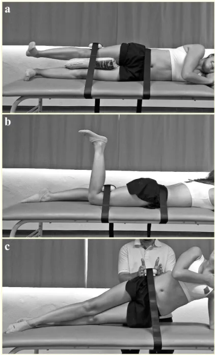
Figure 1 Test position for the evaluation of hip abductor strength (a), hip extensor strength (b) and trunk lateral muscles strength (c).
LISTA DE TABELAS
Table 1 Demographic characteristics of the subjects
Table 2 Pearson correlation coefficients (r) and p-value (p) among hip extensor, hip abductor and lateral trunk strength and initial contact kinematic
SUMÁRIO
CONTEXTUALIZAÇÃO ... 15
REFERÊNCIAS BIBLIOGRÁFICAS ... 20
TEMA DE INTERESSE ... 23
ESTUDO ... 24
1. INTRODUCTION ... 25
2. MATERIALS AND METHODS ... 28
2.1 Subjects ... 28
2.2 Procedures ... 28
2.3 Kinematic Assessment ... 29
2.4 Strength Assessment ... 30
2.5 Data Reduction ... 32
2.6 Statistical Analysis ... 33
3. RESULTS ... 35
4. DISCUSSION ... 37
5. CONCLUSION ... 40
6. REFERENCES ... 41
APÊNDICE A – TERMO DE CONSENTIMENTO LIVRE E ESCLARECIDO.. 45
APÊNDICE B – GUIA PARA COLOCAÇÃO DE MARCADORES...48
ANEXO A – COMPROVANTE DE SUBMISSÃO DO MANUSCRITO ... 50
CONTEXTUALIZAÇÃO
Nos últimos anos tem sido crescente o número de mulheres no esporte (Myer et
al., 2004). Com o crescimento da participação esportiva tem se observado uma maior
predisposição de mulheres para algumas lesões, principalmente da articulação do joelho, quando comparadas a homens nas mesmas atividades. Dentro das atividades esportivas a articulação do joelho é uma das articulações mais acometidas por lesões (Powers, 2010) e dentre essas lesões ganham importância nessa população a ruptura do Ligamento Cruzado Anterior (LCA), onde as mulheres são de duas a oito vezes mais propensas à lesão do LCA do que os homens em uma mesma atividade esportiva (Hertel et al., 2006),
e a Dor Patelofemoral (DPF), principal lesão por sobrecarga em centros de ortopedia (Taunton et al., 2002), onde as mulheres são 2,2 vezes mais acometidas (Boling et al.,
2010).
Estima-se que a incidência de ruptura do LCA seja de 30 lesões para cada 100.000 pessoas (Lobb et al., 2012), atingindo mais de 120.000 atletas (Hewett et al., 2013) e
sendo realizadas mais de 250.000 cirurgias de reconstrução de LCA anualmente nos Estados Unidos da América (Harris et al., 2014), sendo que a maioria das lesões em
mulheres atletas ocorre na ausência de um contato direto entre atletas (Agel et al., 2005).
Por sua vez, a DPF foi observada em aproximadamente 25% das lesões do joelho em centros de reabilitação esportiva da Austrália (Baquie & Brukner, 1997).
et al., 2010; Myer et al., 2010; Nakagawa et al.,2012a; 2012b) dentre outros. O valgo
dinâmico do joelho é um movimento composto pela adução e rotação medial do quadril e pela abdução e rotação lateral do joelho durante a execução de uma tarefa (Zazulak et
al., 2005).
O aumento do ângulo de abdução do joelho durante a fase de aterrissagem de um salto vertical foi visto como preditor da primeira lesão do LCA (HEWETT et al., 2005),
assim como da segunda lesão do LCA em pacientes já submetidos à reconstrução cirúrgica (PATERNO et al., 2010). Durante a aterrissagem de um salto também foi
observado que uma maior abdução do joelho contribuiria para o aumento da incidência da DPF em mulheres atletas (Myer et al., 2010). Ainda em mulheres com DPF têm-se
observado maior adução de quadril e abdução de joelho durante o agachamento unipodal (Nakagawa et al.,2012a; 2012b). Essas alterações no plano frontal e transverso
aumentariam o ângulo do quadríceps (ângulo Q) e, consequentemente, a magnitude do vetor lateralizante que atua sobre a patela, aumentando o estresse patelofemoral lateral (Powers, 2003; Souza et al., 2010) e, assim, predispondo ou agravando a DPF.
Ainda observando alterações no plano frontal, a inclinação ipsilateral do tronco (inclinação do tronco para o lado do membro inferior em apoio) pode estar correlacionada a estas lesões. Quando, durante uma aterrissagem de um salto, ocorre uma queda pélvica contralateral ao membro inferior de apoio (Sinal de Trendelenburg) uma compensação comum é a inclinação ipsilateral do tronco. Esse alinhamento faz com que o vetor de força de reação do solo (FRS) passe lateralmente em relação ao centro da articulação do joelho, criando um momento abdutor no joelho (Nakagawa et al., 2012b; Powers, 2010)
podendo ocasionar o valgo dinâmico e suas consequências em tarefas semelhantes. Esta hipótese parece plausível uma vez que Hewett et al. (2009) encontraram que, no momento
maior inclinação ipsilateral do tronco e abdução do joelho quando comparadas aos homens que também lesaram o LCA e às mulheres que realizaram movimentos semelhantes, mas que não lesaram esse ligamento, e também quando Nakagawa et al. (2012b) verificaram que durante o agachamento unipodal indivíduos com DPF apresentaram maior inclinação ipsilateral do tronco com maior queda pélvica contralateral em comparação a sadios.
Considerando o plano sagital, um outro fator que pode se correlacionar com a ocorrência da ruptura do LCA e da DPF seria uma postura em menor flexão de tronco durante a aterrissagem. A extensão do tronco move o vetor resultante da FRS posteriormente à articulação do joelho aumentando o momento externo flexor. Consequentemente, a demanda sobre os músculos extensores do joelho (Shimokochi et al., 2013; Blackburn & Pádua, 2009; Kulas et al., 2012) aumenta, levando a um maior risco de lesão do LCA por aumentar a força de cisalhamento anterior da tíbia em relação ao fêmur (Souza & Powers, 2009) e da DPF, por aumentar o estresse patelofemoral (Teng et al., 2014). Esses pressupostos teóricos condizem com os resultados de estudos prévios uma vez que uma menor flexão do tronco durante a aterrissagem aumentou o risco de lesão sem contato do LCA em jogadores de futebol (Alentorn-Geli et al.,2009) e a corrida com extensão do tronco aumentou significativamente o momento interno extensor do joelho e, por consequência, também aumentou estresse patelofemoral (Teng et al., 2014).
estar associada ao excessivo movimento do tronco, quadril e joelho nos planos sagital, frontal e transverso.
Têm-se observado a associação entre o déficit de força e ativação dos músculos abdutores do quadril e as alterações na posição do tronco e joelho. Em indivíduos com DPF se têm observado fraqueza principalmente dos músculos abdutores, rotadores laterais e extensores do quadril (Rathleff et al., 2014). Nakagawa et al. (2012a) relataram que homens e mulheres com DPF apesentaram menor ativação do músculo glúteo médio e maior inclinação ipsilateral do tronco e abdução do joelho durante a descida anterior de degrau que homens e mulheres sadios. Além disso, as mulheres com DPF apresentaram menor força abdutora do quadril que os demais grupos estudados. Em outro estudo, Nakagawa et al. (2012b) encontraram que sujeitos com DPF possuíam menor força excêntrica abdutora do quadril, maior inclinação ipsilateral do tronco e abdução do joelho durante o agachamento unipodal que sujeitos sadios. Além disso, as mulheres com DPF apresentaram menor ativação do músculo glúteo médio durante o agachamento unipodal quando comparadas às mulheres sadias (Nakagawa et al., 2012b).
Em uma revisão sobre os déficits musculares em sujeitos com DPF observou-se que em mulheres com DPF um achado comum é a fraqueza dos músculos extensores do quadril (Rathleff et al., 2014). Também em sujeitos com lesão do LCA esse têm sido um achado. Comparando sujeitos com lesão do LCA a sadios após um protocolo de caminhada em esteira (20 minutos em velocidade de 3,5 mph) com incrementos na inclinação, Dalton et al (2011) observaram que a força extensora do quadril diminuiu significativamente nos indivíduos com reconstrução. Uma compensação comum a fraqueza dos extensores do quadril é a extensão do tronco (Perry, 1992), pois dessa forma durante uma aterrissagem o vetor da FRS se aproxima da articulação do quadril diminuindo a demanda dessa musculatura por diminuir o momento externo flexor do quadril (Powers, 2010).
REFERÊNCIAS BIBLIOGRÁFICAS
AGEL, J.; ARENDT, E.A.; BERSHADSKY, B. Anterior cruciate ligament injury in national collegiate athletic association basketball and soccer: a 13-year review. Am J Sports Med. 2005; 33(4): 524-530
ALENTORN-GELI, E.; MYER, G.D.; SILVERS, H.J.; SAMITIER, G.; ROMERO, D.; LÁZARO-HARO, C.; CUGAT, R. Prevention of non-contact anterior cruciate ligament injuries in soccer players. Part 1: Mechanisms of injury and underlying risk factors. Knee Surg Sports Traumatol Arthrosc. 2009 17(7):705-29
BAQUIE, P.; BRUKNER, P. Injuries presenting to an Australian sports medicine centre: a 12-month study. Clin J Sport Med. 1997 Jan;7(1):28-31
BLACKBURN, J.T.; PADUA, D.A. Sagittal-Plane Trunk Position, Landing Forces, and Quadriceps Electromyographic Activity. J Athl Train. 2009; 44(2):174–179 BOLING, M.C.; PADUA, D.A.; MARSHALL, S.W.; GUSKIEWICZ, K.; PYNE, S.; BEUTLER, A. A prospective investigation of biomechanical risk factors for patellofemoral pain syndrome: the Joint Undertaking to Monitor and Prevent ACL Injury (JUMP-ACL) cohort. Am J Sports Med. 2009; 37(11):2108–16
BOLING, M.C.; PADUA, D.A.; MARSHALL, S.W.; GUSKIEWICZ, K.; PYNE, S.; BEUTLER, A. Gender differences in the incidence and prevalence of patellofemoral pain syndrome. Scand J Med Sci Sports.2010; 20(5):725-30.
GRIFFIN, L.Y. American Academy of Orthopaedic Surgeons. Prevention of Noncontact ACL Injuries. Rosemont, III: American Academy of Orthopaedic Surgeons, 2001
HARRIS, J.; ABRAMS, G.; BACH, B.; WILLIAMS, D.; HEIDLOFF, D.; BUSH-JOSEPH, C.; VERMA, N.; FORSYTHE, B.; COLE, B. Return to Sport After ACL Reconstruction. Orthopedics. 2014; 37(2): 103-108
HERTEL, J.; WILLIAMS, N.I.; OLMSTED-KRAMER, L.C.; LEIDY, H.J.; PUTUKIAN, M. Neuromuscular performance and knee laxity do not change across the menstrual cycle in female athletes. Knee Surg Sports Traumatol Arthrosc. 2006; 14(9):817–822
HEWETT, T.E.; DI STASI, S.L.; MYER, G.D. Current Concepts for Injury Prevention in Athletes after Anterior Cruciate Ligament Reconstruction. Am J Sports Med. 2013; 41(1):216-224
Knee Predict Anterior Cruciate Ligament Injury Risk in Female Athletes. A Prospective Study. Am J Sports Med. 2005; 33(4):492-501
HEWETT, T.E.; TORG, J. S.; BODEN, B. P. Video analysis of trunk and knee motion during non-contact anterior cruciate ligament injury in female athletes: Lateral trunk and knee abduction motion are combined components of the injury mechanism. Br J Sports Med. 2009, 43(6):417-422
KULAS, A.S.; HORTOBÁGYI, T.; DeVITA, P. Trunk position modulates anterior cruciate ligament forces and strains during a single-leg squat. Clin Biomech. 2012; 27(1):16-21
LOBB, R.; TUMILTY, S.; CLAYDON, L. S. A review of systematic reviews on anterior cruciate ligament reconstruction rehabilitation. Phys Ther Sport. 2012. 4(13):270-278
MYER, G. D.; FORD, K. R.; HEWETT, T. E. Rationale and Clinical Techniques for Anterior Cruciate Ligament Injury Prevention Among Female Athletes. J Athl Train. 2004; 39(4), 352–364
MYER, G.D.; FORD, K.R.; BARBER FOSS, K.D.; GOODMAN, A.; CEASAR, A.; RAUH, M.J.; DIVINE, J.G.; HEWETT, T.E. The incidence and potential pathomechanics of patellofemoral pain in female athletes. Clin Biomech. 2010; 25(7):700-7
NAKAGAWA, T. H.; MACIEL, C. D.; MORIYA, E. T. U.; SERRAO, F. V. Frontal Plane Biomechanics in Males and Females with and Without Patellofemoral Pain. Med Sci Sports Exer. 2012a; 44(9):1747-1755
NAKAGAWA, T. H.; MORYIA, E. T. U.; MACIEL, C. D.; SERRAO, F. V. Trunk, Pelvis, Hip, and Knee Kinematics, Hip Strength, and Gluteal Muscle Activation During a Single-Leg Squat in Males and Females With and Without Patellofemoral Pain Syndrome. J Orthop Sports Phys Ther. 2012b; 42(6): 491-501
PATERNO, M.V.; SCHMITT, L.C.; FORD, K. R.; RAUH, M.J.; MYER, G.D.; HUANG, B.; HEWETT, T. E. Biomechanical measures during landing and postural stability predict second anterior cruciate ligament injury after anterior cruciate ligament reconstruction and return to sport. Am J Sports Med, 2010; 38(10): 1968-1978
PERRY, J. Gait Analysis: Normal and Pathological Function. Thorofare, NJ: Slack Inc. 1992.
POWERS, C. M. The influence of abnormal hip mechanics on knee injury: A biomechanical perspective. J Orthop Sports Phys Ther. 2010; 40(2): 42-51
extension in persons with lateral subluxation of the patella: a preliminary study. J Orthop Sports Phys Ther. 2003; 33(11):677-85
RATHLEFF, M.S.; RATHLEFF, C.R.; CROSSLEY, K.M.; BARTON, C.J. Is hip strength a risk factor for patellofemoral pain? A systematic review and meta-analysis.Br J Sports Med. 2014; 48(14):1088.
SHIMOKOCHI, Y.; AMBEGAONKAR, J.P.; MEYER, E.G.; LEE, S.Y.; SHULTZ, S.J. Changing sagittal plane body position during single-leg landings influences the risk of non-contact anterior cruciate ligament injury. Knee Surg Sports Traumatol Arthrosc. 2013; 21(4):888-97
SHIMOKOCHI, Y.; SHULTZ, S.J. Mechanisms of noncontact anterior cruciate ligament injury. J Athl Train. 2008, 43(4): 396-408
SOUZA, R.B.; DRAPER, C.E.; FREDERICSON, M.; POWERS, C.M. Femur rotation and patellofemoral joint kinematics: a weight-bearing magnetic resonance imaging analysis. J Orthop Sports Phys Ther. 2010; 40(5):277–85
SOUZA, R.B.; POWERS, C.M. Differences in hip kinematics, muscle strength, and muscle activation between subjects with and without patellofemoral pain. J Orthop Sports Phys Ther. 2009; 39(1):12–9
TAUNTON, J. E.; RYAN, M. B.; CLEMENT, D. B.; MCKENZIE, D. C.; LLOYD-SMITH, D.R.; ZUMBO, B. D. A retrospective case-control analysis of 2002 running injuries. Br J Sports Med. 2002; 36:95-101
TENG, H.L.; POWERS, C.M. Sagittal plane trunk posture influences patellofemoral joint stress during running. J Orthop Sports Phys Ther. 2014; 44(10):785-92
ZAZULAK, B. T.; HEWETT, T. E.; REEVES, N. P.; GOLDBERG, B.; CHOLEWICKI, J. Deficits in neuromuscular control of the trunk predict knee injury risk. A prospective Biomechanical-Epidemiologic study. Am J Sports Med. 2007; (35)7: 1123-1130
TEMA DE INTERESSE
ESTUDO
Relationship of hip and trunk strength with three-dimensional trunk,
hip, and knee kinematics during a jump-landing task
O artigo foi submetido ao periódico:
Knee Surgery, Sports Traumatology, Arthroscopy
1.
INTRODUCTION
Of the lower extremity joints, the knee sustains the highest percentage of injuries, particularly among physically active individuals [33]. In sports requiring pivoting and jumping, women are two to eight times more likely to have noncontact anterior cruciate ligament (ACL) injury than men [16]. In addition, the incidence rate of patellofemoral pain (PFP) in women is 2.2 times greater than in men [9].
It is believed that poor dynamic control of the hip and knee may be related to ACL rupture and PFP development [33]. In turn, poor dynamic control may be the result of hip muscle weakness. For example, the excessive hip adduction during weight-bearing activities may occur due to gluteus medius weakness. Ford et al. [13] demonstrated a positive correlation between hip adduction movement and the amount of knee abduction excursion, and in vitro studies reported that knee abduction increases stress on the ACL
[23]. Furthermore, an excessive hip adduction and knee abduction increase the quadriceps angle (Q angle) and, consequently, lateral patellofemoral stress [33]. Gluteus maximus weakness can also result in altered movements of the frontal and transversal planes. Gluteus maximus weakness can result in excessive hip adduction because its upper portion can abduct the hip [22]. In addition, apart from being a strong hip extensor, the gluteus maximus is the most powerful external rotator of the hip [29] and, consequently, its weakness can cause excessive hip internal rotation during weight-bearing activities. Increased internal rotation of the hip results in a decrease in the patellofemoral contact area and, consequently, increases patellofemoral stress [35].
ipsilateral trunk lean (toward the support limb), which can cause a lateral dislocation of the ground reaction force vector with respect to the knee joint center, creating an abduction moment at the knee [28, 33]. In addition to the gluteus medius weakness, excessive ipsilateral trunk lean could also be a result of lateral trunk muscle weakness. Excessive ipsilateral trunk lean is an important aspect, since prospective studies have shown that the knee abduction moment during a jump-landing task is a predictor of ACL injury [17] and PFP [25] in female athletes. In the sagittal plane, a common compensation to gluteus maximus weakness is the trunk extension, increasing the knee extensor moment and, consequently, increasing ACL stress (especially with the knee near full extension) [19] and patellofemoral stress [41].
Previous studies have evaluated the association between hip and trunk muscle strength with trunk and lower-limb movement during weight-bearing activities. However, these studies evaluated only the trunk and lower-limb movement in the frontal plane (using the frontal plane projection angle for the lower-limb alignment) [3, 2, 40] or sagittal plane [42] during single-leg squat, step-down, and running. To the best of the
authors’ knowledge, only Boling & Padua [8] evaluated the frontal and transverse plane
This study’s purpose was to evaluate the correlation among isometric strength of
2.
MATERIALS AND METHODS
2.1 Subjects
30 female recreational athletes were recruited and 7 were excluded from the study (exclusion reasons: 4 volunteers did not complete the tests, 2 were considered irregularly active and 1 presented pain in order to not complete the tests). Thus, 23 female recreational athletes between 18 and 35 years of age who practiced different physical activities involving jump movements participated in the research. The sample size calculation was performed a priori
according to Gatsonis and Sampson [14]. Thus, we used as parameters for correlation
analyses a significance level α = 0.05, β = 0.2, and estimated r = 0,5, which resulted in a
priori sample size of 23 participants. Recreational athletes were considered women who
practiced physical activity at least three times a week, [5] and the activity level was evaluated using the short form International Physical Activity Questionnaire [10, 30].
Potential participants were screened by a licensed physical therapist, who evaluated the following inclusion criteria: recreational athletes, who did not suffer any injuries in the lumbar spine or lower limbs in the last 12 months, had no pain or injury that makes evaluation impossible, and had no neurological vestibular or visual disorders that prevented participation [20, 21]. All participants signed a written informed consent form(APÊNDICE A), and the study was approved by the University Ethics Committee for Human Investigations (ANEXO B).
2.2 Procedures
evaluation order was randomly selected. All the participants wore a T-shirt, shorts, and athletic shoes (Asics Gel-Equation 5) provided by the examiner.
2.3 Kinematic Assessment
For the kinematic assessment, the subjects were instructed to perform a single-leg drop vertical jump. For the execution of the single-leg drop vertical jump, the participants were positioned on a 31-cm box [20, 21] and were instructed to fold their arms across their chest [20, 21]; not to obstruct the pelvis markers; step off the box without jumping up, stepping down, or losing balance; and land with the dominant leg. Immediately after the initial contact, the participants performed a maximal effort single-leg vertical jump [20, 21]. No verbal or visual clues were given on landing techniques at any time [20, 21]. Five validated trials of the single-leg drop vertical jump were considered for analysis. A valid trial was accepted when the subject landed without losing balance, with arms in the correct position, and without touching the ground with the nonassessed leg [20, 21].
calcaneus, and at lateral side of foot (on both feet but at different distances). The reflective markers at different heights or distances were used to differentiate the two sides (right and left thigh, leg, or foot) for the system. After this preparation, each participant were positioned inside the assessment area staying in a neutral position and with her arms crossed over the trunk [20, 21] looking forward. A static measurement was performed to align the subject with the global coordinates system and to act as a reference for further analysis.
2.4 Strength Assessment
Strength assessment was made by manual dynamometry using a Lafayette Manual Muscle Test System (Lafayette Instruments, Lafayette, IN, USA), and the maximal voluntary isometric strength (MVIS) was performed for the following muscle groups: hip abductors, hip extensors, and lateral trunk muscles. Inelastic straps were used to stabilize the participants, and the dynamometer was used to avoid tester strength external influence on evaluation. The evaluation order was randomly selected.
For MVIS evaluation of the hip abductors, the participants were positioned as described by Nakagawa et al. [28] and as shown in Figure 1a. The participants stayed in lateral decubitus with the dominant leg up and in neutral position supported by pillows [6]. An adjustable inelastic strap was placed around the examination table and proximal to iliac crest to stabilize the hip, and the dynamometer was positioned over the femoral condyle with a second strap positioned over the dynamometer and around the table
resisting the abduction. The researcher instructed the participant to “push trying to move
Figure 1 Test position for the evaluation of hip abductor strength (a), hip extensor strength (b) and trunk lateral muscles strength (c).
For the MVIS evaluation of the extensors hip, the participants were positioned as described by Scattone Silva et al. [37] and Nakagawa et al. [28] (Figure 1b). The participants were positioned in the prone position, lying with dominant leg at 90º knee flexion and the hip in neutral position. The first strap was positioned around the
volunteer’s pelvis and the examination table to stabilize the hip. The dynamometer was
positioned immediately up the popliteal fossa, and a second strap was positioned over the
trying to move your foot toward the ceiling” with maximal effort and offered encouraging
words during the test [37].
For MVIS evaluation of the lateral trunk muscles, we used the side bridge test. The participants were positioned as described by McGill et al. [24] (Figure 1c). Participants were positioned in lateral decubitus with the nondominant side down. The dynamometer was positioned on the iliac crest, and a strap was positioned over the dynamometer and
around the examination table. The researcher asked the participant to “make every effort
to remove the hip of the examination table” and used encouraging words during the test
[27].
Before the test, three submaximal and one maximal trials were made to familiarize the patient with the test [28]. In all evaluations, we recorded the peak value (in kilograms-force) during five seconds. There was a two-minute rest between each trial. For statistical analysis, we considered the three repetitions that show variability less than 10% on average. When a difference greater than 10% occurred between trials, a fourth trial was carried out [7].
2.5 Data Reduction
The results of maximal isometric strength were transformed into Newtons (strength
in kilograms’ force multiplied by 9.81) and normalized by body mass [18].
as the midpoint between the medial and lateral malleoli and the medial and lateral
epicondyles, respectively. The hip center was estimated by the Bell’s method [4]. The
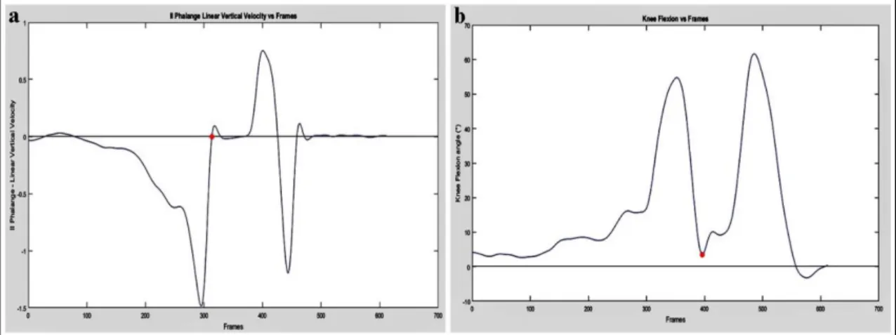
kinematic data were filtered by a second-order zero lag Butterworth 12 Hz low-pass filter. Analysis for determining the kinematic variables was performed by a custom program in Matlab (Mathworks, Natick, MA, USA). The kinematic variables of interest included the angles at initial contact and the peak angles during the landing phase. The kinematic angles of interest included the following: hip flexion, hip internal rotation, hip abduction, knee flexion, knee abduction, trunk flexion, and ipsilateral trunk lean. The landing phase was considered to be from the initial contact to toe-off. The initial contact was defined as when the vertical velocity of the marker that was fixed on the second toe was zero [21] (Figure 2a), and the toe-off was determined by the knee extension peak after support phase [11] (Figure 2b). By convention, positive kinematic values represented flexion, abduction, internal rotation, and ipsilateral trunk lean angles.
2.6 Statistical Analysis
All statistical analyses were performed using the SPSS software version 19 (SPSS, Inc., Chicago, IL, USA). Initially, the statistical distribution and homoscedasticity of the data were checked with the Shapiro-Wilk test and Levene’s test, respectively. Pearson's
3.
RESULTS
The demographic data are shown in table 1.
Table 1 Demographic characteristics of the subjects
n= 23 female athletes Age (years)
Height (cm) Body Mass (kg)
Body Mass Index (kg/cm²) Sportive frequency (times/week) Practice time (months)
Physical Activity Level (mets)
22.9 ± 3.7 163.9 ± 0.06 59.30 ± 8.43 21.84 ± 2.86 4.73 ± 1.05 47.30 ± 66.49 7557.74 ± 5735.18
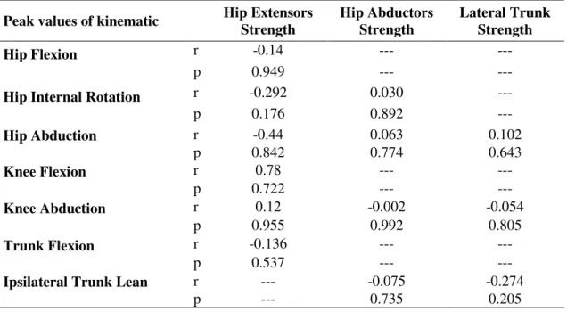
The results of the correlation analysis among the strength data and the angles at initial contact are reported in Table 2. Greater hip extensor strength was found to be significantly associated with greater trunk flexion (r=0.628; p=0.001) at initial contact. No significant correlations were found among the strength data and the peak values of kinematic data during the landing phase, as shown in Table 3.
Table 2 Pearson correlation coefficients (r) and p-value (p) among hip extensor, hip abductor and lateral trunk strength and initial contact kinematic
Initial Contact Kinematic Hip Extensors Strength Hip Abductors Strength Lateral Trunk Strength
Hip Flexion r 0.094 --- ---
p 0.669 --- ---
Hip Internal Rotation r 0.019 -0.357 ---
p 0.930 0.103 ---
Hip Abduction r 0.211 0.301 0.12
p 0.333 0.163 0.957
Knee Flexion r -0.098 --- ---
p 0.655 --- ---
Knee Abduction r 0.075 0.224 -0.129
p 0.733 0.304 0.557
Trunk Flexion r 0.628* --- ---
p 0.001 --- ---
Ipsilateral Trunk Lean r --- 0.057 -0.226
p --- 0.796 0.301
Table 3 Pearson correlation coefficients (r) and p-value (p) among hip extensor, hip abductor and lateral trunk strength and peak values of kinematic.
Peak values of kinematic Hip Extensors Strength Hip Abductors Strength Lateral Trunk Strength
Hip Flexion r -0.14 --- ---
p 0.949 --- ---
Hip Internal Rotation r -0.292 0.030 ---
p 0.176 0.892 ---
Hip Abduction r -0.44 0.063 0.102
p 0.842 0.774 0.643
Knee Flexion r 0.78 --- ---
p 0.722 --- ---
Knee Abduction r 0.12 -0.002 -0.054
p 0.955 0.992 0.805
Trunk Flexion r -0.136 --- ---
p 0.537 --- ---
Ipsilateral Trunk Lean r --- -0.075 -0.274
4.
DISCUSSION
The most important result of this study was the positive correlation among the hip extensor strength with trunk flexion at the initial contact in female athletes. Although we did not find a correlation between hip extensor strength and the peak trunk flexion during the landing phase, for a better comprehension of these results, we correlated the trunk flexion at the initial contact with the peak trunk flexion during the landing phase and found a significant correlation between them (r= 0.628, p= 0.007). These results may indicate that women with lower trunk flexion at the initial contact tend to have a lower trunk flexion during the entire landing phase. Thus, having more strength in the hip extensor muscles can help increase trunk flexion during landing.
sports. This favorable clinical outcome was accompanied by an increase in peak trunk flexion, an increase in hip extensor moment, a decrease in knee extensor moment, and a 26% decrease in patellar tendon force during jump landing measured at 8 weeks. Thus, increased hip extensor strength and better trunk posture (greater flexion) during landing may be important strategies in preventing and rehabilitating ACL injury, PFP, and patellar tendinopathy.
Contrary to the initial hypothesis, no correlation occurred among the hip abductor and hip extensor muscle strength with the trunk, hip, and knee kinematics in transverse and frontal planes.However, excessive movement of these joints in these planes has been related to some knee injuries. An excessive ipsilateral trunk lean [28, 26], hip adduction and internal rotation [28, 26, 39], and knee abduction [28, 26] were observed in PFP subjects. This has been related to increased patellofemoral stress [34, 35]. In addition, dynamic knee valgus (movement composed of hip adduction and internal rotation and knee abduction [44]) was found to be a predictor of ACL injury [17] and second ACL injury in patients with ACL reconstruction [31]. Considering that in a sample of healthy female recreational athletes our results differ from these studies it is possible that the occurrence of these alterations related to hip or trunk weakness are so damaging that this correlation occurs only in injured individuals. In this way, future research investigating these relationships in injured female athletes are needed for a better comprehension of this factors.
[28] found that women with PFP showed greater hip internal rotation and lower gluteus medius activation during single-leg squat than healthy women. Thus, future research is needed to verify whether a relationship exists among trunk and hip muscle activation and trunk and lower-limb kinematics during jumping-landing tasks.
5.
CONCLUSION
6. REFERENCES
1. Alentorn-Geli E, Myer GD, Silvers HJ, Samitier G, Romero D, Lázaro-Haro C, Cugat R (2009) Prevention of non-contact anterior cruciate ligament injuries in soccer players. Part 1: Mechanisms of injury and underlying risk factors. Knee Surg Sports Traumatol Arthrosc 2009 17(7):705-29
2. Almeida GP, Carvalho E Silva AP, França FJ, Magalhães MO, Burke TN, Marques AP (2015) Does anterior knee pain severity and function relate to the frontal plane projection angle and trunk and hip strength in women with patellofemoral pain? J Body Mov Ther 19(3):558-64.
3. Almeida GP, Silva AP, França FJ, Magalhães MO, Burke TN, Marques AP (2016) Relationship between frontal plane projection angle of the knee and hip and trunk strength in women with and without patellofemoral pain. J Back Musculoskelet Rehabil 29(2):259-266
4. Bell AL, Pederson DR, Brand RA (1990) A comparison of the accuracy of several hip joint center location prediction methods. J Biomech 23:617–621
5. Blackburn JT, Padua DA (2009) Sagittal-Plane Trunk Position, Landing Forces, and Quadriceps Electromyographic Activity J Athl Train.; 44(2):174–179
6. Bolgla LA, Malone TR, Umberger BR, Uhl TL (2008) Hip strength and hip and knee kinematics during stair descent in females with and without patellofemoral pain syndrome. J Orthop Sports Phys Ther 38:12-18
7. Bolgla LA, Malone TR, Umberger BR, Uhl TL (2010) Reliability of electromyographic methods used for assessing hip and knee neuromuscular activity in females diagnosed with patellofemoral pain syndrome. J Electromyogr Kinesiol 20(1): 142-147.
8. Boling M, Padua D (2013) Relationship between hip strength and trunk, hip, and knee kinematics during a jump-landing task in individuals with patellofemoral pain. Int J Sports Phys Ther 8(5):661-9
9. Boling MC, Padua DA, Marshall SW, Guskiewicz K, Pyne S, Beutler A (2010) Gender differences in the incidence and prevalence of patellofemoral pain syndrome. Scand J Med Sci Sports 20(5):725–30
10. Craig CL, Marshall AL, Sjöström M, Bauman AE, Booth ML, Ainsworth BE (2003) International physical activity questionnaire: 12-country reliability and validity. Med Sci Sports Exerc 35(8):1381–1395
12. Ford KR, Myer GD, Hewett TE (2003) Valgus knee motion during landing in high school female and male basketball players. Med Sci Sports Exerc 35(10):1745-1750 13. Ford KR, Myer GD, Smith RL, Vianello RM, Seiwert SL, Hewett TE (2006) A comparison of dynamic coronal plane excursion between matched male and female athletes when performing single leg landings. Clin Biomech 21(1):33–40
14. Gatsonis C, Sampson AR (1989) Multiple correlation: exact power and sample size calculations. Psychol Bull 106(3):516-24
15. Griffin LY (2001) American Academy of Orthopaedic Surgeons. Prevention of Noncontact ACL Injuries. Rosemont, III: American Academy of Orthopaedic Surgeons 16. Hertel J, Williams NI, Olmsted-Kramer LC, Leidy HJ, Putukian M (2006) Neuromuscular performance and knee laxity do not change across the menstrual cycle in female athletes. Knee Surg Sports Traumatol Arthrosc 14(9):817–822
17. Hewett TE, Myer GD, Ford KR, Heidt RS, Colosimo AJ, Mclean SG, Van Den Bogert AJ, Paterno MV, Succop P (2005) Biomechanical Measures of Neuromuscular Control and Valgus Loading of the Knee Predict Anterior Cruciate Ligament Injury Risk in Female Athletes. A Prospective Study. Am J Sports Med 33(4):492-501
18. Ireland ML, Willson JD, Ballantyne BT, Davis IM (2003) Hip strength in females with and without patellofemoral pain. J Orthop Sports Phys Ther 33(11):671–676. 19. Kulas AS, Hortobágyi T, DeVita P (2012) Trunk position modulates anterior cruciate ligament forces and strains during a single-leg squat. Clin Biomech 27(1):16-21 20. Lessi GC, Dos Santos AF, Batista LF, de Oliveira GC, Serrão FV (2016) Effects of fatigue on lower limb, pelvis and trunk kinematics and muscle activation: Gender differences. J Electromyogr Kinesiol. 9;32:9-14
21. Lessi GC, Serrão FV (2015) Effects of fatigue on lower limb, pelvis and trunk kinematics and lower limb muscle activity during single-leg landing after anterior cruciate ligament reconstruction Knee Surg Sports Traumatol Arthrosc Doi:10.1007/s00167-015-3762-x
22. Lyons K, Perry J, Gronley JK, Barnes L, Antonelli D (1983) Timing and relative intensity of hip extensor and abductor muscle action during level and stair ambulation. An EMG study. Phys Ther 63:1597-1605
23. Markolf KL, Burchfield DM, Shapiro MM, Shepard MF, Finerman GA, Slauterbeck JL (1995) Combined knee loading states that generate high anterior cruciate ligament forces. J Orthop Res 13(6):930–5
25. Myer GD, Ford KR, Barber Foss KD, Goodman A, Ceasar A, Rauh MJ, Divine JG, Hewett TE (2010) The incidence and potential pathomechanics of patellofemoral pain in female athletes. Clin Biomech 25(7):700-7
26. Nakagawa TH, Maciel CD, Moriya ETU, Serrao FV (2012 Sept) Frontal Plane Biomechanics in Males and Females With and Without Patellofemoral Pain. Med Sci Sports Exer 44(9):1747-1755
27. Nakagawa TH, Maciel CD, Serrão FV (2014) Trunk biomechanics and its association with hip and knee kinematics in patients with and without patellofemoral pain. Man Ther 20(1):189-193
28. Nakagawa TH, Moriya ETU, Maciel CD, Serrão FV (2012 jun) Trunk, pelvis, hip, and knee kinematics, hip strength, and gluteal muscle activation during a single-leg squat in males and females with and without patellofemoral pain syndrome. J Orthop Sports Phys Ther 42(6):491-501
29. Neumann DA (2002) Kinesiology of the Musculoskeletal System St Louis, MO: Mosby Inc.
30. Pardini R, Araújo T, Matsudo V, Andrade E, Braggion G (2001) Validação do questionário internacional de nível de atividade física (IPAQ- versão 6): estudo piloto em adultos jovens brasileiros. Rev Bras Ciên Mov 9(3): 45–51
31. Paterno MV, Schmitt LC, Ford KR, Rauh MJ, Myer GD, Huang B, Hewett TE (2010) Biomechanical measures during landing and postural stability predict second anterior cruciate ligament injury after anterior cruciate ligament reconstruction and return to sport. Am J Sports Med 38(10): 1968-1978
32. Perry J (1992) Gait Analysis: Normal and Pathological Function. Thorofare, NJ: Slack Inc
33. Powers CM (2010) The influence of abnormal hip mechanics on knee injury: A biomechanical perspective. J Orthop Sports Phys Ther 40(2):42–51
34. Powers CM, Ward SR, Fredericson M, Guillet M, Shellock FG (2003) Patellofemoral kinematics during weight-bearing and non-weight-bearing knee extension in persons with lateral subluxation of the patella: a preliminary study. J Orthop Sports Phys Ther. 33(11):677-85
35. Salsich GB, Perman WH (2007) Patellofemoral joint contact area is influenced by tibiofemoral rotation alignment in individuals who have patellofemoral pain. Orthop Sports Phys Ther 37(9):521-8
37. Scattone Silva R, Nakagawa TH, Ferreira AL, Garcia LC, Santos JE, Serrão FV (2016) Lower limb strength and flexibility in athletes with and without patellar tendinopathy. Phys Ther Sport 20:19-25
38. Shimokochi Y, Shultz SJ (2008) Mechanisms of noncontact anterior cruciate ligament injury. J Athl Train 43(4):396-408
39. Souza RB, Powers CM (2009) Differences in hip kinematics, muscle strength, and muscle activation between subjects with and without patellofemoral pain. J Orthop Sports Phys Ther 39(1):12–9.
40. Stickler L, Finley M, Gulgin H (2015) Relationship between hip and core strength and frontal plane alignment during a single leg squat. Phy Ther Sport 66-71
41. Teng HL, Powers CM (2014) Sagittal plane trunk posture influences patellofemoral joint stress during running. J Orthop Sports Phys Ther 44(10):785-92 42. Teng HL, Powers CM (2016) Hip-Extensor Strength, Trunk Posture, and Use of the Knee-Extensor Muscles During Running. J Athl Train 51(7):519-24
43. Wu G, Siegler S, Allard P, Kirtley C, Leardini A, Rosenbaum D, Whittle M,
D’Lima DD, Cristofolini L, Witte H, Schmid O, Stokes I (2002) ISB recommendation on
definitions of joint coordinate system of various joints for the reporting of human joint motion—part I: ankle, hip, and spine. J Biomech 35:543–548