Arch Bone Jt Surg. 2015;3(4):264-268. http://abjs.mums.ac.ir
the online version of this article abjs.mums.ac.ir
Satish Shervegar, MD; Prashanth Nagaraj, MD; Amit Grover, MD; Niranthara Ganesh DJ, MD; Abdul Ravoof, MD Research performed at MS Ramaiah Medical College , Bangalore , Karnataka , India
Introduction
A
rthroscopic anterior cruciate ligament reconstruction has remained a treatment of choice for anterior cruciate ligament deficient knees, since majority of non-operative procedures have resulted in functionally unacceptable outcomes (1). The incidence of ACL deficient knees due to trauma is reported as 1 in 3,500 people, resulting in 95,000 new ACL ruptures per year in few studies (2,3).Biomechanics of injury
ACL is the primary (85%) restraint to limit anterior
translation of the tibia. In full extension, ACL also serves as a secondary restraint to tibial external rotation. The average tensile strength of ACL is 2160N which is half as strong as medial collateral ligament and slightly less than posterior cruciate ligament (2, 3).
Factors influencing the clinical outcomes after ACL
reconstruction
There are many factors which influence the clinical outcomes of ACL reconstruction like type of graft used, method of fixation, placement of graft and post operative rehabilitation programme (4). Nevertheless
Corresponding Author: Amit Grover, MS Ramaiah Medical College, MSR Nagar, BEL Road, Bangalore, Karnataka , India- 560054
E-mail: [email protected]
Received: 8 January 2015 Accepted: 29 April 2015
Functional Outcome Following
Arthroscopic ACL Reconstruction with Rigid Fix: A
Retrospective Observational Study
Abstract
Background: No uniform consensus exists to decide type of ixation for arthroscopic anterior cruciate ligament
reconstruction.
Hypothsis: There is similar functional outcome after rigid ix compared to other methods of ixation which has been published.
Study design: Retrospective observational study.
Methods: A total of 50 patients underwent arthroscopic anterior cruciate ligament reconstruction with hamstring
tendons using femoral Rigid ix cross-pin and interference screw tibial ixation. The evaluation methods were clinical examination, IKDC scores, Lysholm and pre injury and post reconstruction Tegner score. Patients were followed up from minimum of 6 months to 4 year seven months.
Results: C In our study of sample size 50 we found that mean age of patients was 30.8 Years with male preponderance.
Mean post operative IKDC and Lysholm score has been 75.6 and 84.4 respectively.Mean Tegner pre-injury score and post reconstruction score has been 5.4 and 4.26 .Box plot comparison of pre injury and post operativeTegner score reveals a statistically signiicant difference with respect to paired t test P<0.001.
Conclusions: Arthroscopic anterior cruciate ligament reconstruction with femoral rigid ix cross pins and tibial
interference screws results in comparable short term to midterm functional results compared to other types of ixation
the competency of the operating surgeon will play a major role in the clinical outcome of arthroscopic ACL reconstruction surgery.
Types of femoral graft fixation
The literature is repute with many ways of graft fixation.The types of graft fixation are mainly divided into cortical, cancellous and cortico-cancellous fixation.
Systems with purely cortical support, the best known of which is the EndoButton®. Endobutton (Smith & Nephew, Andover, MA, USA) is a fixation method in which a device is placed against the anterolateral cortex of femur, suspending the graft inside the femoral tunnel (5). However, correct positioning of the button on the lateral cortex is not easy to achieve, and requires accurate calculation of the total length of the femoral tunnel. Another mechanical disadvantage is the increase in the total length of the graft that will be subjected to strain.
Rigidfix (DePuy Mitek, Raynham, MA) is a transcondylar fixation system which uses horizontal bars that cross the graft and femoral tunnel and create a bulge in graft. In this fixation, resistance is distributed along the surface between bone and depends on bone quality and lever arm length (5).
Aperfix (Cayenne Medical, Inc, Scottsdale, Arizona) is a system for femoral fixation in which the device in cancellous bone is pulled alongside the shaft and prevents graft from moving back (5).
Materials and methods
The aim of this study was to review a group of fifty patients treated with arthroscopic ACL reconstruction using rigid fix for the femoral component and interference screw to secure tibial component of the graft.
Our aim was to compare functional outcome after arthroscopic ACL reconstruction using Rigid fix for the femoral component and interference screw for the tibial component with other methods of fixation.
Methods
We conducted a retrospective study of fifty patients with ACL deficient knees treated in a single centre with arthroscopic ACL reconstruction using rigid fix for the femoral component and interference fix to secure tibial component of the graft. Patients who were admitted with ACL injury confirmed by clinical records and MRI scan during the period 2007 to 2011 were included in the study based on inclusion criterion.
Indications for surgery were clinically and radio logically confirmed cases of anterior cruciate ligament deficient knees.
The inclusion criterion were • Age between 18 to 55 years
• Clinically ACL deficient knee examined by single surgeon
• Radiological ACL deficient knee confirmed by MRI • Associated menisci injuries
The exclusion criterion were • Infection
• Patients who were lost in the follow up • Associated PCL tear
• Associated tibial plateau fractures
• Patients not willing to involve in the study • Bilateral knee injuries
Using the Orthopedic departmental trauma database 65 suitable patients was identified for inclusion in the study. Five patients were unwilling to participate; 10 were non contactable. The remaining 50 patients were followed up by means of case note and operative record analysis, radiographic assessment and functional questionnaire.
Outcome assessment
Case notes were used to establish all demographic details including mechanism of injury, time to surgery,post-operative immobilization and intra-operative details. Complications were also recorded and confirmed with the patients.
The functional questionnaire was both telephone based and in-person format methods. The functional scoring system utilized was IKDC subjective functional scoring system [Table 1]. The Lysholm score and Tegner prep and post operative scoring system were also considered. The study mainly looked at Tegner pre injury and post reconstruction score [Table 2]. We Table 1. Basic characteristics of the study population
N=50 Mean Std. Deviation Minimum Maximum
Age 30.86 9.69 18 54
Ikdc subjective score 75.60 17.36 18.39 98.8
Lysholm score 84.42 13.24 48 100
Tegner pre injury score 5.44 1.51 3 9
Tegner post reconstruction score 4.26 2.00 0 9
Table 2. Comparison of tegner scores before and after procedure
N=50 Mean Std. Deviation P value
Tegner pre injury score 5.44 1.51
have tried to correlate the post operative Tegner and Lysholm score with mean duration of 2 years in our study.
Statistical methods
Categorical variables and baseline demographic data are described using frequencies and percentages. Continuous variables with a symmetric distribution are presented using means and standard deviations (SD) and continuous variables with a skewed distribution are presented using the median and inter-quartile range (IQR). All tests were
two-sided with the level of significance set at 0.01 to take account of multiple testing.
Surgical procedure
The surgery was performed by single surgeon.
Tourniquet was used in all cases. Standard anteromedial (AM) and anterolateral (AL) portals were used to perform the arthroscopic inspection of the knee joint to confirm the diagnosis and look for associated meniscal or chondral lesions . Then torn ACL is carefully removed with special attention to the anatomic footprints of the 2 ACL bundles.
A transtibial technique was used to create femoral tunnel for ACL double-bundle reconstruction. The guide wire was passed through the tibial tunnel and tip of the guide wire was placed on the femoral footprint of the AM bundle. AM femoral tunnel was drilled to a depth of 35 mm to 40 mm. The far cortex of the AM femoral tunnel was breached with a 4.5-mm drill bit. Once the femoral socket has been fashioned with concentric reaming, a marking hook, firmly mounted on the femoral guide, was introduced. The hook (which matches the socket diameter) is pushed in the socket. A guide pin was drilled through the distal femur, from lateral to medial. The guide pin was them replaced by Table 3. Lysholm score categories
Score Category Frequency Percent
98 - 100 10 20.0
93 - 97 10 20.0
82 - 92 12 24.0
66 - 81 13 26.0
≤ 65 5 10.0
Total 50 100.0
Table 4. Distribution of type of injuries
Injuries Frequency Percent
Lateral meniscal tear 1 2.0
Lateral meniscus tear 1 2.0
Medial and lateral meniscus tear 1 2.0
Medial meniscus bucket handle tear 1 2.0
Medial meniscus complete tear + lateral meniscus patial tear 1 2.0
Medial meniscus tear 3 6.0
Mm + lm tear 1 2.0
Mm bucket handle tear 1 2.0
Mm+lm tear 1 2.0
No injurues 39 78.0
Total 50 100.0
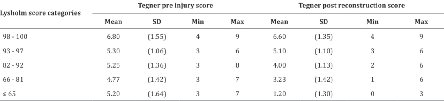
Table 5. Comparison of TEGNER pre and post scores between various lysholm score categories
Lysholm score categories
Tegner pre injury score Tegner post reconstruction score
Mean SD Min Max Mean SD Min Max
98 - 100 6.80 (1.55) 4 9 6.60 (1.35) 4 9
93 - 97 5.30 (1.06) 3 6 5.10 (1.10) 3 6
82 - 92 5.25 (1.36) 3 8 4.00 (1.13) 2 6
66 - 81 4.77 (1.42) 3 7 3.23 (1.42) 1 6
a guide wire, which is brought out through the tibial tunnel. The graft was loaded into the wire loop. Rigidfix (DePuy Mitek, Raynham, MA) was slided over the wire, through the axilla of the graft, and impacted into the lateral femoral cortex. First, the tendon grafts were trimmed and the diameters of the double-stranded grafts were adjusted. The AM tendon graft was trimmed such that the diameter of the double-stranded graft is 8 mm to 9 mm; the PL graft was trimmed such that the diameter of the double stranded graft is 7 mm to 8 mm.A Beath pin with a long looped suture attached to the eyelet wass passed through the accessory anteromedial portal and out through the PL femoral tunnel. The looped suture was visualized within the joint and retrieved with an arthroscopic suture grasper through the PL tibial tunnel. On the tibial side, we used a bioabsorbable interference screw fixation. The PL bundle graft was tensioned and fixed between 0° to 10° of flexion. The AM bundle graft was tensioned and fixed between 60° and 90° of flexion.
Postoperatively, the patients were put on a knee brace for 4-6 weeks but range of motion exercises were started immediately after surgery. The patients were allowed full weight bearing as tolerated. Return to full activity level was usually allowed at about 6 months postoperatively.
Results & discussion
The Mean IKDC subjective score post reconstruction was 75.6 with a standard deviation of 17.36. The mean post operative Lysholm score was 84.42 with standard deviation of 13.24. The pre injury and post reconstruction Tegner score was 5.44 and 4.26 respectively. 13% of patients had a Lysholm score of 66-81 with male to female ratio of 80:20 [Table 3]. 39 patients did not had any concomitant injuries.7 patients had medial meniscal and lateral meniscal injuries were found in 2 cases. The Box plot analysis revealed that the Tegner pre injury score was higher compared to the post scores. This difference in Tegner scores were found to be statistically significant on paired t test (P<0.001)
There has been many articles with reported complications of transfix like lateral or medial pin slip, breakage of bioabsorbable cross pin,ilotibial band friction syndrome, femoral tunnel widening ,intraarticular pin translation and chondral lesions of lateral femoral condyle and tibial condyle region. Most of these complications resulted due to technical error in placement of screws. They manifested within 3-6 months after surgery. In our study with average follow up of 2 years our patients did well subjectively with improved performance oriented physical activity. Using MRI, Studeler et al. found fractured cross pins in 35 patients (17%), breach of the posterior femoral cortex in 57 patients (28%), and migration of fractured pin fragments in 12 patents (6%) (6). Again, there was no correlation between fractured pins and graft nor was there any clinical correlation between fractured pins and instability.
Choi et al. analysed 31 patients who underwent ACL reconstruction using Rigid Fix bioabsorbable cross-pin
fixation. Using MRI, they found that 38.7% of the Rigid Fix implants fractured 6 months postoperatively (7). In our study we did not find any case of implant breakage or migration in our follow up of average 2 years.
Asik et al. analysed mid to long-term results of anterior cruciate ligament reconstruction with hamstring tendons using Transfix technique (8). Anterior cruciate ligament (ACL) reconstruction with four-strand hamstring tendon was performed with Transfix technique on 271 patients with anterior cruciate ligament ruptures. When compared with Tegner activity scale, 189 (70%) patients scored <6 preoperatively and only 24 (8%) postoperatively.Our functional results were found to be satisfactory in more than 90% of patients. In our study of 50 patients mean preoperative Tegner score was 5.44 compared to post reconstruction score of 4.26 with P-value less than
0.001. Comparitively our sample size is small but in our study patient’s satisfaction remains more than 90% comparable to previous studies. 24-26% of patients had a post operative Lysholm score between 66-92 [Table 3]. Most of our patients did not have any other ligament injuries [Table 4] and right sided ACL tears were more common. It was found in our study that at lower post operative Lysholm score < 65 correlated well with the lower post reconstruction Tegner score.Post operative Higher Lysholm scores >93 correlated positively with increase of pre injury Tegner scores with respect to post injury scores. On average patients with pre injury Tegner scores between 4.7-5.25 demonstrated significant improved post reconstruction Tegner scores of 3.23 and 4.00 respectively which correlated well with Lysholm score in the range between 81-92 [Table 5].
Seo et al. compared the short term clinical results of anterior cruciate ligament reconstruction with autologous hamstring tendon between Rigid-fix and PINN-ACL Cross Pin for femoral side fixation and reported that there was no resultant difference between the employment of PINN-ACL Cross Pin and Rigid-fix as femoral graft fixation for ACL reconstruction with hamstring tendon (9). However, PINN-ACL Cross Pin led to complications with extensive operation times.
Musil D et al. in their study on anterior cruciate ligament reconstruction compared patellar bone-tendon-bone and hamstring tendon graft methods (10). They intended to analyse short-term evaluation of the hamstring tendon graft technique with use of the Rigidfix system compared with B-T-B graft .They concluded that ACL reconstruction with a hamstring tendon autograft fixed with the Rigidfix system is a suitable alternative technique to a patellar B-T-B graft. It is suitable for patients who have contraindications for the B-T-B technique and in persons practicing little or no sports. The main drawback was this study compared the results between two types of grafts used rather than types of fixation used.
This study concludes that the reconstruction of ACL with hamstring tendons and rigid fix technique is reasonably safe with less complications and good functional outcome. The accurate placement of graft in the tunnel and preparation of graft are important to obtain optimal results. Further studies with reference to long term follow up , radiological parameters including MRI, relevant subjective scores, double blind prospective trials comparing the effectiveness of different methods of graft fixation are however required to provide more clarity on the use of femoral fixation systems.
Satish Shervegar MD Amit Grover MD
Niranthara Ganesh DJ MD
MS Ramaiah Medical College, Bangalore, Karnataka, India Prashanth Nagaraj MD
Sparsh Hospitals, Bangalore, Karnataka, India Abdul Ravoof MD
Adichunchungiri Institute of Medical Sciences, Bellur, Bangalore, Karnataka, India
References
7. Choi NH, Lee JH, Victoroff BN. Do broken cross-pins compromise stability after anterior cruciate ligament reconstructions with hamstring tendons?. Arthroscopy. 2007; 23(12):1334-40.
8. Asik M, Sen C, Tuncay I, Erdil M, Avci C, Taser OF. Comparison between Rigidfix and bio-Transfix The mid- to long-term results of the anterior cruciate ligament reconstruction with hamstring tendons using Transfix technique. Knee Surg Sports Traumatol Arthrosc. 2007; 15(8):965-72.
9. Seo SS, Kim CW, Nam TS, Choi SY. ACL Reconstruction with Autologous Hamstring Tendon: Comparison of Short Term Clinical Results between Rigid-i x and PINN-ACL Cross Pin. Korea Knee Surg Relat Res. 2011;23(4):208-12
10. Musil D, Sadovský P, Stehlí�k J.BTB allograft for revision surgery of the anterior cruciate ligament - part 2. Acta Chir Orthop Traumatol Cech. 2005;72(5):297-303.
11. Monaco E, Labianca L, Speranza A, Agrò AM, Camillieri G, D’Arrigo C, et al. Biomechanical evaluation of different anterior cruciate ligament fixation techniques for hamstring graft. J Orthop Sci. 2010;15(1):125-31.
1. Fu FH ,Bennet CH,Latterman C and MA CB. Current trends in anterior cruciate ligament reconstruction. Part 1: Biology and biomechanics of reconstruction. Current concepts. Am J Sports Med. 1999; 27(6):821-30.
2. Lyman S, Koulouvaris P, Sherman S, Do H, Mandl LA, Marx RG. Epidemiology of anterior cruciate ligament reconstruction: trends, readmissions, and subsequent knee surgery. J Bone Joint Surg Am. 2009; 91(10):2321-8.
3. Frank CB, Jackson DW. Current concepts review. J Bone Joint Surg Am. 1998; 79(10);1556-75
4. Johnson RJ, Beynnon BD, Nicholas CE, Renstrom PA. The treatment of injuries to antrerior cruciate ligament current concepts review. J Bone Joint Surg Am. 1992; 74(1);140-51
5. Eajazi A, Madadi F, Madadi F, Boreiri M. Comparison of Different Methods of Femoral Fixation Anterior Cruciate Ligament Reconstruction. Acta Medica Iranica. 2013; 51(7):444-8.