Contents lists available atScienceDirect
Journal of the Mechanical Behavior of
Biomedical Materials
journal homepage:www.elsevier.com/locate/jmbbm
The e
ff
ect of zoledronate-containing primer on dentin bonding of a universal
adhesive
Walter Zenobi
a, Victor Pinheiro Feitosa
a,b,⁎, Maria Elisa Martins Moura
a, Camillo D
’
arcangelo
c,
Lidiany Karla de Azevedo Rodrigues
a, Salvatore Sauro
a,daPost-Graduate Program in Dentistry, Dental School, Department of Restorative Dentistry, Federal University of Ceará, Rua Monsenhor Furtado S/N, Bairro Rodolfo Teófilo, 60430-325 Fortaleza, Brazil
bPaulo Picanço School of Dentistry, Fortaleza, Brazil
cUnit of Restorative Dentistry, Department of Oral Science, Nano and Biotechnology,
“G. D'Annunzio”University of Chieti, Italy
dDental Biomaterials, Departmento de Odontologia, Facultad de Ciencias de la Salud, CEU-Cardenal Herrera University, Alfara del Patriarca, 46115 Valencia, Spain
A R T I C L E I N F O
Keywords: Bond strength Dental adhesive Zoledronic acid Mechanical stress
A B S T R A C T
Objectives:To evaluate the bonding ability and nanoleakage of a universal adhesive applied to dentin pre-treated using a zoledronate-containing primer (zol-primer) before and after mechanical load cycling.
Materials and Methods:Flat dentin surfaces obtained from human molars were assigned to one of the following adhesion procedures (n=6): 1-Single Bond Universal(SBU) applied in etch-and-rinse mode; 2- SBU applied as etch-and-rinse after the application of zol-primer; 3- SBU applied in self-etch strategy; 4- SBU applied as self-etch after the use of zol-primer. Half of the specimens were processed for microtensile bond strength test after 24 h, while the other half part was submitted to 200,000 mechanical cycles. Further specimens were silver-im-pregnated and assessed for interface nanoleakage by SEM. Data were analyzed with two-way ANOVA and Tukey's test (p < 0.05).
Results:At 24 h evaluation, the four groups presented similar bond strengths, whilst both groups bonded with etch-and-rinse technique showed significant bond strength reduction after mechanical load (p < 0.05), with the highest drop in bond strength for the specimens pre-treated with the zol-primer. No negative effects were found for self-etch strategy (p > 0.05) in microtensile test. Lower nanoleakage expression was observed for etch-and-rinse specimens treated with zol-primer. However, noteworthy reduction of adhesive layer thickness was ob-served when combining the zol-primer with the self-etch bonding approach.
Conclusion:It can be concluded that zol-primer should not be used along with a universal adhesive in etch-and-rinse mode, but its application before self-etch application may provide less degradation of the resin-dentin interface.
1. Introduction
The mechanism of bonding to enamel and dentin is essentially based on an exchange process in which minerals removed are replaced by resin monomers, that upon polymerization become micromechanically interlocked (Cardoso et al., 2011). Conversely, impregnation of syn-thetic resin monomers into demineralized dentin is challenging due to the humidity, permeability and physiologic hydrostatic pulpal pressure (Feitosa et al., 2012a). After adhesive polymerization, exposed collagen is easily detected and represents an area prone to degradation. Collagen degradation is accelerated by proteolytic enzymes, the so-called matrix metalloproteinases (MMPs) and cysteine cathepsins (CC) (Scaffa et al.,
2012).
Several MMP inhibitors have been investigated (Breschi et al., 2008; Scaffa et al., 2012) such as galardin, batimastat and chlorhexidine, some have already been incorporated into experimental adhesives (Almahdy et al., 2012; Yiu et al., 2012). Synthetic MMP inhibitors should contain a functional group capable of chelating the zinc ions, which binds to the active site in the MMP molecule (Feitosa et al., 2012a; Tezvergil-Mutluay et al., 2014). A polyphosphonic acid such as zoledronate may provide additional inhibition of MMP due to the a potential bond which may create between zoledronate and proteins (Murphy et al., 2014). Moreover, it has been adovacated that the use of zolendric acid may reduce the activity of MMPs via dentin
http://dx.doi.org/10.1016/j.jmbbm.2017.09.015
Received 27 July 2017; Received in revised form 5 September 2017; Accepted 11 September 2017
⁎Corresponding author at: Post-Graduate Program in Dentistry, Dental School, Department of Restorative Dentistry, Federal University of Ceará, Rua Monsenhor Furtado S/N, Bairro Rodolfo Teófilo, 60430-325 Fortaleza, CE, Brazil.
E-mail addresses:[email protected],[email protected](V.P. Feitosa).
Available online 14 September 2017
1751-6161/ © 2017 Elsevier Ltd. All rights reserved.
remineralization and reduce the degradation of resin-dentin interface especially when associated with ion-releasing adhesives ( Tezvergil-Mutluay et al., 2014).
Recent multi-mode self-etch adhesives (so-called universal ad-hesives) may be applied in etched or non-etched enamel and dentin. The longevity and bond strength of these materials have been studied, but these remain questionable (Hanabusa et al., 2012; Perdigão and Loguercio, 2014). We hypothesize that zoledronate could improve the durability of dentin bonds created with universal adhesives, especially when dentin-etching is undertaken. Furthermore, zoledronate might also improve the resistance of partially demineralized collagenfibrils to mechanical stress. However, to our knowledge, this combination (zo-ledronate+universal adhesive) has never been investigated so far. Therefore, the objective of this study was to assess the effect of dentin pre-treatment using an experimental primer containing 7% zoledronate (Zol-primer), applied prior to the application bonding a universal ad-hesive in etch-and-rinse or self-etch mode, on the microtensile bond strength (μTBS) before and after mechanical cycling load challenge.
2. Materials and methods
2.1. Preparation of experimental zol-primer
The formulation of the zol-primer used has been described by Tezvergil-Mutluay et al. (2014), and was formulated with deionized water (50 vol%) saturated (7 mg/mL) with zoledronate [1-hydroxy-2-(1H-imidazol-1-yl) ethane-1,1-diyl]bis-phosphonic acid (MW 290; Santa Cruz Biotechnology, Santa Cruz, USA) and absolute ethanol (50 vol%) (pH adjusted to 6.8 with 0.1 M NaOH).
2.2. Sample preparation
Twenty-four third molars recently extracted for surgical reasons in the service of Dentistry and Maxillofacial Surgery Hospital General Universitario de Valencia under a protocol approved by the Biomedical Research Ethics Committee of the University and Polytechnic La Fe Hospital in Valencia, with registration No. 2014/00487/PI and. stored in deionized water (pH 7.4) at 4 °C no longer than 3 months,. Middle dentin specimens were obtained by removing the roots 2 mm below cemento–enamel junction (CEJ) and with a parallel cut at 2 mm above CEJ using a slow-speed water-cooled diamond saw (Isomet, Buehler, Lake Bluff, USA). The dentin surface was wet-polished with 600-grit SiC papers for 1 min to create a standard smear layer prior to bonding procedures (Feitosa et al., 2012b; Hamouda et al., 2011).
2.3. Experimental design and bonding procedures
Dentin specimens were divided randomly by using Microsoft Excel (Windows) randomization into two principal groups (n=12) based on the bonding technique used: self-etch and etch-and-rinse. Subsequently, the specimens from each main group were divided into subgroups (n=6), regarding the use or not of Zol-primer before adhesive appli-cation. Specimens of each principal group were divided into further sub-groups (n=3), based on the challenge test: Control: water immer-sion for 24 h and MCL: mechanical-cycling load.
The universal adhesive employed in this study was the Single Bond Universal (3 M-ESPE, St. Paul, USA). The composition and application procedures are listed inTable 1. Resin composite build-ups were con-structed in 3 horizontal layers (2-mm thick) up to 6 mm with Spectrum ® TPH® resin composite (Dentsply, Petropolis-RJ, Brazil) and light-cured for 30 s each layer.
Light-curing procedures were performed using the LED-curing unit DB85 (Dabi Atlante, Ribeirao Preto, Brazil). The output intensity was monitored with a Demetron Radiometer (Model 100, Demetron Research, Danbury, USA) to maintain a minimal light output intensity of 1000 mW/cm2throughout all experiments. All materials were used
following the manufacturers’recommendations.
2.4. Mechanical-cycling challenge
The resin-bonded specimens were submitted to the mechanical cy-cling load executed using Chewing Simulator CS-4 (SD Mechatronik, Feldkirchen-Westerham, Germany) which has a stainless steel tip of 4 mm in diameter in contact with the central part of the restored spe-cimens. All resin-bonded specimens were submitted to 200,000 me-chanical cycles under a load of 30 N, at a rate of 2 Hz for one week (Ulker et al., 2010).
2.5. Microtensile bond strength (µTBS) and fracture type analysis
After 24 h immersion in distilled water (Control) or MCL, resin-bonded specimens were sectioned in resin-dentin sticks (0.9 mm × 0.9 mm) for microtensile bond strength testing. Sticks from the most peripheral area presenting remaining enamel were excluded. The sticks were attached to a jig with a cyanoacrylate cement (Super Bonder gel, Loctite, Henkel Corp., Rocky Hill, USA) and tested to tensile failure in a universal testing machine (DL2000, EMIC, Sao Jose do Rio Preto, Brazil) with a 500-N load cell and 0.5 mm/min cross-head speed. The exact cross-sectional area of each tested stick was measured with a digital caliper. The µTBS results were calculated and expressed in MPa. The µTBS values obtained from the sticks of the same resin-bonded tooth were averaged. Mean bond strength of each individual tooth was used as one unit for statistical analysis. Three resin-bonded teeth (n = 3) were evaluated for each sub-group. The µTBS data were statistically analyzed with two-way ANOVA (presence of zol-primer and aging re-gimen) and Tukey's post-hoc test at 5% significance level. Subsequent to the µTBS testing, the mode of failure of each fractured stick was de-termined using a stereomicroscope (Olympus SZ 40–50; Tokyo, Japan) at 100X magnification. The fractures were classified as adhesive, mixed, cohesive in composite or cohesive in dentin.
2.6. Nanoleakage analysis
Three resin-dentin sticks were selected from each bonded tooth and storage condition during the cutting procedure. These sticks were im-mersed in 50 wt% ammoniacal silver nitrate (AgNO3(aq)) solution in complete darkness for 24 h (Tay et al., 2002). Subsequently, the spe-cimens were rinsed with distilled water to remove the excess of silver nitrate and immersed in photo-developing solution for 8 h under light to reduce silver ions into metallic silver grains. The silver-impregnated sticks were embedded in epoxy resin and polished using 600-, 1200-, 2000-grit SiC papers and diamond pastes (Buehler, Lake Bluff, IL, USA) with 1 and 0.25 µm particle sizes, and ultrasonically cleaned of 15 min after each abrasive/polishing step. Specimens were finally air-dried, dehydrated overnight in silica gel under vacuum, coated with carbon and analyzed using SEM (Inspect 50, FEI, Amsterdam, Netherlands) and observed in backscattered electron mode at 20 kV.
3. Results
3.1. Microtensile bond strength testing (µTBS)
analysis were adhesive for all groups, especially after mechanical load aging.
3.2. Scanning electron microscopy (SEM)
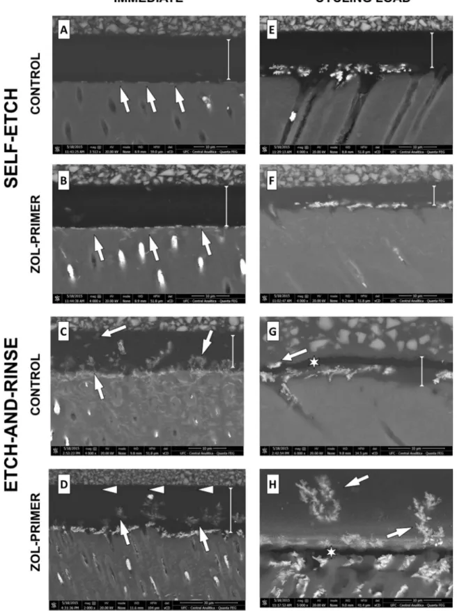
The nanoleakage results of the samples analyzed by SEM are shown inFig. 1. Comparing the 24 h group with and without zol-primer ap-plication, sparse silver impregnation was found only at the bottom of hybrid layer. When zoledronate was employed in etch-and-rinse mode, water channels were detected up to the adhesive-composite interface (Fig. 1C). However, a silver-free zone at the top of adhesive layer was found when zol-primer was used (Fig. 1D). After mechanical load cy-cling in self-etch mode, anincrease of nanoleakage was observed at hybrid layer and bottom of adhesive layer, which was accompanied by a reduction of adhesive layer thickness only for zol-primer treated specimens (Fig. 1F). This reduction of adhesive layer thickness was also detected when etch-and-rinse specimens without zol-primer were sub-jected to load cycling (Fig. 1G). In addition, gaps were found at the top of adhesive layer. When zol-primer was combined with etch-and-rinse technique after load cycling (Fig. 1H), some gaps were also observed and located at the hybrid layer. Water trees were observed along the entire interface of cycled etch-and-rinse specimens regardless the ap-plication of zoledronate.
4. Discussion
The results of microtensile bond strength test after 24 h storage in water, in both adhesion technique (self-etch and etch-and-rinse) de-picted that the application of zol-primer caused no significant inter-ference with the initial bond strength, but demonstrating full compat-ibility with the universal adhesive (Single Bond Universal, 3M-ESPE, St. Paul, USA). Conversely, the results obtained after load cycling showed that the pre-treatment with zol-primer was only effective in self-etch strategy.
The application mode (self-etch or etch-and-rinse) did not affect the initial dentin bond strength of universal adhesives, so that our out-comes are in agreement with those reported in previous investigations (Muñoz et al., 2015; Kenshima et al., 2006). The demineralization in-duced by phosphoric-acid etchings or acidic functional monomers plays a fundamental role on smear layer demineralization/modification and demineralization of underlying dentin in order to create spaces for resin monomers infiltration (Camps and Pashley, 2000; Carrilho et al., 2007).
Host-derived matrix metalloproteinases (MMPs), found both in saliva and in dentin, have been shown to be involved on the degrada-tion of the unprotected collagen fibrils within the hybrid layer (Chaussain-Miller et al., 2006; Hannas et al., 2007). The durability of dentin bonding agents is affected by the degradation of the resin compounds occurring via hydrolysis of sub-optimally polymerized hy-drophilic resins and degradation of collagen matrices byhydrolysis and
matrix metalloproteinases (MMPs) and cysteine cathepsins (Tjäderhane et al., 2013). Nevertheless, current attempts to extend the longevity of resin–dentin bonds via incorporation of matrix metalloproteinase inhibitors (Tersariol et al., 2010) lack to address the mineral phase re-deposition in demineralized dentin collagen, thereby providing fossili-zation of MMPs (Brackett et al., 2011). Yet, when calcium phosphate formation is produced, MMP-2 and MMP-9 form complexes CaP:MMP with high molecular weight and restricted mobility (Makowski et al. (2004);
Toledano et al., 2012), which impairs the activity of MMPs.
The use of bioactive materials in order to remineralize collagenfi -brils may provide a feasible strategy to extend the longevity of re-sin–dentin interface (Profeta et al., 2013). A poly(phosphonic acid) such as zoledronate may provide MMP inhibition (Sulkala et al., 2001) due to its chelation ability (Tezvergil-Mutluay et al., 2014). The com-bination of these two agents (bioactive materials and MMP inhibitiors) might represent a further method for the inhibition of endogenous proteases within the resin-dentin interface as recently demonstrated by Tezvergil-Mutluay and collaborators (2014) when using a zoledronate-containing primer and ion-releasing resins. Regarding the activation of MMPs, when dentin is completely demineralized, a severe release of the calcium and phosphates necessary for MMP activation is warranted (Tezvergil-Mutluay et al., 2014). Phosphoric acid etching in etch-and-rinse adhesion strategy cause great exposure of collagenfibrils, which would hypothetically promote a more suitable substrate for zoledronate to infiltrate and interact with the substrate.
Nevertheless, according to results in Table 2, the use of the zol-primer cannot be indicated in association with the universal adhesive tested in this study in etch-and-rinse mode due to the striking decrease of bond strength after mechanical load cycling. Contrariwise, zol-primer does not impair the bond strength when used prior to the self-etch strategy. This limitation may due to the fact that zoledronate could bind to collagenfibrils (Ryan et al., 1996) jeopardizing the monomers infiltration within the acid-etched dentin. Such results are reported herein for thefirst time, since the present study is thefirst to survey the combination of zoledronate-primer with universal adhesives in terms of bonding performance. Moreover, the acid nature of zoledronate
(Tezvergil-Mutluay et al., 2014) might have interfered with the cam-phorquinone/amine light-initiation;amine coinitiator may act as a Lewis base donating electrons to zoledronate and reducing its reaction with cam-phorquinone, thereby decreasing the adhesive polymerization. After load cycling, this negative effect was more noteworthy due to the breakdown of resin-sparse demineralized fibrils. Lower concentration of zole-dronate or the use of other etch-and-rinse adhesives could surpass this effect, thereby demonstrating the advantages of zol-primer over longer time period of aging.
When comparing the images of nanoleakage (Fig. 1), when self-etch Table 1
Materials, compositions and application procedures.
Materials Composition Application
Composite Resin
THP Spectrum®Dentsply SHADE A3
Matrix: BisGMa, UDMA
Filler: Barium Glass, Aluminium Boron Silicate, Pyralytic Silonized Silicia
Apply in 1–2 mm increments and light cure for 40 s
Adhesive
Single Bond Universal™
6-MHP Phosphate Monomer, Dimethacrylate resins, HEMA, Vitrebond™ Copolymer, Filler, Ethanol, Water, Initiators, Silane
Self-etch or Etch-and-rinse technique
Experimental Zol-primer
Zoledronate [1-hydroxy−2-(1H-imidazol−1-yl) ethane− 1,1-diyl]bis-phosphonic acid, ethanol (50 vol%)
Apply one coat of zol primer for 15 s with gentle agitation. Gently air dry for 10 s
BiSGMA: bisphenol A diglycidylmethacrylate; UDMA: urethane dimethacrylate; MDP: 10-methacryloyloxi decyl phophate; HEMA: hydroxyethylmethacrylate.
Table 2
Means (standard deviations)μTBS of each group in MPa.
µTBS (MPa) Control Load Cycled
Self-etch 26.4 (1.9) A,a 26.1 (4.1) A,a
Self-etch + Zol 29.7 (5.8) A,a 22.5 (4.6) AB,a Etch-and-rinse 26.5 (4.4) A,a 18.8 (4.1) B,b Etch-and-rinse + Zol 28.1 (4.2) A,a 8.0 (2.1) C,b
technique was used, low silver infiltration located only at the bottom of hybrid layer was found, regardless the presence of zoledronate (im-mediate images).Overall, in self-etch mode, zoledronate induced a neutral performance, which may indicate its application in combination with self-etch adhesives with minor alterations and likely therapeutic effects.After load cycling, both self-etch groups depicted increased nanoleakage in the hybrid layer, and particularly for zoledronate treated specimens, the thickness of adhesive layer had a notable reduction, suggesting a lower degree of polymerization and a lower modulus of elasticity. In-deed, this might result in higher degradation over time. One may speculate that the high chelating activity of zol-primer is responsible for the capture of ions H+during amine co-initiating photo-activation of camphoroquinone which may alter the degree of conversion (Feitosa et al., 2012a). Indeed, a potential correlation of nanoleakage and bond strength results for self-etch technique demonstrates the minor influence of zoledronate in the universal adhesive tested.
For etch-and-rinse technique, we observed greater nanoleakage than in self-etch groups thanks to the action of phosphoric acid etching resulting in a greater degree of demineralization and removal of smear plugs, thereby increasing dentin permeability (Sauro et al., 2011; Tay et al., 1996). We believe that when the experimental zoledronate-con-taining primer was used, it could bind mineralized tissue in peritubular dentin and partially occlud dentin tubules, so reducing permeability. In fact, decreasing the permeability, overall nanoleakage is reduced; this may explain why silver nitrate was not detected in the upper portion of the adhesive layer of zol-primer treated specimens. Furthermore, the presence of zoledronate on etched-dentin may have interfered with the infiltration of adhesive monomers around collagen fibrils. Therefore, after load cycling, the area with higher presence of water (weak zone) was at the hybrid layer (Figs. 1D and H); this may explain the gaps in this part of the interface (Fig. 1H).Yet, hydroxyl functionalities of zole-dronate might increase the water sorption, which can further explain such results obtained in this study.
By observing the specimens created by etch-and-rinse technique without zol-primer treatment (control), the presence of water (nano-leakage) at the top of adhesive layer was found (Fig. 1C).In this case, in the absence of zoledronate of etch-and-rinse mode resulted in more perme-able adhesive layer prone to osmosis,explaining theflow of water through almost the entire thickness of adhesive layer (water trees)(Van Landuyt et al., 2007). Clearly, hydrolysis as well as incomplete polymerization may have occurred due to water seepage, turning this part of the in-terface (top of adhesive layer) more susceptible to degradation and fractures, in particular after load cycling (Fig. 1G); thismay explain the reduction of adhesive layer thickness.
In the present study, important adverse effects on the adhesive resin were noted after mechanical stress when etch-and-rinse strategy was employed, especially when adjunctive use of zol-primer was under-taken. However, several investigations (Brackett et al., 2011; Feitosa et al., 2012a) demonstrated the barriers of etch-and-rinse technique
especially with two-step adhesives, such as lack of optimal solvent eva-poration, adequate polymerization and impaired dentin sealing (Sauro et al., 2010). Nevertheless, current literature (Muñoz et al., 2015) suggests higher durability of dentin bonds for the self-etch application of universal adhesives, which may favour the adjuctive use of zol-primer. Indeed, future studies might focus on reduced concentrations of zoledronate in the primer as well as the combination with more dentin bonding agents. Certainly, the incorporation of MMP inhibitors (i.e. zoledronate) into dental adhesives may be one of the suitable strategies to improve the durability of resin-dentin bonds.
5. Conclusion
Zoledronate-containing primer does impair the bond durability of a universal adhesive applied in etch-and-rinse mode after mechanical load cycling. However, minor alterations on water impregnation in the interface suggest reduced dentin permeability. Moreover, zoledronate
treatment had no adverse effects when the adhesive is applied in self-etch technique.
Acknowledgments
This research was supported by CAPES and CNPq-Brazil (grant 457931/2014-0). This work was also supported by the research grant INDI16/34 and INDI1527B, Programa de Consolidación de Indicadores: Fomento Plan Estatal CEU-UCH 2014-2017 to S.S (principal in-vestigator [PI]) The authors have nofinancial or commercial conflicts of interest in any of the products used in this study.
References
Almahdy, A., Koller, G., Sauro, S., Bartsch, J.W., Sherriff, M., Watson, T.F., Banerjee, A., 2012. Effects of MMP inhibitors incorporated within dental adhesives. J. Dent. Res. 91, 605–611.
Brackett, M.G., Li, N., Brackett, W.W., Sword, R.J., Qi, Y.P., Niu, L.N., Pucci, C.R., Dib, A., Pashley, D.H., Tay, F.R., 2011. The critical barrier to progress in dentine bonding with the etch-and-rinse technique. J. Dent. 39, 238–248.
Breschi, L., Mazzoni, A., Ruggeri, A., Cadenaro, M., Di Lenarda, R., De Stefano Dorigo, E., 2008. Dental adhesion review: aging and stability of the bonded interface. Dent. Mater. 24, 90–101.
Camps, J., Pashley, D.H., 2000. Buffering action of human dentin in vitro. J. Adhes. Dent. 2, 39–50.
Cardoso, M.V., Neves, De. Almeida, Mine, A., Coutinho, A., Van Landuyt, K, E., De Munck, J., Van Meerbeek, B., 2011. Current aspects on bonding effectiveness and stability in adhesive dentistry. Aust. Dent. J. 56, 31–44.
Carrilho, M.R.O., Geraldeli, S., Tay, F., de Goes, M.F., Carvalho, R.M., Tjäderhane, L., Reis, A.F., Hebling, J., Mazzoni, A., Breschi, L., Pashley, D., 2007. In vivo preserva-tion of the hybrid layer by chlorhexidine. J. Dent. Res. 86, 529–533.
Chaussain-Miller, C., Fioretti, F., Goldberg, M., Menashi, S., 2006. The role of matrix metalloproteinases (MMPs) in human caries. J. Dent. Res. 85, 22–32.
Feitosa, V.P., Leme, A.A., Sauro, S., Correr-Sobrinho, L., Watson, T.F., Sinhoreti, M.A., Correr, A.B., 2012a. Hydrolytic degradation of the resin-dentine interface induced by the simulated pulpal pressure, direct and indirect water ageing. J. Dent. 40, 1134–1143.
Feitosa, V.P., Sauro, S., Watson, T.F., Correr, A.B., Osorio, R., Toledano, M., Correr-Sobrinho, L., Sinhoreti, M.A.C., 2012b. Evaluation of the micro-mechanical strength of resin bonded-dentin interfaces submitted to short-term degradation strategies. J. Mech. Behav. Biomed. Mater. 15, 112–120.
Hamouda, I.M., Samra, N.R., Badawi, M.F., 2011. Microtensile bond strength of etch and rinse versus self-etch adhesive systems. J. Mech. Behav. Biomed. Mater. 4, 461–466. Hanabusa, M., Mine, A., Kuboki, T., Momoi, Y., Van Ende, A., Van Meerbeek, B., De
Munck, J., 2012. Bonding effectiveness of a new“multi-mode”adhesive to enamel and dentine. J. Dent. 40, 475–484.
Hannas, A.R., Pereira, J.C., Granjeiro, J.M., Tjäderhane, L., 2007. The role of matrix metalloproteinases in the oral environment. Acta Odontol. Scand. 65, 1–13. Kenshima, S., Francci, C., Reis, A., Loguercio, A.D., Filho, L.E.R., 2006. Conditioning
effect on dentin, resin tags and hybrid layer of different acidity self-etch adhesives applied to thick and thin smear layer. J. Dent. 34, 775–783.
Makowski, G.S., Ramsby, M.L., 2004. Differential effect of calcium phosphate and cal-cium pyrophosphate on binding of matrix metalloproteinases tofibrin: comparison to afibrin-binding protease from inflammatory jointfluids. Clin. Exp. Immunol. 136, 176–187.
Muñoz, M., Luque-Martinez, I., Malaquias, P., Hass, V., Reis, A., Campanha, N., Loguercio, A., 2015. In vitro longevity of bonding properties of universal adhesives to dentin. Oper. Dent. 40, 282–292.
Murphy, C.M., Schindeler, A., Gleeson, J.P., Yu, N.Y.C., Cantrill, L.C., Mikulec, K., Peacock, L., O’Brien, F.J., Little, D.G., 2014. A collagen–hydroxyapatite scaffold al-lows for binding and co-delivery of recombinant bone morphogenetic proteins and bisphosphonates. Acta Biomater. 10, 2250–2258.
Perdigão, J., Loguercio, A.D., 2014. Universal or multi-mode adhesives: why and how? J. Adhes. Dent. 16, 193–194.
Profeta, A.C., Mannocci, F., Foxton, R., Watson, T.F., Feitosa, V.P., De Carlo, B., Mongiorgi, R., Valdré, G., Sauro, S., 2013. Experimental etch-and-rinse adhesives doped with bioactive calcium silicate-based micro-fillers to generate therapeutic resin-dentin interfaces. Dent. Mater. 29, 729–741.
Ryan, M.E., Ramamurthy, S., Golub, L.M., 1996. Matrix metalloproteinases and their inhibition in periodontal treatment. Curr. Opin. Periodontol. 3, 85–96.
Sauro, S., Di Renzo, S., Castagnola, R., Grande, N.M., Plotino, G., Foschi, F., Mannocci, F., 2011. Comparison between water and ethanol wet bonding of resin composite to root canal dentin. Am. J. Dent. 24, 25–30.
Sauro, S., Toledano, M., Aguilera, F.S., Mannocci, F., Pashley, D.H., Tay, F.R., Watson, T.F., Osorio, R., 2010. Resin-dentin bonds to EDTA-treated vs. acid-etched dentin using ethanol wet-bonding. Dent. Mater. 26, 368–379.
Scaffa, P.M.C., Vidal, C.M.P., Barros, N., Gesteira, T.F., Carmona, A.K., Breschi, L., Pashley, D.H., Tjaderhane, L., Tersariol, I.L.S., Nascimento, F.D., Carrilho, M.R., 2012. Chlorhexidine inhibits the activity of dental cysteine cathepsins. J. Dent. Res. 91, 420–425.
2001. The effects of MMP inhibitors on human salivary MMP activity and caries progression in rats. J. Dent. Res. 80, 1545–1549.
Tay, F.R., Gwinnett, A.J., Pang, K.M., Wei, S.H., 1996. Resin permeation into acid-con-ditioned, moist, and dry dentin: a paradigm using water-free adhesive primers. J. Dent. Res. 75, 1034–1044.
Tay, F.R., King, N.M., Chan, K., Pashley, D.H., 2002. How can nanoleakage occur in self-etching adhesive systems that demineralize and infiltrate simultaneously? J. Adhes. Dent. 4, 255–269.
Tersariol, I.L., Geraldeli, S., Minciotti, C.L., Nascimento, F.D., Pääkkönen, V., Martins, M.T., Carrilho, M.R., Pashley, D.H., Tay, F.R., Salo, T., Tjäderhane, L., 2010. Cysteine cathepsins in human dentin-pulp complex. J. Endod. 36, 475–481.
Tezvergil-Mutluay, A., Seseogullari-Dirihan, R., Feitosa, V.P., Tay, F.R., Watson, T.F., Pashley, D.H., Sauro, S., 2014. Zoledronate and ion-releasing resins impair dentin collagen degradation. J. Dent. Res. 93, 999–1004.
Tjäderhane, L., Nascimento, F.D., Breschi, L., Mazzoni, A., Tersariol, I.L.S., Geraldeli, S.,
Tezvergil-Mutluay, A., Carrilho, M., Carvalho, R.M., Tay, F.R., Pashley, D.H., 2013. Strategies to prevent hydrolytic degradation of the hybrid layer—a review. Dent. Mater. 29, 999–1011.
Toledano, M., Yamauti, M., Ruiz-Requena, M.E., Osorio, R., 2012. A ZnO-doped adhesive reduced collagen degradation favouring dentine remineralization. J. Dent. 40, 756–765.
Ulker, M., Ozcan, M., Sengün, A., Ozer, F., Belli, S., 2010. Effect of artificial aging regi-mens on the performance of self-etching adhesives. J. Biomed. Mater. Res. B. Appl. Biomater. 93, 175–184.
Van Landuyt, K.L., Snauwaert, J., De Munck, J., Coutinho, E., Poitevin, A., Yoshida, Y., Suzuki, K., Lambrechts, P., Van Meerbeek, B., 2007. Origin of interfacial droplets with one-step adhesives. J. Dent. Res. 86, 739–744.