UNIVERSIDADE FEDERAL DO CEARÁ
FACULDADE DE FARMÁCIA, ODONTOLOGIA E ENFERMAGEM PROGRAMA DE PÓS-GRADUAÇÃO EM ODONTOLOGIA
MESTRADO EM CLÍNICA ODONTOLÓGICA
DEBORAH CAVALCANTE BEZERRA MAGALHÃES
EFEITO DA REMOÇÃO DO COLÁGENO E DA TERMOCICLAGEM NA RESISTÊNCIA DE UNIÃO DE DOIS SISTEMAS ADESIVOS CONVENCIONAIS DE DOIS PASSOS À DENTINA
DEBORAH CAVALCANTE BEZERRA MAGALHÃES
EFEITO DA REMOÇÃO DO COLÁGENO E DA TERMOCICLAGEM NA RESISTÊNCIA DE UNIÃO DE DOIS SISTEMAS ADESIVOS CONVENCIONAIS DE DOIS PASSOS À DENTINA
Dissertação de Mestrado apresentada à Coordenação do Curso de Pós-Graduação em Odontologia, da Universidade Federal do Ceará, como requisito parcial para obtenção do Título de Mestre em Odontologia. Área de
concentração: Clínica Odontológica.
Orientador: Prof. Dr. Vicente de Paulo Aragão Sabóia
FORTALEZA
Dados Internacionais de Catalogação na Publicação
Universidade Federal do Ceará
Biblioteca de Ciências da Saúde
M165e Magalhães, Deborah Cavalcante Bezerra.
Efeito da remoção do colágeno e da termociclagem na resistência de união de dois sistemas adesivos convencionais de dois passos à dentina. / Deborah Cavalcante Bezerra Magalhães. – 2014.
50 f.: il. color., enc.; 30 cm.
Dissertação (mestrado) – Universidade Federal do Ceará; Centro de Ciências da Saúde; Faculdade de Farmácia, Odontologia e Enfermagem; Departamento de Odontologia; Programa de Pós-Graduação em Odontologia; Mestrado em Odontologia; Fortaleza, 2014.
Área de concentração: Clínica Odontológica.
Orientação: Prof. Dr. Vicente Paulo Aragão Sabóia.
1. Dentina. 2. Adesivos Dentinários. 3. Hipoclorito de Sódio. 4. Colágeno. I. Título.
DEBORAH CAVALCANTE BEZERRA MAGALHÃES
EFEITO DA REMOÇÃO DO COLÁGENO E DA TERMOCICLAGEM NA RESISTÊNCIA DE UNIÃO DE DOIS SISTEMAS ADESIVOS CONVENCIONAIS DE DOIS PASSOS À DENTINA
Dissertação de Mestrado apresentada ao Programa de Pós-Graduação em Odontologia, da Universidade Federal do Ceará, como requisito parcial para a obtenção do Título de Mestre em Odontologia. Área de concentração: Clínica Odontológica.
Orientador: Prof. Dr. Vicente de Paulo Aragão Sabóia
Aprovada em: ___/____/____
BANCA EXAMINADORA
_____________________________________
Prof. Dr. Vicente de Paulo Aragão Sabóia (Orientador) Universidade Federal do Ceará
______________________________________ Dr. Victor Pinheiro Feitosa
Universidade Federal do Ceará
____________________________________ Prof. Dr. Marcelo Barbosa Ramos
A Deus,
que me carregou em seus braços nos momentos difíceis e que não desiste de completar a sua obra em minha vida.
A meus pais Sérgio e Káthia,
Pelo imenso carinho que me deram, por me terem ensinado o valor das coisas mais simples da vida e pelo apoio incondicional, sem o qual não teria chegado tão longe, muito obrigada.
A meu irmão Daniel,
AGRADECIMENTOS
Ao Professor Doutor Vicente de Paulo Aragão Sabóia, por ter-me oferecido a oportunidade de compartilhar seus conhecimentos, por suas orientações e pela paciência despendidas, as quais me têm permitido crescer como pessoa e profissional.
Ao Doutor Victor Pinheiro Feitosa, por ter-me ajudado pessoalmente na realização de parte do trabalho. Obrigada pelos conselhos, por ter monitorado o andamento do trabalho no laboratório e por suas considerações, durante e após a defesa. Sua participação foi fundamental à execução desse trabalho.
Ao namorado e amigo Ronaldo Felipe Rolim Nogueira, por ter-me oferecido seu apoio, amor e confiança, por ter embarcado neste projeto de vida comigo, sem esperar compensações além do amor que sentimos um pelo outro, sem desprezar os nossos sonhos pessoais.
À minha família, por ter acreditado em mim, ter me dado a graça de sua companhia em muitos momentos da vida em que precisei, por perdoar meus momentos de renúncia ao convívio familiar e por me prover as ferramentas para chegar até aonde não imaginava poder. Nada disso seria possível sem vocês.
À Universidade Federal do Ceará, por me acolher duas vezes, na Graduação e no Mestrado, formando-me como profissional.
Ao Programa de Pós-Graduação em Odontologia e ao seu corpo docente, por ter-me proporcionado os conhecimentos que me capacitam como profissional.
Às queridas companheiras de Mestrado Fabianni Apolônio, Lidiane Costa e Livia Barros, pelos ensinamentos transmitidos, pela colaboração durante o experimento e pela amizade sincera.
Aos acadêmicos Ronaldo Cabral, Rafael Monteiro, Mara Assef, Marcella Lima, Alanna Ratis e Yara Farias, pela colaboração, a dedicação durante a realização deste experimento e a amizade a mim dedicadas durante a confecção deste estudo.
À turma de Mestrado, especialmente às amigas Mirella, Élvia, Carolina, Cecília, Fabíola, Rebeca, Emmanuelle, Catarina, Richelle, Luisa, Ximena, e aos amigos Guilherme, Mário e Felipe, por todas as experiências vividas em conjunto, o auxílio mútuo, as críticas e as sugestões compartilhadas ao longo desse tempo de convivência juntos.
Ao técnico de laboratório David Queiroz, por todo o suporte dado durante a realização deste experimento.
“Veni, vidi, vici”
RESUMO
Introdução: O aumento da infiltração do adesivo no substrato dentinário pode diminuir as falhas na interface adesiva. O hipoclorito de sódio (NaOCl) tem sido empregado para remoção do colágeno da dentina desmineralizada transformando-a em um substrato mais permeável e facilitando a difusão do adesivo. Objetivo: Avaliar o efeito da remoção do colágeno da dentina por meio da aplicação do NaOCl 10% por 60 s na resistência de união de dois sistemas adesivos convencionais simplificados a esse substrato, de forma imediata ou após envelhecimento por termociclagem. Metodologia: Quarenta terceiros molares recém-extraídos tiveram a dentina superficial exposta e foram divididos em quatro grupos de acordo com o solvente do sistema adesivo utilizado (etanol/água - Adper Single Bond 2 ou acetona - Stae) e o tipo de tratamento da dentina (condicionamento ácido seguido ou não da remoção de colágeno). Platôs foram construídos de forma incremental com a resina composta Z100 (3M-ESPE). Após 24 horas de armazenamento em água destilada a 37ºC, os dentes foram seccionados para o teste de microtração. Os palitos de cada grupo foram subdivididos em dois subgrupos: teste imediato e teste após 10.000 ciclos térmicos. Os dados foram submetidos aos testes de ANOVA two-way e de Tukey (p<0.05). As falhas foram analisadas em lupa e classificadas como coesivas em dentina, coesivas em resina e mistas/adesivas. Resultados: O uso do NaOCl resultou em significante diminuição da resistência de união imediata e após termociclagem para o Single Bond 2 enquanto que o Stae não foi afetado pelo tratamento da dentina ou pela termociclagem. Conclusão: A remoção do colágeno através da aplicação do hipoclorito de sódio, bem como a termociclagem, mostraram efeito adverso para o adesivo convencional de dois passos à base de etanol/água. Tais tratamentos não tiveram influência na resistência de união dos adesivos à base de acetona à dentina.
ABSTRACT
Introduction: The increase of the adhesive infiltration in the dentin may reduce the gaps in bond interface. The sodium hypochlorite (NaOCl) has been employed for removing the demineralized dentin collagen, transforming it into a more permeable substrate, thus facilitating the diffusion of the adhesive resins intra and intertubular level. Objective: To assess the effect of dentin collagen removal by 10% NaOCl for 60 s on microtensile bond strength of two simplified conventional adhesive systems to this substrate, immediately after or after thermocycling. Methods: Forty freshly extracted third molars had the dentin surface exposed and were divided into four groups according to the type of adhesive solvent used (water/ethanol - Adper Single Bond 2 or acetone - Stae) and the proposed dentin surface treatment (acid-etch or acid-etched + 10% NaOCl). Then, the restoration incrementally with resin Z100 (3M ESPE) was performed. After 24 hours of storage in distilled water at 37 ºC all teeth were sectioned for microtensile test. The specimens in each group were subdivided into two subgroups: immediate test and test after 10,000 cycles. µTBS data were analyzed by two-way ANOVA and Tukey´s test (p<0.05). Failure mode was analyzed and classified as cohesive in dentin, cohesive in resin and mixed/adhesive. Results: The use of NaOCl resulted in a significant decrease on immediate and long-term microtensile bond strength for Single Bond 2 (p<0.001), while Stae was unaffected by the deproteinization and the thermocycling (p=0.194). Conclusion: The collagen removal promoted by 10% NaOCl for 60 s as well as the thermocycling, showed an adverse effect for the water/ethanol-based adhesive. These treatments have no influence on the microtensile bond strength of acetone-based adhesive to dentin.
SUMÁRIO
1) INTRODUÇÃO GERAL... Página 13
2) PROPOSIÇÃO... Página 18
2.1) OBJETIVO GERAL
2.2) OBJETIVOS ESPECÍFICOS
3) CAPÍTULO ... Página 20
4) CONCLUSÃO GERAL... Página 37
5) REFERÊNCIAS... Página 39
1. Introdução Geral
Diversos estudos têm sido realizados durante os últimos anos a fim de desenvolver uma técnica capaz de promover maior durabilidade às restaurações em resina composta. Sabe-se que a união desse material ao esmalte é confiável, no entanto, à adesão em superfícies dentinárias são menos previsíveis (YAMAZAKI, BEDRAN-RUSSO & PEREIRA, 2007; RAVINSHANKER &
CHAITANYA, 2012). Isso ocorre devido à composição química da dentina com alto conteúdo orgânico, água e devido a sua estrutura tubular com presença de fluido intratubular dentinário (SHINOHARA et al., 2004).
Outra dificuldade na obtenção de uma adequada união à dentina é a presença da smear layer, camada superficial formada por detritos gerados durante
o preparo cavitário (ARIAS, BEDRAN-DE-CASTRO & PIMENTA, 2004). O
condicionamento com ácido fosfórico promove a remoção dessa camada, desmineraliza superficialmente a dentina, expõe os túbulos dentinários aumentando a luz tubular e a rede de fibrilas de colágeno (AGUILERA et al., 2012;
RAVINSHANKER & CHAITANYA, 2012). A união à dentina é prioritariamente
micromecânica (LAHMOUZI et al., 2012; CORRER et al., 2006; SHINOHARA et al., 2004), uma vez que o adesivo aplicado sobre a superfície contém solventes e
monômeros hidrofílicos e hidrofóbicos que infiltram nos túbulos dentinários e nas irregularidades criadas na dentina intertubular, englobando as fibrilas colágenas. Essa zona de transição entre a resina polimerizada e a dentina, formada por uma mistura de colágeno e monômeros resinosos presentes no adesivo foi primeiramente descrita por Nakabayashi e denominada camada híbrida (NAKABAYASHI, KOJIMA & MASUHARA, 1982).
O tipo de solvente contido no adesivo é um fator importante que pode afetar as propriedades de união ao substrato dentinário, tais como resistência de união, nanoinfiltração e capacidade de infiltração do adesivo (JUNEJA et al., 2014;
RIBEIRO et al., 2011; ABO et al., 2006; SILVA et al., 2006). Os solventes mais
acetona possuem melhor capacidade de difusão devido à sua maior pressão de vapor e elevada volatilidade, o que contribui para um deslocamento mais significativo da água presente na dentina (RIBEIRO et al., 2011; VAN LANDUYT et
al., 2007; SHETTY, B & B, 2007). Porém, a maioria dos adesivos comercializados
tem o etanol, associado ou não, como solvente, o que é justificado por este ser um solvente orgânico, barato, possuir grande disponibilidade no mercado e ser biocompatível (VAN LANDUYT et al., 2007).
Contudo, a necessidade da manutenção do colágeno e da camada híbrida no processo de união à dentina tem sido questionada (GONÇALVES et al., 2009;
BASEGGIO et al., 2009; ENHARDT et al., 2008). Às fibrilas de colágeno são
atribuídas a dificuldade e o desafio da união a esse substrato visto que elas necessitam estar umedecidas para manter sua configuração espacial (YAMAZAKI, BEDRAN-RUSSO & PEREIRA, 2008). Caso haja secagem excessiva da dentina após a lavagem para a remoção do condicionamento ácido, a rede de colágeno perde sua sustentação e entra em colapso (FAWZY, 2010), sendo, dessa forma, uma barreira física para a completa infiltração do adesivo. Em contrapartida, o excesso de umidade pode também dificultar esse processo de hibridização da dentina (PRATI, CHERSONI & PASHLEY, 1999), uma vez que o solvente pode
não ser capaz de remover toda a quantidade de água do substrato e substituí-lo por monômeros resinosos. Isso resulta na formação de uma camada híbrida com água residual e parte de fibras de colágeno não encapsuladas pelo monômero, o que facilita a degradação hidrolítica, levando à deterioração da interface de união (FARIA-E-SILVA et al., 2012; BASEGGIO et al., 2009; TJÄDERHANE et al., 2013;
BRESCHI et al., 2008). Dessa forma, o controle de umidade é um passo
fundamental da técnica convencional, tornando o protocolo de união à dentina um procedimento bastante sensível (YAMAZAKI, BEDRAN-RUSSO & PEREIRA, 2008).
FARIA-E-SILVA et al., 2012; AGUILERA et al., 2012; PRASANSUNTTIPORN et
al., 2011). Tem sido relatado o uso de concentrações de 1, 5 e 10% de NaOCl
durante 10, 2 e 1 minuto, respectivamente (YAMAZAKI, BEDRAN-RUSSO & PEREIRA, 2008; DE SOUSA et al., 2011; GONÇALVES et al., 2009; SABOIA et
al., 2008; SABOIA et al., 2002; RIBEIRO et al., 2011; PRATI, CHERSONI &
PASHLEY, 1999). Porém, tempos superiores a 60 segundos acabam por inviabilizar clinicamente a técnica (RIBEIRO et al., 2011). O NaOCl é um agente
proteolítico não específico capaz de remover componentes orgânicos na dentina, aumentando a porosidade da superfície dentinária e a penetração dos monômeros adesivos por meio da remoção da camada superficial do colágeno (SAURO et al.,
2009; YAMAZAKI, BEDRAN-RUSSO & PEREIRA, 2008; SABOIA et al., 2008)
Além disso, a técnica se tornaria menos sensível uma vez que não haveria necessidade de um rígido controle de umidade na superfície do substrato (ENHARDT et al., 2008; GONÇALVES et al., 2009; FARIA-E-SILVA et al., 2012).
Aparentemente, a superfície da dentina desmineralizada e desproteinizada torna-se similar àquela obtorna-servada em esmalte condicionado, com pretorna-sença abundante de cristais de hidroxiapatita e alta energia de superfície (RAVINSHANKER et al.,
2012; MOUNTOURIS et al., 2004; SABOIA et al., 2002), o que facilita a
molhabilidade resultando em aumento da resistência de união inicial (SAURO et
al., 2009; DUARTE et al., 2007; CORRER et al., 2006).
Porém, o uso do NaOCl e sua habilidade de promover aumento na resistência de união ainda é controverso (SABOIA et al., 2008). Autores atribuem
ao poder oxidante do NaOCl o insucesso da técnica, visto que o oxigênio liberado na decomposição da molécula de NaOCl pode ficar retido na dentina, dificultando a penetração e polimerização dos monômeros resinosos (SABOIA et al., 2008;
VAN LANDUYT et al., 2007; DE SOUZA et al., 2011; UCEDA-GOMEZ et al.,
2007). Também, dependendo da metodologia testada e/ou composição específica de cada adesivo, a remoção do colágeno tem apresentado resultados contraditórios em relação à resistência de união dentina/resina (AGUILERA et al.,
2012; BASEGGIO et al., 2009). Estudos demonstraram que os adesivos à base de
aplicados em superfícies de dentina desproteinizada (SILVA et al., 2006; RIBEIRO
et al., 2011; SABOIA et al., 2002; ARIAS et al., 2004). Inversamente, adesivos à
base de água e etanol têm mostrado redução significativa na resistência de união (SANTOS et al., 2005; GONÇALVES et al., 2009; SABOIA et al., 2008;
BASEGGIO et al., 2009; ARIAS et al., 2004) ou não têm sido influenciados pela
técnica (SARACENI et al., 2013; FARIA-E-SILVA et al., 2013; AGUILERA et al.,
2012; UCEDA-GOMEZ et al., 2007; SABOIA et al., 2002; SABOIA et al., 2006).
Uma possível vantagem da técnica da remoção do colágeno seria a manutenção da união à dentina após certo tempo de uso clínico uma vez que, na ausência do colágeno na interface de união dentina/resina, o início da degradação dessa interface de união seria teoricamente postergado (NAGPAL et al., 2007). No
entanto, poucas pesquisas foram desenvolvidas visando a elucidar essa hipótese (GONÇALVES et al., 2009; SABOIA et al., 2002; TORRES et al., 2014) o que
sugere a necessidade de mais trabalhos que associem a remoção de colágeno a métodos de envelhecimento, como ciclagem térmica, mecânica e de pH (AMARAL
et al., 2007). Foi sugerido que 10.000 ciclos térmicos correspondem a 1 ano de
função clínica (GALE & DARVELL, 1999). No entanto isso ainda é controverso visto que desafios mecânicos, como a ciclagem oclusal e os desafios bioquímicos, como a ciclagem de pH e a exposição aos produtos bacterianos precisam ser incorporados à técnica da ciclagem térmica para que haja uma simulação mais fidedigna do que ocorre em meio bucal (AMARAL et al., 2007).
2. Proposição
O presente trabalho tem como objetivos:
2.1- Objetivo Geral:
- Avaliar in vitro o efeito da remoção do colágeno com NaOCl 10% por 60 s
na resistência de união da interface formada por dois sistemas adesivos convencionais de dois passos e pela dentina, imediatamente após a restauração ou após o envelhecimento por termociclagem.
2.2 - Objetivos específicos
- Comparar a resistência de união de um adesivo convencional de dois passos à base de etanol, com e sem remoção do colágeno, após 24 horas ou após envelhecimento por termociclagem.
- Comparar a resistência de união de um adesivo convencional de dois passos à base de acetona, com e sem remoção do colágeno, após 24 horas ou após envelhecimento por termociclagem.
3. Capítulo
Esta dissertação está baseada no Artigo 46 do Regimento Interno do Programa de Pós-Graduação em Odontologia da Universidade Federal do Ceará, que regulamenta o formato alternativo para dissertações de Mestrado e teses de Doutorado, e que permite a inserção de artigos científicos de autoria ou coautoria do candidato. Por se tratar de estudos que envolvem seres humanos, ou parte deles, o projeto de pesquisa foi submetido à apreciação do Comitê de Ética em Pesquisa da Universidade Federal do Ceará, tendo sido aprovado (Anexo 1). Desse modo, esta dissertação é composta de um artigo científico que será submetido ao periódico The Journal of Adhesive Dentistry, conforme descrito
abaixo:
EFFECT OF DEPROTEINIZATION AND THERMOCYCLING ON THE BOND STRENGTH OF TWO-STEP ETCH-AND-RINSE ADHESIVES TO DENTIN
Effect of deproteinization and thermocycling on microtensile bond strength of two etch-and-rinse adhesive systems on dentin
Short title: Dentin deproteinization and adhesive systems
D.C.B. MAGALHAES1, V.P. FEITOSA2, V.P.A. SABOIA3
1 MS student, Department of Restorative Dentistry, Federal University of Ceará –
Fortaleza, Brazil
2 DDS, MS, PhD – Associate Reseacher, Department of Restorative Dentistry -
Faculty of Pharmacy, Dentistry and Nursing, Federal University of Ceará, Fortaleza, Ceará, Brazil
3 DDS, MS, PhD - Full Professor, Department of Restorative Dentistry - Faculty of
Pharmacy, Dentistry and Nursing, Federal University of Ceará, Fortaleza, Ceará, Brazil
CORRESPONDING AUTHOR
Vicente de Paulo Aragão Sabóia, Department of Restorative Dentistry, College of Pharmacy, Dentistry and Nursing, Federal University of Ceará – Fortaleza, Ceará, Brazil
Adress: Rua Gilberto Studart, 770/901, Cocó, Fortaleza, CE, Brazil
Zip Code: 60190-750
Tel: +55 85 88074623
Email: vpsaboia@yahoo.com
ABSTRACT
Purpose: This study evaluated the effect of deproteinization on the microtensile bond strength of a two-step etch-and-rinse ethanol-based adhesive and a two-step etch-and-rinse acetone-based adhesive to dentin, after 24 hours or 10,000 thermal cycles. Materials and Methods: Prepared occlusal dentin surfaces of forty extracted human third molars was distributed into four groups according to the adhesive solvent (Adper Single Bond 2 - ethanol or Stae - acetone) and the dentin pre-treatment (with or without deproteinization using 10% sodium hypochlorite for 60 s). Composite build-ups were constructed incrementally with Z100 (3M-ESPE). Bonded teeth were cut into sticks which were subdivided into two subgroups: tested after 24h and tested after 10,000 cycles. Data from each adhesive were submitted to two-way repeated measures ANOVA and Tukey´s test (p<0.05). Failure modes were classified as cohesive in dentin, cohesive in resin and mixed/adhesive. Results: The use of NaOCl resulted in a significant decrease on immediate and long-term microtensile bond strength for Single Bond 2 (p<0.001), while the Stae was unaffected by the deproteinization and the thermocycling (p=0.194). The analysis of the failure modes revealed mostly mixed/adhesive failures for all tested groups. Conclusion: The collagen removal promoted by 10% NaOCl for 60 s as all as the thermocycling, showed an adverse effect for the ethanol-based adhesive. These treatments have no influence on the microtensile bond strength of acetone-based adhesive to dentin.
INTRODUCTION
layer, first described by Nakabayashi27. Yamazaki et al.50,51 and Santos et al.37
affirm that collagen is very important in the bond strength (BS) between composite and dentin surface.
However, it has been pointed out that the hybrid layer is not uniform and the presence of porosities, voids and other types of defects could be identified as weak and degradable zones, highly susceptible to water degradation5,7,8,18. Besides the exposed collagen network not encapsulated by resin, which represents a delicate zone in the bonded interface31, is prone to hydrolytic degradation, leading to reduction in BS32,44 and contributing to the sensitivity of bonding procedure5,14, 26. In this context, researchers have done extensive work on the subject of BS to dentin with the use of different adhesives and dentinal surface treatments, in an attempt to enhance bonding to dentin substrate33,38.
In order to improve the dentin adhesion and overcome the limitations associated with the use of total-etch systems, researches have been directed towards techniques that alter the chemical composition of dentin by partially removing the collagen fibrils from previously etched surfaces to increase resin bonding to this substrate10,11,26,35,36,49. Based on that, removal of the demineralized collagen matrix with NaOCl, a well-known non-specific deproteinizing agent, has been proposed as an adjunctive procedure following the etch-and-rinse technique2,10,11,14,18,29,30,35,36.
Collagen-depleted substrate may benefit the dentin-adhesive interaction due to the increase in dentin wettability8,12,39,40. This protocol exposes a subsurface dentin layer with nearly the same characteristics as that of the etched enamel, with a larger presence of hydroxyapatite crystals and high surface energy that increases adhesive strength and improves adhesion12,25,31,32,35,39,40,42.
applied on deproteinized dentin surfaces4,18,26,33,35,38,41, as opposed to what happened with water/ethanol-based adhesives, which showed significant reduction in bond strength4,18,36-38. Furthermore, data about the stability of the dentin bonding after thermocycling is also important, to predict the restorations long-term behavior. Hence, the aim of this in vitro study was to evaluate the effect of
deproteinization on the microtensile bond strength of a two-step etch-and-rinse ethanol-based adhesive and a two-step etch-and-rinse acetone-based adhesive to dentin, after 24 hours or 10,000 thermal cycles. The null hypothesis is that the deproteinization and thermocycling do not influence the microtensile bond strength of resin to dentin.
MATERIALS AND METHODS
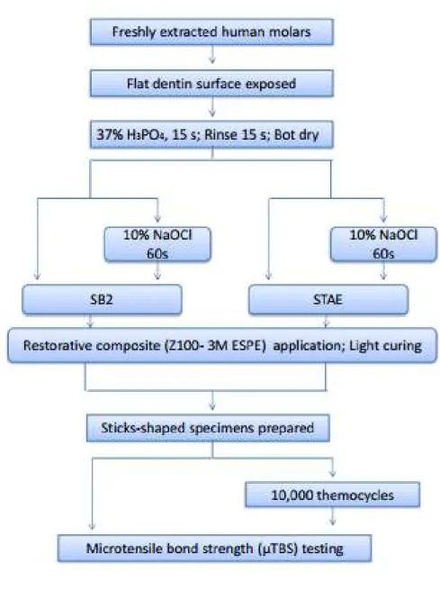
Forty freshly extracted human non-carious third molars were used in this study after obtaining the patients informed consent for their use, under a protocol approved by the local Ethics Committee. The teeth were stored in 0.01% thymol solution at 4°C for no more than 1 month. A flat dentin surface was exposed on each tooth after wet grinding of the occlusal enamel on 100- and 400-grit SiC paper. Dentin surfaces were exposed and inspected under ×80 magnification to ensure that no enamel remnants were left (Leica S8AP0 – Leica Microsystems, Cingapure). The exposed dentin surfaces were further polished on wet 600-grit silicon-carbide paper for 20 s to produce a standardized smear layer. Each tooth was individually fixed to a cutting machine (Isomet 1000, Buehler, Lake Bluff, USA) and sectioned perpendicular to its longitudinal axis using a diamond blade under water cooling, in order to remove the roots and to obtain dentin discs of 4 mm thick. All materials, compositions and manufacturers are presented in Table 1.
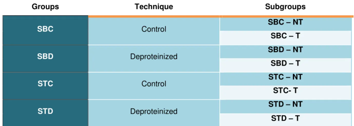
and the adhesive employed: Single Bond 2 - control (SBC), Single Bond 2 - deproteinized (SBD), Stae - control (STC) and Stae - deproteinized (STD)(Table 2).
For groups SBC and STC the adhesives were applied in accordance with manufacturer´s instructions. For groups SBD and STD, 10% NaOCl solution was applied for 60 s after dentin acid etching, rinsed for 60 s, then blot dried before application of the adhesive. Subsequently, a 4-mm layer of composite Z100 (3M-ESPE) was built-up incrementally.
Each bonded tooth was longitudinally sectioned in both “x” and “y” directions, across the bonded interface, using a diamond saw to obtain sticks with cross-sectional area of approximately 0.81 mm2, in accordance with “non-trimming” technique. The sticks of each bonded tooth were randomly subdivided in two subgroups (Table 2) for the microtensile test: immediate (test after 24 h in distilled water at 37ºC) and after thermocycling (Figure 1). For this procedure, the resin-dentin sticks were placed in a thermocycling machine (THE-1100 Thermocycler; SD Mechatronik Gmbh, Feldkirchen-Westerham, Germany) in distilled water baths for 10,000 cycles of 5°C and 55°C with a dwell time of 60 s in each bath.
The specimens were measured individually with digital caliper (Absolute Digimatic, Mitutoyo, Tokyo, Japan) and subjected to a tensile force in a universal testing machine (EMIC, DL2000, São José dos Pinhais, Brazil), fixed with cyanoacrylate glue (Loctite Super Bonder Gel, Diadema, Brazil) and tested under tension at a crosshead speed of 0.5 mm/minute.
Statistical Analysis
All data were expressed as means with their respective standard deviations (SD). Bond strengths data from the 8 experimental groups were statistically analyzed by two-way ANOVA (surface treatment vs. thermocycling) for each
adhesive, with statistical significance set at α= 0.05. Post-hoc multiple comparisons
Failure mode
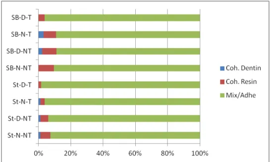
The dentin side of each fractured stick was analyzed using stereoscopic light microscopy (Leica S8AP0 – Leica Microsystems, Cingapure) at 80X magnification and classified according to the failure mode as mixed/adhesive (M/A), cohesive in dentin (CD) or cohesive in composite (CC). Premature failures were recorded but not included in the statistical analysis.
RESULTS
The µTBS means and standard deviations expressed in MPa are reported in Table 3, for Single Bond 2, and in Table 4, for Stae.
ANOVA showed no significant interaction between the two factors for Single Bond 2 (p=0.643) and for Stae (p=0.572). Tukey´s test showed that dentin deproteinization by 10% NaOCl (p<0.001) and thermocycling (p<0.001) significantly decreased the µTBS of Single Bond 2. On the other hand, for Stae the bond strength were unaffected by dentin surface treatment (p= 0.558) and thermocycling (p= 0.194).
The distribution of failure modes for each group is summarized in Figure 2. The analysis of the failure modes revealed mostly mixed/adhesive failures for all groups.
DISCUSSION
The current in vitro study evaluated the influence of thermocycling on the
effectively displace the water that remains into the intertubular dentin after deproteinization, delaying the process of adhesive infiltration on dentin28,49. It is also conceivable that the relatively short dwell time, recommended by the manufacturer, may have been insufficient to allow fully diffusion of the adhesive resin into the porous dentin substrate6,49. Besides, it has been claimed that the high viscosity of Single Bond 2 may compromise its penetration into the collagen mesh and that lower hydrophilicity may result in poor dentin wetting, interfering with in the adhesive infiltration14.
On the other hand, the current experiment showed that deproteinization did not influenced immediately µTBS for the Stae group. This result is in agreement with studies that presented that acetone-based adhesives have greater performance with residual dentinal humidity compared to those diluted with water or ethanol, which show more sensitivity to moisture14, 34, 41, 43, 46, 47 .Besides, the outcome achieved may be due to the higher vapor pressure, the lower viscosity and high volatility of acetone, which is probably sufficient to displace water and allow infiltration of the adhesive monomers into the demineralized and the deproteinized dentin11,14.
Thermocycling aging method decreased the µTBS value only for Single Bond 2 while it has no significant influence on Stae. The maintenance of values shown by Stae after thermocycling can be observed with and without deproteinization. This may be attributed to the thinner adhesive layer using acetone as a solvent which could be less affected by thermocycling. Besides it is possible that when the specimens were submitted to thermocycling under wet conditions, voids presented in the interface between the resin and dentin of Single Bond 2 may lead to a propagation of small cracks and might have significantly reduced the bond strength3.
results. Nevertheless, a relation between the decrease in µTBS associated with deproteinization and the type of adhesive solvent seen in the present study has been described in the literature1,4,10,24,31,33,37. However, this issue is rather controversial and needs to be elucidated once some works have demonstrated that deproteinization had no effect on dentin/resin µTBS 2, 5,34,47.
These results were obtained through different methodologies and materials - such as adhesive compositions, mechanical tests, aging method and deproteinization protocol (time application and concentration of NaOCl)2,19,37,43,45, which may have influenced the outcomes and the comparison among them should be done carefully.
Corroborating with the current results, Saboia et al.34, at a 2-year recall study, reported that the treatment with 10% sodium hypochlorite to remove exposed collagen after acid-etching did not affect the clinical performance of non-carious cervical lesions composite restorations, using both acetone or water/ethanol-based adhesive systems.
It has been searched a new protocol that could enhance the µTBS to dentin leading to restorations more reliable and long lasting. Some studies inhibited the MMP expression through MMP inhibitors instead of removing the collagen by NaOCl application and got better results on improvement of BS28,39. Besides, Feitosa et al.17 tested the meta-phosphoric acid rather than ortho-phosphoric acid
Moreover, the unaffected µTBS values for Stae cannot be associated only with the collagen-depleted treatment, once both no deproteinized and deproteinized groups maintained the µTBS.
Therefore, the use of this technique represents an additional clinical procedure that increases technical complexity and working time. and should be avoided, as there is a little evidence of its effectiveness.
CONCLUSION
The deproteinization showed no positive improvements on the bonding of two-step etch-and-rinse adhesives tested, demonstrating worse results when NaOCl is associated with water/ethanol adhesive.
CLINICAL RELEVANCE
The use of 10% NaOCl for 60 s after dentin demineralization did not improved the bond strength to dentin either immediately or after 10,000 thermal cycles.
REFERENCES
1. Abo T, Asmussen E, Uno S, Tagami J. Short- and long-term in vitro study of the bonding of eight commercial adhesives to normal and deproteinized dentin. Acta Odontol Scand. 2006 Aug;64(4):237-43.
2. Aguilera FS, Osorio R, Osorio E, Moura P, Toledano M . Bonding efficacy of an acetone/based etch-and-rinse adhesive after dentin deproteinization. Med Oral Patol Oral Cir Bucal. 2012 Jul 1;17 (4):e649-54.
4. Arias VG, Bredan-de-Castro AKB, Pimenta LA. Effects of sodium hypochlorite gel and sodium hypochlorite solution on dentin bond strength. J Biomed Mater Res Part B: Appl Biomater 2005 Feb 15;72(2):339-44.
5. Baseggio W, Consolmagno EC, De Carvalho FLN, Ueda JK, Schmitt VL, Formighieri LA, Naufel FS. Effect of deproteinization and tubular occlusion on microtensile bond strength and marginal microleakage of resin composite restorations. J Appl Oral Scie 2009; 17(5):462-6.
6. Borges BC, Souza-Junior EJ, Brandt WC, Loguercio AD, Montes
MA, Puppin-Rontani RM, Sinhoreti MA.
Degree of conversion of simplified contemporary adhesive systems as
influenced by extended air-activated or passive solvent volatilization modes. Oper Dent. 2012 May-Jun;37(3):246-52. doi: 10.2341/11-248-L.
7. Breschi L, Mazzoni A, Ruggeri A, Cadenaro M, Di Lenarda R, De Stefano
Dorigo E. Dental adhesion review: aging and stability of
the bonded interface. Dent Mater. 2008 Jan;24(1):90-101.
8. Correr GM, Alonso RCB, Grando MF, Bonges AFS, Puppin-Rontani RM. Effect of Sodium Hypocholite on primary dentin – a scanning eletron microscopy (SEM) evaluation. J Dent 2006; 34: 454-459
9. De Paula AB, Duque C, Correr-Sobrinho L, Puppin-Rontani RM. Effect of restorative technique and thermal/mechanical treatment on marginal adaptation and compressive strength of esthetic restorations. Oper Dent. 2008 Jul-Aug;33(4):434-40
10. De Souza FB, Delfino CS, Turbino ML, Braz R. Deproteinizes dentin: a favorable substrate to self-bonding resin cements? J Biomed Mater Res Part B: Appl Biomater 2011, 98B: 387–394.
microTBS and SEM. J Biomed Mater Res Part B: Appl Biomater 2005;75B:158–167.
12. Duarte PBPC, Silva EM. Nanoleakage phenomenon on deproteinized human dentin J Appl Oral Sci. 2007;15(4):285-91
13. Erhardt MC, Osorio E, Aguilera FS, Proença JP, Osorio R, Toledano M. Influence of dentin acid-etching and NaOCl-treatment on bond strengths of self-etch adhesives. Am J Dent. 2008 Feb;21(1):44-8.
14. Faria-e-silva AL, Araújo JE, Rocha GP, Oliveira AS, Moraes RR. Solvent content and dentin bond strengths using water-wet, ethanol-wet and deproteinization bonding techniques. Acta Odonto Scand. 2013 May-Jul;71(3-4):710-5.
15. Fawzy AS, Amer MA, El-Askary FS. Sodium Hypocholite as dentin pretreatment for etch-and-rinse single-bottle and two-step self-etching adhesives: atomic force microscope and tensile bond strenght evaluation. J Adhe Res 2008 10-2: 135-144.
16. Fawzy AS. Variations in collagen fibrils network structure and surface dehydration of acid demineralized intertubular dentin: effect of dentin depth and air-exposure time. Dent Mater 2010; 26:35–43.
17. Feitosa VP, Bazzocchi MG, Putignano A, Orsini G, Luzi AL, Sinhoreti MA, Watson TF, Sauro S. Dicalcium phosphate (CaHPO4 _2H2O) precipitation through ortho- or meta-phosphoric acid-etching: Effects on the durability and nanoleakage/ ultra-morphology of resin–dentine interfaces. J Dent. 2013 Nov;41(11):1068-80.
18. Gonçalves LS, Consani S, Sinhoreti MAC, Schneider LFJ, Saboia VPA. Effect of storage and compressive cycles on the bond strength after collagen removal. Oper Dent 2009; 34-36:681-687.
19. Juneja R, Duhan J, Tewari S, Sangwan P, Bhatnagar N.
20. Lahmouzi J, Farache M, Umana M, Compere P, Nyssen-Behets C, Samir N. Influence of Sodium Hypochlorite on Er:YAG Laser-Irradiated Dentin and its Effect on the Quality of Adaptation of the Composite Restoration Margins. Photomed Laser Surg. 2012; 30(11):655-62.
21. Lai SC, Mak YF, Cheung GS, Osorio R, Toledano M, Carvalho RM, et al. Reversal of compromised bonding to oxidized etched dentin. Journal of Dental Research 2001;80:1919–24.
22. Marshall, G.W., Yücel, N., Balooch, M., Kinney, J.H., Habelitz, S, and Marshall, S.J. Sodium hypochlorite alterations of dentin and dentin collagen. Surf. Sci. 2001; 491, 444– 455.
23. Mazzitelli C, Monticelli F, Toledano M, Ferrari M, Osorio R. Dentin treatment effects on the bonding performance of selfadhesive resin cements. Eur. J. Oral. Sci. 2010; 118(1):80-86.
24. Monticelli F, Toledano M, Silva AS, Osorio E, Osorio R. Sealing effectiveness of etch-and-rinse vs self-etching adhesives after water aging: influence of acid etching and NaOCl dentin pretreatment. J Adhes Dent 2008; 10: 183-188.
25. Mountouris, G., Silikas, N., and Eliades, G. Effect of sodium hypochlorite treatment on the molecular composition and morphology of human coronal dentin. J. Adhes. Dent. 2004; 6, 175–182
26. Nagpal R, Tewari S, Gupta R. Effect of various surface treatments on the microleakage and ultrastructure of resin-tooth interface. Oper Dent. 2007; 32(1):16-23.
27. Nakabayashi N, Kojima K, Masuhara E. The promotion of adhesion by the infiltration of monomers into tooth substrates. J Biomed Mater Res 1982; 16:265–273.
28. Pashley DH, Carvalho RM, Tay FR, Agee KA, Lee KW. Solvation of dried dentin matrix by water and other polar solvents. Am J Dent 2002;15(2):97–
29. Prasansuttiporn T, Nakajima M, Kunawarote S, Foxton RM, Tagami J. Effect of reducing agents on bond strenght to NaOCl-treated dentin. Journal of Dental Materials 2011; 27: 229-234.
30. Prasansuttiporn T, Nakajima M, Kunawarote S, Foxton RM, Tagami J. Scrubbing Effect of Self-etching Adhesives on Bond Strength to NaOCl-treated Dentin. J Adhes Dent 2012; 14: 121–127.
31. Prati C, Chersoni S, Pashley DH. Effect of removal of surface collagen fibrils on resin-dentin bonding. Dent. Mater. 1999; 15:323–331.
32. Ravishanker P, Chaitanya K. In vitro evaluation of the effect of deproteinization on the marginal leakage of resin restorations using three bonding agents. Dent Res J (Isfahan). 2012; 9(4):452-9.
33. Ribeiro AIAM, Dantas DCRE, Guênes GMT, Araújo RKP, Cyrillo C, Braz R. Ação dos agentes desproteinizantes e antioxidantes sobre a resistência de união à microtração de sistemas adesivos convencionais. RGO (Porto Alegre) abr.-jun. 2011; 59(2):227-221.
34. Saboia VP, Almeida PC, Rittet AV, Swift EJ Jr, Pimenta LA. 2-year Clinical evaluation of sodium hypochlorite treatment in the restoration of non-carious cervical lesions: a pilot study. Oper Dent. 2006; 31(5):530-5.
35. Saboia VP, Pimenta LA, Ambrosano GM. Effect of collagen removal on microleakage of resin composite restorations. Oper Dent. 2002; 27(1):38-43.
36. Saboia VPA, Nato F, Mazzoni A, Orsini G, Putignano A, Giannini M, Breschi L. Adhesion of a two-step etch-and-rinse adhesive on collagen-depleted dentin. J Adhes Dent 2008; 10: 419-422.
38. Saraceni CH, Liberti E, Navarro RS, Cassoni A, Kodama R, Oda M. Er:YAG-Laser and Sodium Hypochlorite Influence on Bond to Dentin Microsc. Res. Tech. 2013; 76:72–78.
39. Sauro S, Mannocci F, Tay FR, Pashley DH, Cook R, Carpenter GH, Watson TF. Deproteinization Effects of NaOCl on Acid-etched Dentin in Clinically-relevant vs Prolonged Periods of Application. A Confocal and Environmental Scanning Electron Microscopy Study. Oper Dent, 2009; 34-2, 166-173.
40. Sauro, S., Mannocci, F., Toledano, M., Osorio, R., Pashley, D.H., Watson,
T.F. EDTA or H3PO4/NaOCl dentin treatments may increase hybrid layers’
resistance to degradation: a microtensile bond strength and confocal-micropermeability study. J Dent. 2009; 37, 279–288.
41. Shetty S, B M, B S. Dentine deproteinization and microleakage around gingival third resin restorations. J Conserv Dent. 2008 Jan;11(1):11-5.
42. Shinohara MS, Bedran-de-Castro AKB, Amaral CM, Pimenta LAF. The effect of Sodium Hypocholite on microleakage of composite resin restorations using three adhesive systems. J Adhe Dent 2004; 6: 123-127.
43. Silva EM, Duarte PB, Poskus LT, Barcellos AA, Guimarães JG. Nanoleakage and microshear bond strength in deproteinized human dentin. J Biomed Mater Res B Appl Biomater. 2007 May;81(2):336-42.
44. Tjäderhane L, Nascimento FD, Breschi L, Mazzoni A, Tersariol IL, Geraldeli S, Tezvergil-Mutluay A, Carrilho M, Carvalho RM, Tay FR, Pashley DH. Strategies to prevent hydrolytic degradation of the hybrid layer-A review. Dent Mater. 2013 Oct;29(10):999-1011.
45. Torres CR, de Araújo MA, Torres AC. Effects of dentin collagen removal on microleakage of bonded restorations. J Adhes Dent. 2004 Spring;6(1):33-42.
deproteinization technique in non-carious cervical lesions. J Dent. 2014 Jul;42(7):816-23.
47. Uceda-Gómez N, Loguercio AD, Moura SK, Grande RHM, Oda M, Reis A. Long-term bond strength of adhesive systems applied to etched and deproteinized dentin. J Appl Oral Sci. 2007; 15(6): 475-479.
48. Van Landuyt KL, Snauwaert J, De Munck J, Peumans M, Yoshida Y, Poitevin A, et al. Systematic review of the chemical composition of contemporary dental adhesives. J of Biomaterials 2007; 28:3757–85.
49. Wakabayashi Y, Kondou Y, Suzuki K, Yatani H, Yamashita A. Effect of dissolution of collagen on adhesion to dentin. Int J Prosthodont 1994; 7:302–306.
50. Yamazaki PCV, Bedran-Russo AKB, Pereira PNR. Importance of the hibrid layer on the bond strengh of restorations subjected to cyclic loading. J Biomed Mater Res Part B: Appl Biomater 2007; 84B: 291-297.
4. Conclusão Geral
Da avaliação dos resultados obtidos neste trabalho, pode-se concluir que:
5. Referências
1. Abo T, Asmussen E, Uno S, Tagami J. Short- and long-term in vitro study of the bonding of eight commercial adhesives to normal and deproteinized dentin. Acta Odontol Scand. 2006 Aug;64(4):237-43.
2. Aguilera FS, Osorio R, Osorio E, Moura P, Toledano M . Bonding efficacy of an acetone/based etch-and-rinse adhesive after dentin deproteinization. Med Oral Patol Oral Cir Bucal. 2012 Jul 1;17 (4):e649-54.
3. Amaral, F.L., Colucci, V., Palma-Dibb, R.G., Corona, S.A. Assessment of in vitro methods used to promote adhesive interface degradation: a critical review. J Esthet Restor Dent 2007;19:340–354.
4. Arias VG, Bredan-de-Castro AKB, Pimenta LA. Effects of sodium hypochlorite gel and sodium hypochlorite solution on dentin bond strength. J Biomed Mater Res Part B: Appl Biomater 2005 Feb 15;72(2):339-44.
5. Baseggio W, Consolmagno EC, De Carvalho FLN, Ueda JK, Schmitt VL, Formighieri LA, Naufel FS. Effect of deproteinization and tubular occlusion on microtensile bond strength and marginal microleakage of resin composite restorations. J Appl Oral Scie 2009; 17(5):462-6.
6. Breschi L, Mazzoni A, Ruggeri A, Cadenaro M, Di Lenarda R, De Stefano Dorigo E. Dental adhesion review: aging and stability of the bonded interface. Dent Mater. 2008 Jan;24(1):90-101.
7. Correr GM, Alonso RCB, Grando MF, Bonges AFS, Puppin-Rontani RM. Effect of Sodium Hypocholite on primary dentin – a scanning eletron microscopy (SEM) evaluation. J Dent 2006; 34: 454-459.
8. De Souza FB, Delfino CS, Turbino ML, Braz R. Deproteinizes dentin: a favorable substrate to self-bonding resin cements? J Biomed Mater Res Part B: Appl Biomater 2011, 98B: 387–394.
10. Erhardt MC, Osorio E, Aguilera FS, Proença JP, Osorio R, Toledano M. Influence of dentin acid-etching and NaOCl-treatment on bond strengths of self-etch adhesives. Am J Dent. 2008 Feb;21(1):44-8.
11. Faria-e-silva AL, Araújo JE, Rocha GP, Oliveira AS, Moraes RR. Solvent content and dentin bond strengths using water-wet, ethanol-wet and deproteinization bonding techniques. Acta Odonto Scand. 2013 May-Jul;71(3-4):710-5.
12. Fawzy AS. Variations in collagen fibrils network structure and surface dehydration of acid demineralized intertubular dentin: effect of dentin depth and air-exposure time. Dent Mater 2010; 26:35–43.
13. Gale MS, Darvell BW. Thermal cycling procedures for laboratory testing of dental materials restorations. J Dent 1999;27:89–99
14. Gonçalves LS, Consani S, Sinhoreti MAC, Schneider LFJ, Saboia VPA. Effect of storage and compressive cycles on the bond strength after collagen removal. Oper Dent 2009; 34-36:681-687.
15. Lahmouzi J, Farache M, Umana M, Compere P, Nyssen-Behets C, Samir N. Influence of Sodium Hypochlorite on Er:YAG Laser-Irradiated Dentin and its Effect on the Quality of Adaptation of the Composite Restoration Margins. Photomed Laser Surg. 2012; 30(11):655-62.
16. Mountouris, G., Silikas, N., and Eliades, G. Effect of sodium hypochlorite treatment on the molecular composition and morphology of human coronal dentin. J. Adhes. Dent. 2004; 6, 175–182
17. Nagpal R, Tewari S, Gupta R. Effect of various surface treatments on the microleakage and ultrastructure of resin-tooth interface. Oper Dent. 2007; 32(1):16-23.
19. Prasansuttiporn T, Nakajima M, Kunawarote S, Foxton RM, Tagami J. Effect of reducing agents on bond strenght to NaOCl-treated dentin. Journal of Dental Materials 2011; 27: 229-234.
20. Prasansuttiporn T, Nakajima M, Kunawarote S, Foxton RM, Tagami J. Scrubbing Effect of Self-etching Adhesives on Bond Strength to NaOCl-treated Dentin. J Adhes Dent 2012; 14: 121–127.
21. Prati C, Chersoni S, Pashley DH. Effect of removal of surface collagen fibrils on resin-dentin bonding. Dent. Mater. 1999; 15:323–331.
22. Ravishanker P, Chaitanya K. In vitro evaluation of the effect of deproteinization on the marginal leakage of resin restorations using three bonding agents. Dent Res J (Isfahan). 2012; 9(4):452-9.
23. Ribeiro AIAM, Dantas DCRE, Guênes GMT, Araújo RKP, Cyrillo C, Braz R. Ação dos agentes desproteinizantes e antioxidantes sobre a resistência de união à microtração de sistemas adesivos convencionais. RGO (Porto Alegre) abr.-jun. 2011; 59(2):227-221.
24. Saboia VP, Almeida PC, Rittet AV, Swift EJ Jr, Pimenta LA. 2-year Clinical evaluation of sodium hypochlorite treatment in the restoration of non-carious cervical lesions: a pilot study. Oper Dent. 2006; 31(5):530-5.
25. Saboia VP, Pimenta LA, Ambrosano GM. Effect of collagen removal on microleakage of resin composite restorations. Oper Dent. 2002; 27(1):38-43. 26. Saboia VPA, Nato F, Mazzoni A, Orsini G, Putignano A, Giannini M, Breschi
L. Adhesion of a two-step etch-and-rinse adhesive on collagen-depleted dentin. J Adhes Dent 2008; 10: 419-422.
27. Santos PH, Sinhoreti MA, Consani S, Sobrinho LC, Adabo GL, Vaz LG. Effect of cyclic compressive loading on the bond strength of an adhesive system to dentin after collagen removal. J Adhes Dent. 2005 Summer;7(2):127-31
29. Sauro S, Mannocci F, Tay FR, Pashley DH, Cook R, Carpenter GH, Watson TF. Deproteinization Effects of NaOCl on Acid-etched Dentin in Clinically-relevant vs Prolonged Periods of Application. A Confocal and Environmental Scanning Electron Microscopy Study. Oper Dent, 2009; 34-2, 166-173.
30. Shetty S, B M, B S. Dentine deproteinization and microleakage around gingival third resin restorations. J Conserv Dent. 2008 Jan;11(1):11-5.
31. Shinohara MS, Bedran-de-Castro AKB, Amaral CM, Pimenta LAF. The effect of Sodium Hypocholite on microleakage of composite resin restorations using three adhesive systems. J Adhe Dent 2004; 6: 123-127.
32. Silva EM, Duarte PB, Poskus LT, Barcellos AA, Guimarães JG. Nanoleakage and microshear bond strength in deproteinized human dentin. J Biomed Mater Res B Appl Biomater. 2007 May;81(2):336-42.
33. Tjäderhane L, Nascimento FD, Breschi L, Mazzoni A, Tersariol IL, Geraldeli S, Tezvergil-Mutluay A, Carrilho M, Carvalho RM, Tay FR, Pashley DH. Strategies to prevent hydrolytic degradation of the hybrid layer- A review. Dent Mater. 2013 Oct;29(10):999-1011.
34. Torres CR, Barcellos DC, Batista GR, Pucci CR, Antunes MJ, de La Cruz DB, Borges AB. Five-year clinical performance of the dentine deproteinization technique in non-carious cervical lesions. J Dent. 2014 Jul;42(7):816-23. doi: 10.1016/j.jdent.2014.04.004. Epub 2014 Apr
35. Uceda-Gómez N, Loguercio AD, Moura SK, Grande RHM, Oda M, Reis A. Long-term bond strength of adhesive systems applied to etched and deproteinized dentin. J Appl Oral Sci. 2007; 15(6): 475-479.
37. Yamazaki PCV, Bedran-Russo AKB, Pereira PNR. Importance of the hibrid layer on the bond strengh of restorations subjected to cyclic loading. J Biomed Mater Res Part B: Appl Biomater 2007; 84B: 291-297.
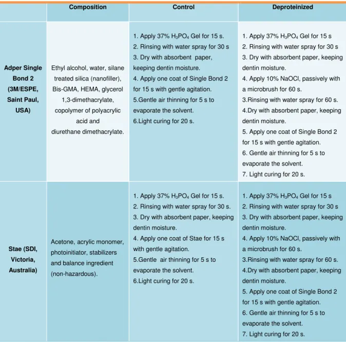
Table 1: Information about the adhesives, their application protocols and strategy of application.
Composition Control Deproteinized
Adper Single Bond 2 (3M/ESPE, Saint Paul,
USA)
Ethyl alcohol, water, silane treated silica (nanofiller), Bis-GMA, HEMA, glycerol
1,3-dimethacrylate, copolymer of polyacrylic
acid and diurethane dimethacrylate.
1. Apply 37% H3PO4 Gel for 15 s. 2. Rinsing with water spray for 30 s 3. Dry with absorbent paper, keeping dentin moisture.
4. Apply one coat of Single Bond 2 for 15 s with gentle agitation. 5.Gentle air thinning for 5 s to evaporate the solvent. 6.Light curing for 20 s.
1. Apply 37% H3PO4 Gel for 15 s 2. Rinsing with water spray for 30 s 3. Dry with absorbent paper, keeping dentin moisture.
4. Apply 10% NaOCl, passively with a microbrush for 60 s.
3.Rinsing with water spray for 60 s. 4.Dry with absorbent paper, keeping dentin moisture.
5. Apply one coat of Single Bond 2 for 15 s with gentle agitation. 6. Gentle air thinning for 5 s to evaporate the solvent. 7. Light curing for 20 s.
Stae (SDI, Victoria, Australia)
Acetone, acrylic monomer, photoinitiator, stabilizers and balance ingredient (non-hazardous).
1. Apply 37% H3PO4 Gel for 15 s. 2. Rinsing with water spray for 30 s. 3. Dry with absorbent paper, keeping dentin moisture.
4. Apply one coat of Stae for 15 s with gentle agitation.
5.Gentle air thinning for 5 s to evaporate the solvent. 6.Light curing for 20 s.
1. Apply 37% H3PO4 Gel for 15 s 2. Rinsing with water spray for 30 s 3. Dry with absorbent paper, keeping dentin moisture.
4. Apply 10% NaOCl, passively with a microbrush for 60 s.
3.Rinsing with water spray for 60 s. 4.Dry with absorbent paper, keeping dentin moisture.
Table 2: Division of specimens on groups according to the treatment of dentin, the adhesive used and aging regimen (n=5).
Groups Technique Subgroups
SBC Control SBC – NT
SBC – T
SBD Deproteinized SBD – NT
SBD – T
STC Control STC – NT
STC- T
STD Deproteinized STD – NT
STD – T
Table 3: Mean and standard deviations of microtensile bond strength of water/ethanol-based system (BS ± SD).
Dentin treatment No thermocycling Thermocycling Mean
No deproteinized 34,7 ± 4,4 25,8 ± 3,1 30,2a
Deproteinized 25,2 ± 4,5 17,9 ± 2,9 21,5b
Mean 29,9A 21,8B
Distinct capital letters represent statistical significant difference between with or without thermocycling (p<0.05). Distinct lowercase letters represent statistical significant difference between dentin treatments (p<0.05).
Table 4: Mean and standard deviations of microtensile bond strength of acetone-based system (BS ± SD).
Distinct capital letters represent statistical significant difference between with or without thermocycling (p<0.05). Distinct lowercase letters represent statistical significant difference between dentin treatments (p<0.05).
Figure 2: Distribution of failure modes among groups.
Dentin treatment No thermocycling Thermocycling Mean
No deproteinized 19,2 ± 6,7 17,2 ± 6,2 18,2a
Deproteinized 19,2 ± 3,7 14,2 ± 6,0 16,7a