279 Radiol Bras. 2013 Set/Out;46(5):279–283
Role of Doppler ultrasonography evaluation of superior
mesenteric artery flow volume in the assessment of Crohn’s
disease activity
*
Contribuição da medida do volume de fluxo da artéria mesentérica superior pelo Doppler na caracterização da atividade inflamatória em pacientes com doença de Crohn
Fabiana Paiva Martins1, Eduardo Garcia Vilela2, Maria de Lourdes Abreu Ferrari2, Henrique Osvaldo da Gama Torres2, Juliana Brovini Leite3, Aloísio Sales da Cunha4
Objective: To investigate superior mesenteric artery flow measurement by Doppler ultrasonography as a means of characterizing inflammatory activity in Crohn’s disease. Materials and Methods: Forty patients were examined and divided into two groups – disease activity and remission – according to their Crohn’s disease activity index score. Mean superior mesenteric artery flow volume was calculated for each group and correlated with Crohn’s disease activity index score. Results: The mean superior mesenteric artery flow volume was significantly greater in the patients with active disease (626 ml/min ± 236 × 376 ml/min ± 190; p = 0.001). As a cut off corresponding to 500 ml/min was utilized, the superior mesenteric artery flow volume demonstrated sensitivity of 83% and specificity of 82% for the diagnosis of Crohn’s disease activity. Conclusion: The present results suggest that patients with active Crohn’s disease have increased superior mesenteric artery flow volume as compared with patients in remission. Superior mesenteric artery flow measurement had a good performance in the assessment of disease activity in this study sample.
Keywords: Doppler ultrasonography; Superior mesenteric artery flow; Crohn’s disease; Inflammatory activity.
Objetivo: Avaliar a medida do volume de fluxo da artéria mesentérica superior pelo Doppler como método para carac-terizar a atividade inflamatória em pacientes com doença de Crohn. Materiais e Métodos: Quarenta pacientes foram submetidos ao exame e divididos em dois grupos – doença em atividade e remissão –, de acordo com o índice de ati-vidade da doença de Crohn. Foram estabelecidas as médias do volume de fluxo da artéria mesentérica superior e feita a correlação com o escore do índice de atividade da doença de Crohn. Resultados: A média do volume de fluxo da artéria mesentérica superior foi significativamente maior nos pacientes com doença em atividade (626 ml/min ± 236 × 376 ml/min ± 190; p = 0,001). Utilizando-se o ponto de corte de 500 ml/min, a medida apresentou sensibilidade de 83% e especificidade de 82% para o diagnóstico de atividade inflamatória. Conclusão: Os resultados sugerem que pacientes com doença de Crohn em atividade apresentam um aumento do volume de fluxo da artéria mesentérica superior. O teste apresentou bom desempenho na avaliação da atividade inflamatória nesta amostra de pacientes.
Unitermos: Ultrassom Doppler; Fluxo da artéria mesentérica superior; Doença de Crohn; Atividade inflamatória. Abstract
Resumo
* Study developed at the School of Medicine, Universidade Federal de Minas Gerais (UFMG), Belo Horizonte, MG, Brazil.
1. Master, Assistant Professor, Department of Anatomy and Imaging, School of Medicine of Universidade Federal de Minas Gerais (UFMG), Belo Horizonte, MG, Brazil.
2. PhDs, Associate Professors, Department of Medical Prac-tice, School of Medicine of Universidade Federal de Minas Ge-rais (UFMG), Belo Horizonte, MG, Brazil.
3. Master, Biochemist, Hospital e Maternidade Terezinha de Jesus, Juiz de Gora, MG, Brazil.
4. Private Docent, Titular Professor of Medical Practice at School of Medicine, Universidade Federal de Minas Gerais (UFMG), Belo Horizonte, MG, Brazil.
Martins FP, Vilela EG, Ferrari MLA, Torres HOG, Leite JB, Cunha AS. Role of Doppler ultrasonography evaluation of superior mesenteric artery flow volume in the assessment of Crohn’s disease activity. Radiol Bras. 2013 Set/Out;46(5):279–283.
well-being sensation, and present poor cor-relation with endoscopic and histopatho-logical signs of disease activity. Biochemi-cal tests may be more objective, but are not more specific than clinical indices. Endo-scopic methods are invasive, costly and time consuming, so their utilization as a routine for all patients is not feasible, par-ticularly in cases where serial examinations are necessary(1,2). Computed tomography
enterography has demonstrated to be effi-cient in the evaluation of inflammatory activity, but, as this method relies on ion-izing radiation, its indication is limited in the context of frequent recurrences(3,4).
though effective in relieving symptoms and in improving the quality of life, is not cura-tive. The evaluation of the inflammatory activity is of paramount importance, as the delay in instituting the treatment may lead to complications.
The ideal marker of inflammatory activ-ity at CD is still to be determined. The avail-able clinical indices rely on subjective symptoms such as abdominal pain and
INTRODUCTION
Crohn’s disease (CD) is a chronic recur-rent inflammatory disorder which affects the gastrointestinal tract. Its treatment,
al-Mailing Address: Dra. Fabiana Paiva Martins. Avenida Pro-fessor Alfredo Balena, 189/803, Centro. Belo Horizonte, MG, Brazil, 30130-100. E-mail: fabpaivamartins@gmail.com.
Magnetic resonance enterography provides results comparable to those of computed tomography enterography(1), however it is
still scarcely available in our country. Microvascular changes characterized by vascular injuries or infarct are observed in small bowel resection specimens from pa-tients with CD, and vary according to the inflammatory reaction intensity, resulting in neovascularization(5,6), which could also be
observed in angiographic studies(7,8),
prob-ably reflecting an increase in blood flow to the affected bowel segments. With the pub-lication of studies demonstrating the viabil-ity of Doppler ultrasonography (US) for the evaluation of the superior mesenteric artery (SMA) flow(9–11), there has been an
increas-ing interest in the splanchnic hemodynam-ics evaluation in CD by means of Doppler US, considering its noninvasiveness, safety, low-cost and capability for quanti-tative analysis.
MATERIALS AND METHODS
Patients
A group of 42 patients previously diag-nosed with CD with small bowel involve-ment, defined according to clinical, endo-scopic, radiological, surgical and histo-pathological(12) criteria, were prospectively
evaluated. All the patients originated from the Bowel Clinic of Instituto Alfa de
Gas-troenterologia – Hospital das Clínicas da Universidade Federal de Minas Gerais (UFMG), recruited between October 2006 and November 2009. The study was ap-proved by the Committee for Ethics in Research of UFMG (ETIC 87/08). All pa-tients signed a term of free and informed consent before their inclusion in the study. The patients were divided into two groups on the basis of the score from the Crohn’s disease activity index – CDAI (Table 1) as a reference standard(13): group
1 – comprising patients with CDAI < 150, characterizing disease in remission; group 2 – comprising patients with CDAI > 150, characterizing active disease. Pregnant pa-tients and those with severe coexisting dis-orders of cardiopulmonary or renal origin were excluded from the study.
The patients were clinically evaluated according to the routine of the outpatient clinic and underwent Doppler US before any change of the medical approach. The scans were blindly performed, with the observer being unaware of the patients’ clinical history/classification.
Once the patients had undergone the scan, their records were reviewed for clini-cal and laboratory data collection. The pa-tients were classified according to their ages at the time of the diagnosis, site and form of disease according to the Vienna classification(14) (Table 2).
Doppler US of SMA
The scans were performed by an expe-rienced radiologist specialized in Doppler US, utilizing a duplex Doppler US ATL 5000 Sono-CT model apparatus (Philips Medical Systems; Best, The Netherlands), equipped with a 2–5 MHz multi-frequency convex transducer.
The patients were examined in the morning, after fasting for eight hours. The SMA was sagittal scanned, with high defi-nition zooming and 5 kHz pulse repetition. The Doppler sampling volume was posi-tioned 2–3 cm distally from the vessel ori-gin, before the emergence of its branches and adjusted to comprise the vascular lu-men, but without touching its walls. A spec-trum of at least five cardiac cycles was obtained during breath hold, and the mean flow velocity was automatically deter-mined by the equipment. The insonation angle was determined on the real time B-mode image and always maintained below 60°. The measurement of the SMA diam-eter was utilized by the equipment for de-termining the vessel area and automatically calculating the SMA flow volume (Figure 1). With the objective of reducing random errors, each measurement was repeated for three times, and the mean value was con-sidered as the final result(15).
After Doppler US, the patients were submitted to total abdominal US studies,
Table 1 Crohn’s disease activity index (CDAI). Variable
1) Number of liquid or liquid-pasty stools in the last seven days... 2) Classification of abdominal pain/colic in the last seven days (0 = no pain; 1 = mild pain; 2 = moderate pain; 3 = intense pain)... 3) Well-being sensation in the last seven days (0 = well; 1 = regular; 2 = bad; 3 = very bad; 4 = lousy)... 4) Afecções relacionadas com a DC...
– arthritis or arthralgia – iritis/uveitis
– erythema nodosum or pyoderma gangrenosum or aphthous stomatitis – anal fissure or fistula or perianal abscess
– other bowel-related fistulae
– fever above 37.8°C during the last seven days
5) Antidiarrheal therapy (0 = none; 1 = yes)... 6) Abdominal mass (0 = none; 2 = questionable; 5 = unquestionable)... 7) Hematocrit...
– male: 47 – Hct – female: 42 – Hct
8) Weight... – standard weight – actual body weight × 100/standard weight
Multiplier factor
2
5
7 20
30 10 6
1
Subtotal
= ___________
= ___________
= ___________ = ___________
= ___________ = ___________ = ___________ Add or subtract
= ___________ Add or subtract
with special attention to the evaluation of thickening of small bowel walls (> 4 mm) and to the presence of fistulas and ab-scesses(16,17).
Statistical analysis
The comparison between the two groups was carried out with the by utiliz-ing the chi-squared test and the Fisher’s exact test for dichotomous variables and the Student’s t-test for continuous vari-ables. The considered significance level was 5% (p < 0.05). The evaluation of the test performance for characterization of in-flammatory activity in CD was carried out by obtaining the receiver operating charac-teristic curve (ROC) and selection of a
cut-off point, with determination of sensitivity, specificity, positive and negative predictive values in relation to the reference standard.
RESULTS
Two patients were excluded from the analysis for not presenting with satisfactory technical conditions to be submitted to Doppler US of SMA, because of excessive abdominal distension. Thus, group 1 com-prised 28 patients (70%) with CD in remis-sion, and group 2 comprised 12 patients (30%) with active disease evaluated ac-cording to the CDAI score.
Abdominal US demonstrated alterations characterized by ileum wall thickening,
presence of entero-enteral fistula and cavi-tary collection in 16 patients (40%), which were the changes most frequently observed in group 2 (p = 0.037). On average, the SMA flow volume was significantly greater in group 2 (626 ml/min ± 236) as compared with group 1 (376 ml/min ± 190), with p = 0.001 (Figure 2).
The area under the ROC curve for the SMA flow volume was 0.833 (p = 0.001). The value of 500 ml/min was adopted as the cutoff point, since such value is consid-ered in literature as the upper limit of nor-mality for healthy individuals18–20) and is
also utilized for evaluation of the test per-formance. In the present study, the mea-surement of the SMA flow volume at Dop-pler US presented a sensitivity of 83% and specificity of 82% for the diagnosis of in-flammatory activity. There was also statis-tically significant association between al-tered result of SMA flow volume measure-ment and the presence of inflammatory activity in CD (p = 0.0001).
DISCUSSION
The presence of hemodynamic changes in patients with CD has already been pre-viously demonstrated by angiography(7,8)
and studies with radionuclides(21),
charac-terized by a hyperdynamic state, probably caused by vascular congestion, stasis and neovascularization. From the physiopatho-logical point-of-view, it would be reason-able to assume that such changes imply an increase in the SMA flow volume at Dop-pler US, a hypothesis already admitted in other studies.
Although some authors assert that sults in the literature are conflicting as re-gards the association between changes ob-served at Doppler US of SMA and presence of inflammatory activity in CD(22,23), the
present study authors believe that such dis-crepancies are mostly due to crucial differ-ences in the utilized method, notably in the parameters adopted for Doppler US of SMA. Undoubtedly, among the already evaluated parameters, the SMA flow vol-ume stands out as that with best correlation with inflammatory activity(24,25).
As previously observed by other au-thors(24–26), the present study demonstrates
that patients with active CD presented
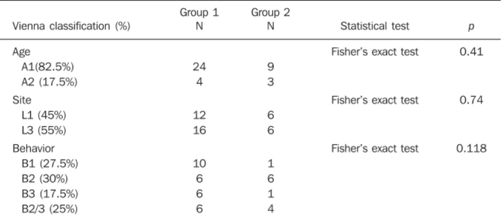
sig-Table 2 Vienna classification in the patient groups with CD in remission (group 1) and active disease (group 2).
Vienna classification (%)
Age A1(82.5%) A2 (17.5%) Site
L1 (45%) L3 (55%) Behavior
B1 (27.5%) B2 (30%) B3 (17.5%) B2/3 (25%)
Group 1 N
24 4
12 16
10 6 6 6
Group 2 N
9 3
6 6
1 6 1 4
Statistical test
Fisher’s exact test
Fisher’s exact test
Fisher’s exact test
p
0.41
0.74
0.118
A1: < 40 years; A2: > 40 years; L1: terminal ileum; L3: ileocolon; B1: non-stenosing, non-penetrating; B2: stenosing; B3: penetrating.
nificantly greater SMA flow volume than patients with disease in remission. Addi-tionally, as the value of 500 ml/min is con-sidered as the cutoff point, the authors of the present study observed sensitivity of 83% and specificity of 82% in the diagnosis of inflammatory activity, values which are similar to those reported by other studies. Some authors(25,27) question the
useful-ness of the SMA flow volume measurement for characterizing inflammatory activity in CD, even after finding significant associa-tion between increased flow and clinical and laboratory signs of activity, due to the existence of an overlapping range of flow volume values between the patients with active CD and patients with the disease in remission, close to the cutoff point of 500 ml/min. Such range was observed by van Oostayen et al.(28,29) and defined as a “gray
zone” of values between 450 and 600 ml/ min, where the test would be less useful.
In the present study, the authors ob-served seven patients with the flow volume within such “gray zone” of values. One might assume that patients presenting with less intense inflammatory activity or those
who normally have a greater flow volume would be in such zone; it is important to remind that CDAI, although being widely utilized, presents many limitations in the evaluation of inflammatory activity(13). In
the present study, there were two patients with false-negative results. One of them had a CDAI score of 180 and flow volume of 469 ml/min (therefore within the “gray zone”) and had been previously submitted to partial ileocolic enterectomy, which can explain the absence of increased SMA flow volume. The other patient had a CDAI score of 206 and the flow volume of 259 ml/min; despite the absence of an explana-tion for such a discrepancy, the patient pre-sented normal C-reactive protein levels, a fact which allows the authors to assume that perhaps the increased CDAI might sult from subjective symptoms and not re-lated to inflammatory activity, an influence which is recognized in the literature(13).
The main limitation of the present study was the sample size, which led to wide confidence intervals in the measurement of the test performance. Additionally, further studies with prospective evaluation of
pa-tients before and after treatment may be useful to elucidate some of the assumptions raised in the discussion.
CONCLUSION
The mean values for SMA flow volume where higher in patients with active CD, and a statistically significant association was observed between the altered results of the test and the inflammatory activity evaluated by the CDAI score. Because of its noninvasiveness, safety and low-cost, Doppler US of SMA can be useful in the evaluation of the inflammatory activity in patients with CD.
Acknowledgements
The authors wish to thank Instituto Hermes Pardini, for having provided the facilities for performing SMA Doppler US studies at no cost to patients and investiga-tors.
REFERENCES
1. Vilela EG, Torres HOG, Martins FP, et al. Evalu-ation of inflammatory activity in Crohn’s disease and ulcerative colitis. World J Gastroenterol. 2012;18:872–81.
2. Nikolaus S, Schreiber S. Diagnostics of inflam-matory bowel disease. Gastroenterology. 2007; 133:1670–89.
3. Booya F, Fletcher JG, Huprich JE, et al. Active Crohn disease: CT findings and interobserver agreement for enteric phase CT enterography. Radiology. 2006;241:787–95.
4. Jaffe TA, Gaca AM, Delaney S, et al. Radiation doses from small-bowel follow-through and abdominopelvic MDTC in Crohn’s disease. AJR Am J Roentgenol. 2007;189:1015–22. 5. Brahme F, Lindstöm C. A comparative
radio-graphic and pathological study of intestinal vaso-architecture in Crohn’s disease and in ulcerative colitis. Gut. 1970;11:928–40.
6. Wakefield AJ, Sawyerr AM, Dhillon AP, et al. Pathogenesis of Crohn’s disease: multifocal gas-trointestinal infarction. Lancet. 1989;2:1057–62. 7. Boijsen E, Reuter SR. Mesenteric angiography in the evaluation of inflammatory and neoplastic disease of the intestine. Radiology. 1966;87: 1028–36.
8. Lunderquist A, Knutsson H. Angiography in Crohn’s disease of the small bowel and colon. Am J Roentgenol Radium Ther Nucl Med. 1967;101: 338–44.
9. Jäger K, Bollinger A, Valli C, et al. Measurement of mesenteric blood flow by duplex scanning. J Vasc Surg. 1986;3:462–9.
10. Moneta GL, Taylor DC, Helton WS, et al. Duplex ultrasound measurement of postprandial intesti-nal blood flow: effect of meal composition. Gas-troenterology. 1988;95:1294–301.
11. Sato S, Ohnishi K, Sugita S, et al. Splenic artery and superior mesenteric artery blood flow: non-Figure 2. Boxplot of the comparison of the SMA flow volume in patients with CD in remission (group 1)
surgical Doppler US measurement in healthy sub-jects and patients with chronic liver disease. Ra-diology. 1987;164:347–52.
12. Gollop JH, Phillips SF, Melton LJ 3rd, et al. Epi-demiologic aspects of Crohn’s disease: a popula-tion based study in Olmsted county, Minnesota, 1943-1982. Gut. 1988;29:49–56.
13. Best WR, Becktel JM, Singleton JW, et al. Devel-opment of Crohn’s disease activity index. Gastro-enterology. 1976;70:439–44.
14. Gasche C, Scholmerich J, Brinskov J, et al. A simple classification of Crohn’s disease: report of the Working Party for the World Congress of Gastroenterology, Vienna 1998. Inflam Bowel Dis. 2000;6:8–15.
15. Perko MJ, Just S. Duplex ultrasound of superior mesenteric artery: interobserver variability. J Ul-trasound Med. 1993;12:259–63.
16. Maconi G, Radice E, Greco S, et al. Bowel ultra-sound in Crohn’s disease. Best Pract Res Clin Gastroenterol. 2006;20:93–112.
17. Tarján Z, Tóth G, Györke T, et al. Ultrasound in Crohn’s disease of the small bowel. Eur J Radiol. 2000;35:176–82.
18. Cooper AM, Braatvedt GD, Qamar MI, et al. Fast-ing and postprandial splanchnic blood flow is reduced by a somatostatin analogue (octreotide) in man. Clin Sci. 1991;81:168–75.
19. Qamar MI, Read AE, Skidmore R, et al. Transcu-taneous Doppler ultrasound measurement of su-perior mesenteric artery blood flow in man. Gut. 1986;27:100–5.
20. Nakamura T, Moriyasu F, Ban N, et al. Quantita-tive measurement of abdominal arterial blood flow using image-directed Doppler ultrasonogra-phy: superior mesenteric, splenic and common hepatic arterial blood flow in normal adults. J Clin Ultrasound. 1989;17:261–8.
21. Giaffer MH, Tindale WB, Senior S, et al. Quanti-fication of disease activity in Crohn’s disease by computer analysis of Tc-99m hexamethyl propy-lene amine oxime (HMPAO) labeled leucocyte images. Gut. 1993;34:68–74.
22. Maconi G, Parente F, Bollani S, et al. Factors af-fecting splanchnic haemodynamics in Crohn’s disease: a prospective controlled study using Doppler ultrasound. Gut. 1998;43:645–50. 23. Maconi G, Parente F, Bollani S, et al. Abdominal
ultrasound in the assessment of extent and activ-ity of Crohn’s disease: clinical significance and implication of bowel wall thickening. Am J Gastroenterol, 1996;91:1604–9.
24. van Oostayen JA, Wasser MN, van Hogezand RA, et al. Activity of Crohn disease assessed by mea-surement of superior mesenteric artery flow with Doppler US. Radiology. 1994;193:551–4.
25. Byrne MF, Farrell MA, Abass S, et al. Assessment of Crohn’s disease activity by Doppler sonography of the superior mesenteric artery, clinical evaluation and the Crohn’s disease activ-ity index: a prospective study. Clin Radiol. 2001;56:973–8.
26. Erden A, Cuhmur T, Ölçer T. Superior mesenteric artery Doppler waveform changes in response to inflammation of the ileocecal region. Abdom Imaging. 1997;22:483–6.
27. Sjekavica I, Barbaric-Babic V, Krznaric Z, et al. Assessment of Crohn’s disease activity by Dop-pler ultrasound of superior mesenteric artery and mural arteries in thickened bowel wall: cross-sec-tional study. Croat Med J. 2007;48:822–30. 28. van Oostayen JA, Wasser MNJM, van Hogezand
RA, et al. Doppler sonography evaluation of su-perior mesenteric artery flow to assess Crohn’s disease activity: correlation with clinical evalua-tion, Crohn’s disease activity index, and alpha-1-antitrypsin clearance in feces. AJR Am J Roentgenol. 1997;168:429–43.