Crohn’s disease activity assessed by doppler
sono-graphy: the role of aortic flow parameters
Thais Guarana´ Andrade,I Homero Soares Fogac¸a,I Celeste Carvalho Siqueira Elia,I Melissa Tassano Pitrowsky,IHeitor Siffert Pereira de SouzaI,II
IUniversidade Federal do Rio de Janeiro, Hospital Universita´rio, Servic¸o de Gastroenterologia and Laborato´rio Multidisciplinar de Pesquisa, Rio de Janeiro/
RJ, Brazil.IIUniversidade Federal do Rio de Janeiro, Departamento de Clı´nica Me´dica, Rio de Janeiro/RJ, Brazil.
OBJECTIVES:Intestinal neovascularization and abnormal abdominal arterial flow rates have been reported in Crohn’s disease. The aim of this study was to evaluate Doppler sonography as a method for assessing Crohn’s disease activity based on changes in splanchnic hemodynamics.
METHODS:Forty-eight patients with Crohn’s disease, 22 healthy volunteers and 12 patients with irritable bowel syndrome were evaluated by Doppler ultrasound for flow parameters of the aorta and superior mesenteric artery. This evaluation included the cross-sectional area, maximum flow volume, peak systolic velocity, end diastolic velocity, resistance and the pulsatility index. Disease activity was classified according to the Crohn’s disease activity index.
RESULTS:Most measurements in the aorta and superior mesenteric artery were significantly different between Crohn’s disease patients and both control groups. Only the aortic maximum flow volume (CC = 0.37,p= 0.009) and aortic peak systolic velocity (CC = 0.30,p= 0.035) showed a significant positive correlation with the Crohn’s disease activity index. The determination of cut-off points for the aortic maximum flow volume and peak systolic velocity measurements increased the sensitivity (80 and 75% for flow volume and velocity, respectively), specificity (57 and 75%), accuracy (67 and 75%) and positive (57 and 68%) and negative (80 and 81%) predictive values. These cut-off values permitted the correct classification of most of the patients with Crohn’s disease with respect to disease activity. None of the superior mesenteric artery measurements were able to discriminate patients in relation to disease activity.
CONCLUSION: The aortic maximum flow volume and peak systolic velocity levels estimated by Doppler sonography reflected disease activity in Crohn’s disease. Doppler sonography of the aorta is therefore a novel noninvasive adjunct method that may be useful in the clinical follow-up of patients with Crohn’s disease.
KEYWORDS: Crohn’s disease; Doppler Sonography; Inflammatory Bowel Diseases; Splanchnic Hemodynamics.
Andrade TG, Fogac¸a HS, Elia CCS, Pitrowsky MT, Souza HSP. Crohn’s disease activity assessed by doppler sonography: the role of aortic flow parameters. Clinics. 2013;68(4):457-462.
Received for publication onOctober 10, 2012;First review completed onNovember 15, 2012;Accepted for publication onDecember 7, 2012 E-mail: [email protected]; [email protected]
Tel.: 55 21 2562-2669
& INTRODUCTION
Crohn’s disease (CD) is characterized by recurrent inflammation of the gastrointestinal tract with inappropriate or delayed healing, and it affects genetically susceptible individuals who are exposed to environmental risk factors (1). Although great advances have been made recently in the understanding of CD etiopathogenesis (2,3), the medical management of CD continues to be challenging. The objective assessment of disease activity and response to
therapy has become progressively more critical for indivi-dualized and optimized care, as the decision of whether and when to initiate novel therapies may alter the natural course of CD (4).
Serial assessment of CD activity is regarded as a fundamental tool in the follow-up of patients and allows the identification of specific conditions that may influence clinical decisions. Several methods have been used to investigate CD activity, but there is no consensus reference method (5,6). Even the Crohn’s disease activity index (CDAI), which is the most used and cited reference method, has limitations because of the subjectivity of some of its criteria (7). Endoscopic methods are commonly used for the diagnosis and follow-up of CD patients; however, these methods are invasive, expose patients to some potential risks, usually require hospitalization, are expensive and still have some limitations depending on the disease location (8). Therefore, a new alternative method should be simple, Copyrightß2013CLINICS– This is an Open Access article distributed under
the terms of the Creative Commons Attribution Non-Commercial License (http:// creativecommons.org/licenses/by-nc/3.0/) which permits unrestricted non-commercial use, distribution, and reproduction in any medium, provided the original work is properly cited.
No potential conflict of interest was reported.
reproducible and noninvasive to monitor disease activity and response to therapy in CD; such a new method could become an invaluable adjunct to clinical practice.
Recent advances in vascular biology have defined a key role for the microcirculation in both the initiation and perpetuation of inflammatory bowel diseases (IBD), includ-ing CD. In particular, angiogenesis has been detected in surgical samples of both the small and large bowel affected by CD (9). In addition to histopathologic changes, CD is also accompanied by remarkable changes in the splanchnic circulation, as demonstrated by imaging studies (10,11).
Doppler sonography allows the assessment of the splanchnic circulation and permits the measurement of various parameters in the mesenteric arterial and venous vasculature. In recent years, several studies with Doppler sonography have been conducted to evaluate splanchnic hemodynamics in CD (12-14). As an adjunct for CD evaluation, preliminary Doppler sonography studies have focused on the potential relationship between disease activity and intestinal blood flow data, typically using flow parameters of the superior mesenteric artery (SMA). However, these studies have produced conflicting results (15-21).
The aim of this prospective study was to evaluate the potential application of Doppler sonography of the abdom-inal aorta and SMA as a method for assessing CD disease activity.
& PATIENTS AND METHODS
The study protocol was approved by the Ethical Committee of the University Hospital of Federal University of Rio de Janeiro and was therefore performed in accordance with the ethical standards described in the 1964 Declaration of Helsinki. All subjects gave their informed consent prior to inclusion in the study.
Study population
Forty-eight consecutive patients with Crohn’s disease (CD) involving the ileum and/or the right colon were enrolled in this study. Clinical diagnosis of CD was confirmed by the combination of radiological, endoscopic and histological criteria. Twelve patients with irritable bowel syndrome (IBS) comprised the control group. A third group composed of 22 healthy individuals, matched for age and gender, was included for comparative analysis.
The patients with CD consisted of 28 females and 20 males, with a mean age of 38 years (range: 18-57 years). According to the Montreal Classification (22), most (80.5%) CD patients are diagnosed before the age of 40 years. The classification of disease activity was determined by a combination of a history, physical examination, laboratory investigation, colonoscopy, barium study and Crohn’s disease activity index (CDAI) for each patient (23). The disease was categorized as active if the CDAI was$150 and inactive if the CDAI was,150. The mean value observed for disease activity was 102.9 (range 16-264). Twenty-four subjects were in remission at the time of the study, whereas the other 24 had moderately to severely active CD (score of
$150); none of the patients had a CDAI score above 450. To establish the most homogeneous CD group and to minimize potential confounding physical factors, only patients with ileum and/or right colon involvement were selected for analysis; patients with any history of prior
extensive surgery involving the small bowel or colon (extensive ileal resection with ileo-transverse-anastomosis and right hemicolectomy) were excluded from this study. The disease location was ileocolonic in 55.3% of the patients, restricted to the right colon in 27.9% and exclusively in the terminal ileum in 16.8%. Regarding disease behavior, the prevalence of predominantly penetrating, stricturing and nonpenetrating nonstricturing forms was 39.6%, 35.4% and 25.0%, respectively. Twelve patients were taking prednisone (10-40 mg/day) in combination with other medications, whereas three patients were taking prednisone only. Seventeen patients were using mesalamine (2-4 g/day) alone or in combination with another group of drugs, and ten patients were taking azathioprine (2-2.5 mg/kg/day). Among the patients with CD, a previous history of intestinal resection was present in 37.5% of the patients with active disease and in 25% of the patients in remission.
The control group consisted of 22 healthy volunteers, with 15 females and 7 males and a median age of 33 years (20-55 years). An additional control group comprised patients with IBS, with 10 females and 2 males and a median age of 46 years (range: 27-65 years). All of the patients presented with chronic functional diarrhea, fulfilled the positive Rome III criteria for IBS and had normal colonoscopies and no histological abnormalities.
Regarding the mean age of the study groups, no significant difference was found between CD patients and the controls (p= 0.885) or IBS (p= 0.159) patients or between
the controls and IBS patients (p= 0.091).
Study Protocol
This cross-sectional study was conducted at a tertiary care setting (University Hospital of the Federal University of Rio de Janeiro) between April 2004 and December 2009. The study population was representative of patients undergoing regular follow-up in the gastroenterology outpatient unit. All of the patients were clinically evaluated and received a CDAI score during the same week of the Doppler measurements. The extent of disease was evaluated using a barium enema and/or colonoscopy. Exclusion criteria were age over 65 years and under 18 years, tobacco smoking, and any chronic disease that could have significant hemodynamic effects, such as advanced renal disease, vascular disease, cardiac failure and cor pulmonale. None of the subjects in the study were taking any cardiovascular medications, such as alpha-or beta-blocking agents, calcium-channel blockers, angioten-sin-converting enzyme inhibitors, angiotensin II receptor antagonists, alpha-2 agonists, hydralazine or nitrates.
Doppler measurements
Color Doppler and gray-scale sonography analyses of the aorta and the superior mesenteric artery (SMA) were performed in all participants involved in this study. Color Doppler sonography was performed using a Philips HDI-3000 ultrasound system with a 3.75 MHz B-mode convex transducer (Andover, Massachusetts, USA). The color Doppler analysis was initiated 15 minutes after rest, in the supine position and following a 12 hour fast to minimize the influence of exercise, postural changes and meals on hemodynamic variables. The same investigator, who was unaware of the clinical status of the patients, examined all of the subjects in the study.
arteries were then examined in the long axis using an insonation angle of less than 60 degrees. For the aortic measurements, the parameters were acquired after the SMA emergence.
The following parameters were measured: cross-sectional area (cm2), peak systolic velocity (cm/s), end diastolic velocity (cm/s) and resistive index. Each parameter was recorded three times from three different cardiac cycles and averaged for each patient to minimize random error. The flow volume (ml/min), pulsatility index and mean velocity (cm/s) were automatically calculated from digitized spec-tral velocity waveform envelopes stored on a computer and analyzed using HDI Laboratory software [ATL (Advanced Technologies Laboratories); Philips Ultrasound].
Statistical Analysis
Statistical analyses were performed using the statistical software SPSS for Windows (Version 10.1, SPSS Inc., 1989-1999, USA). Significant differences among the experimental groups were evaluated using the non-parametric Kruskal-Wallis ANOVA ranks test, whereas the Mann-Whitney test was applied for pairwise comparisons. Correlations between sonographic measurements and the CDAI were assessed using the Spearman rank correlation coefficient. Values were expressed as medians with 95% confidence intervals. Selected sonographic parameter-related sensitiv-ities and specificsensitiv-ities with positive and negative predictive values and overall accuracy rates for CD activity were calculated. The level of significance was set atp,0.05.
& RESULTS
Doppler measurements and potential clinical utility for CD
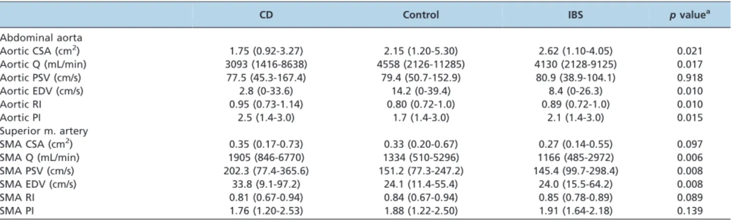
Doppler sonography measurements were successfully obtained from all individuals referred to the study. The sonographic parameters described were calculated for each of the 82 subjects examined. The median values (with 95% confidence intervals) for each Doppler measurement in the CD group, healthy volunteers and IBS group are summar-ized in Table 1. Most measurements in the aorta and in the superior mesenteric artery were significantly different among the three groups. For the aortic measurements, the
cross-sectional area (CSA), maximum flow volume (Q), end diastolic velocity (EDV) and pulsatility index (PI) were significantly lower, whereas the resistive index (RI) was higher in the CD patients than in both control groups (Table 1). For the SMA measurements, the maximum flow volume (Q), peak systolic velocity (PSV) and the end diastolic velocity (EDV) were higher in the CD patients than in the control groups (Table 1). No significant difference was detected between healthy volunteers and IBS patients (data not shown).
Doppler measurements and assessment of CD activity
To determine Doppler sonographic parameters for the assessment of Crohn’s disease activity, the relationship between the measurements obtained for CD patients and the CDAI scores was analyzed. Significant correlations were found between the CDAI and aortic maximum flow volume (Q) (CC = 0.374, p= 0.009) and aortic peak systolic velocity
(PSV) (CC = 0.305,p= 0.035). Of all the parameters analyzed,
only the aortic Q and aortic PSV were significantly different, with lower values in the active CD patients than in the inactive CD patients (Tables 2 and 3). Of note, none of the patients in the active group scored higher than 317.
Next, we analyzed the two variables in the aorta to distinguish between active and inactive CD and also determined cut-off points for stratification of patients.
Table 1 -Doppler sonography evaluation of the abdominal aorta and superior mesenteric artery of patients with CD, healthy volunteers and IBS patients.
CD Control IBS pvaluea
Abdominal aorta
Aortic CSA (cm2) 1.75 (0.92-3.27) 2.15 (1.20-5.30) 2.62 (1.10-4.05) 0.021
Aortic Q (mL/min) 3093 (1416-8638) 4558 (2126-11285) 4130 (2128-9125) 0.017
Aortic PSV (cm/s) 77.5 (45.3-167.4) 79.4 (50.7-152.9) 80.9 (38.9-104.1) 0.918
Aortic EDV (cm/s) 2.8 (0-33.6) 14.2 (0-39.4) 8.4 (0-26.3) 0.010
Aortic RI 0.95 (0.73-1.14) 0.80 (0.72-1.0) 0.89 (0.72-1.0) 0.010
Aortic PI 2.5 (1.4-3.0) 1.7 (1.4-3.0) 2.1 (1.4-3.0) 0.015
Superior m. artery
SMA CSA (cm2) 0.35 (0.17-0.73) 0.33 (0.20-0.67) 0.27 (0.14-0.55) 0.097
SMA Q (mL/min) 1905 (846-6770) 1334 (510-5296) 1166 (485-2972) 0.006
SMA PSV (cm/s) 202.3 (77.4-365.6) 151.2 (77.3-247.2) 145.4 (99.7-298.4) 0.008
SMA EDV (cm/s) 33.8 (9.1-97.2) 24.1 (11.4-55.4) 24.0 (15.5-64.2) 0.008
SMA RI 0.81 (0.67-0.94) 0.84 (0.67-0.94) 0.85 (0.78-0.89) 0.089
SMA PI 1.76 (1.20-2.53) 1.88 (1.22-2.50) 1.91 (1.64-2.18) 0.139
CD, Crohn’s disease; IBS, irritable bowel syndrome; SMA, superior mesenteric artery; CSA, cross-sectional area; Q, maximum flow volume; PSV, peak systolic velocity; EDV, end diastolic velocity; RI, resistive index; PI, pulsatility index. The values presented are the medians with 95% confidence intervals. aKruskal-Wallis H test.
Table 2 - Doppler sonography evaluation of the abdominal aorta of patients with active and quiescent Crohn’s disease based on the CDAI.
Active CD Quiescent CD pvaluea
Aortic CSA (cm2) 1.55 (1.00-3.30) 1.90 (0.90-3.20) 0.166 Aortic Q (mL/min) 2685 (1362-6182) 3751 (1733-9095) 0.010 Aortic PSV (cm/s) 69.4 (54.2-143.4) 90.7 (45.2-169.5) 0.036 Aortic EDV (cm/s) 1.70 (0-16.1) 3.95 (0-35.9) 0.394 Aortic RI 0.99 (0.77-1.20) 0.90 (0.73-1.00) 0.155 Aortic PI 2.85 (1.60-3.00) 2.35 (1.40-3.00) 0.314
Arbitrary cut-off values of aortic Q,4500 mL/min and an aortic PSV,70 cm/s were chosen so that the maximum possible number of CD patients was correctly classified according to disease activity. The overall sensitivity, specificity, accuracy, positive predictive value and negative predictive value were calculated for both parameters (Table 4).
& DISCUSSION
In the current study, we demonstrated the technical feasibility and potential clinical utility of Doppler sonogra-phy for patients with CD. The sonographic parameters analyzed showed that most measurements in the aorta and in the superior mesenteric artery were significantly different in the CD patients relative to either healthy subjects or patients with IBS. For assessment of disease activity, only the aortic measurements of maximum flow volume and aortic peak systolic velocity were significantly correlated with CDAI, with lower values in active CD. In addition, after selecting these two parameters, we determined cut-off points to accurately classify patients with regard to disease activity. The results indicate that the stratification of aortic maximum flow volume and peak systolic velocity values allowed a simple and accurate distinction to be made between active and inactive CD patients.
Despite advances in clinical techniques, the optimal therapy for CD still depends on correct classification of disease activity. Although substantial efforts have been made to develop new methods for evaluating CD activity, the CDAI is still the most widely used method in clinical trials and in routine practice, including the analysis of the response to treatment. In the last decade, abdominal Doppler ultrasound has been presented as a potential alternative for the evaluation of CD activity and is predominantly based on measurements obtained in the SMA. In addition, neovascularization and hyperemia of inflamed areas of the gut have also been investigated with
the use of color Doppler and power Doppler (24,25) as well as pulsed Doppler spectral analysis (26,27). In this study, in addition to comparing SMA measurements with data from previous studies, we demonstrated for the first time the simultaneous acquisition of SMA and abdominal aorta measurements.
We demonstrated in this study that most of the parameters analyzed by Doppler ultrasound examination were significantly different between patients with CD and control individuals. Overall, our data appear to agree with previous studies that also detected differences in splanchnic hemodynamics in CD (14,28-30). Furthermore, the opposing results for the aortic and SMA parameters in the same individuals support the presence of different levels of macrovascular control in the splanchnic circulation and possibly the existence of pathologic compensatory mechan-isms in CD hemodynamics. Although Doppler sonography is not required for the diagnosis of CD and is not being proposed as a new diagnostic tool, the results presented here support the general hypothesis that vascular abnorm-alities are constitutive elements in the pathogenesis of CD (31,32).
With regard to CD activity, prior studies on Doppler sonography have presented conflicting results. In a recent study, Doppler sonography for the evaluation of the SMA by segments in gray scale was presented as an excellent method for assessing CD activity (33). Other studies on the analysis of specific SMA measurements have suggested that either the resistance index (17,18) or the maximum flow volume (13,29) is the best parameter for differentiating CD patients. In contrast, a similar study failed to correlate the hyperdynamic mesenteric circulation detected in CD patients with the clinical or biochemical activity of the disease (14). In agreement with these results, our Doppler measurements obtained in the SMA confirmed the presence of hyperdynamic circulation in CD; however, these mea-surements could not distinguish disease activity among patients. In contrast, the significant differences in aortic measurements were correlated with disease activity. The most accurate Doppler parameter for the evaluation of CD activity was the estimated intensity of the maximum flow volume and peak systolic velocity; both of these parameters were shown to be lower in active CD.
Previous studies questioned the reliability of results obtained by Doppler sonography of small size vessels for the evaluation of CD activity (15,17). Similarly, we present superior results from the aorta relative to the SMA; this difference is likely associated with the dimensions and particularly the diameter of the vessels involved. Previous major abdominal surgeries could also impose technical difficulties and affect the overall quality of results (28). To minimize this potentially confounding factor in our study, we only examined patients who had never been submitted to any abdominal surgery and patients who underwent only Table 3 -Doppler sonography evaluation of the superior
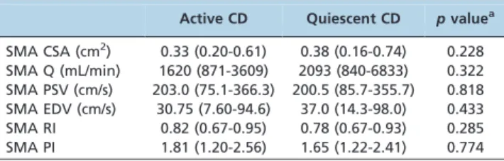
mesenteric artery of patients with active and quiescent Crohn’s disease based on the CDAI.
Active CD Quiescent CD pvaluea
SMA CSA (cm2) 0.33 (0.20-0.61) 0.38 (0.16-0.74) 0.228 SMA Q (mL/min) 1620 (871-3609) 2093 (840-6833) 0.322 SMA PSV (cm/s) 203.0 (75.1-366.3) 200.5 (85.7-355.7) 0.818 SMA EDV (cm/s) 30.75 (7.60-94.6) 37.0 (14.3-98.0) 0.433 SMA RI 0.82 (0.67-0.95) 0.78 (0.67-0.93) 0.285 SMA PI 1.81 (1.20-2.56) 1.65 (1.22-2.41) 0.774
CD, Crohn’s disease; SMA, superior mesenteric artery; CSA, cross-sectional area; Q, maximum flow volume; PSV, peak systolic velocity; EDV, end diastolic velocity; RI, resistive index; PI, pulsatility index. The values presented are medians with 95% confidence intervals.aMann-Whitney test.
Table 4 -Critical analysis of selected Doppler sonography parameters for detecting active Crohn’s disease based on the CDAI.
Variable (s) Sensitivity Specificity Accuracy Positive Predictive Value Negative Predictive Value
Aortic Q,4500 mL/min 16 of 20 (80) 16 of 28 (57) 32 of 48 (67) 16 of 28 (57) 16 of 20 (80) Aortic PSV,70 cm/s 15 of 20 (75) 21 of 28 (75) 36 of 48 (75) 15 of 22 (68) 21 of 26 (81)
minor surgical procedures, such as an inguinal hernia repair, cholecystectomy or appendectomy.
Of note, this study inadvertently selected consecutive CD patients with a relatively low intensity of disease based on the CDAI. In contrast to most studies where patients with a CDAI.450 have been categorized as having a high degree of activity, we only analyzed patients with a CDAI between 9 and 317. This factor may explain some differences between our results and those of other studies, especially concerning the SMA measurements. Moreover, the narrow range of disease activity may not have allowed a clear discrimination among the CD subjects enrolled in this study. Therefore, further studies with a higher number of both patients and control subjects are necessary to confirm the findings and the utility of Doppler sonography as an adjunctive tool for the evaluation of CD.
In conclusion, as a feasible and accurate technique, Doppler sonography of the aorta is a novel noninvasive adjunct method that may be useful in the clinical follow-up of patients with CD.
& ACKNOWLEDGMENTS
The authors wish to thank the Brazilian research foundations CNPq and FAPERJ for financial support. The authors are grateful to the medical staff of the Gastroenterology Division of the University Hospital for their assistance with patient selection. The authors are also grateful to the D’Or Laboratory for their support and for the opportunity to use their equipment for this study.
& AUTHOR CONTRIBUTIONS
Andrade TG was involved with the study design, patient selection and follow-up, data collection and interpretation and manuscript preparation. Fogac¸a H was involved with the study design, ultra-sonographic examinations, patient selection and critical review of the manuscript. Elia C was involved with data interpretation, obtained funding and provided a critical review of the manuscript. Pitrowsky M was involved with the patient selection and follow-up, data collection and interpretation and statistical analysis. Souza HS obtained funding and was involved with data interpretation, statistical analysis, critical review of the manuscript and manuscript writing.
& REFERENCES
1. Podolsky DK. Inflammatory bowel disease. N Engl J Med. 2002;347(6):417-29.
2. Xavier RJ, Podolsky DK. Unravelling the pathogenesis of inflammatory bowel disease. Nature. 2007;448(7152):427-34, http://dx.doi.org/10. 1038/nature06005.
3. Strober W, Fuss I, Mannon P. The fundamental basis of inflammatory bowel disease. J Clin Invest. 2007;117(3):514-21, http://dx.doi.org/10. 1172/JCI30587.
4. D’Haens GR, Fedorak R, Le´mann M, Feagan BG, Kamm MA, Cosnes J, et al. Endpoints for clinical trials evaluating disease modification and structural damage in adults with Crohn’s disease. Inflamm Bowel Dis. 2009;15(10):1599-604, http://dx.doi.org/10.1002/ibd.21034.
5. Sandborn WJ, Feagan BG, Hanauer SB, Lochs H, Lo¨fberg R, Modigliani R, et al. A review of activity indices and efficacy endpoints for clinical trials of medical therapy in adults with Crohn’s disease. Gastroenterology. 2002;122(2):512-30, http://dx.doi.org/10.1053/gast. 2002.31072.
6. Lichtenstein GR, Hanauer SB, Sandborn WJ. Practice Parameters Committee of American College of Gastroenterology (2009). Management of Crohn’s disease in adults. Am J Gastroenterol. 2009;104:465-83, http://dx.doi.org/10.1038/ajg.2008.168.
7. Freeman HJ. Use of the Crohn’s disease activity index in clinical trials of biological agents. World J Gastroenterol. 2008;14(26):4127-130. 8. Terheggen G, Lanyi B, Schanz S, Hoffmann RM, Bo¨hm SK, Leifeld L, et al.
Safety, feasibility, and tolerability of ileocolonoscopy in inflammatory bowel disease. Endoscopy. 2008;40(8):656-63, http://dx.doi.org/10. 1055/s-2008-1077445.
9. Wakefield AJ, Sawyerr AM, Dhillon AP, Pittilo RM, Rowles PM, Lewis AA, Pounder RE. Pathogenesis of Crohn’s disease: multifocal gastrointestinal
infarction. Lancet. 1989;2(8671):1057-62, http://dx.doi.org/10.1016/S0140-6736(89)91078-7.
10. Lunderquist A, Knutsson H. Angiography in Crohn’s disease of the small bowel and colon. Am J Roentgenol Radium Ther Nucl Med. 1967;101(2):338-44, http://dx.doi.org/10.2214/ajr.101.2.338.
11. Giaffer MH, Tindale WB, Senior S, Barber DC, Holdsworth CD. Quantification of disease activity in Crohn’s disease by computer analysis of Tc-99m hexamethyl propylene amine oxime (HMPAO) labelled leucocyte images. Gut. 1993;34(1):68-74, http://dx.doi.org/10. 1136/gut.34.1.68.
12. Maconi G, Imbesi V, Bianchi Porro G. Doppler ultrasound measurement of intestinal blood flow inflammatory bowel disease. Scand J Gastroenterol. 1996;31(6):590-3.
13. Erden A, Cumhur T, Olcer T. Superior mesenteric artery Doppler waveform changes in response to inflammation of the ileocecal region. Abdom Imaging. 1997;22(5):483-6, http://dx.doi.org/10.1007/s002619900243. 14. Maconi G, Parente F, Bollani S, Imbesi V, Ardizzone S, Russo A, Bianchi
Porro G. Factors affecting splanchnic haemodynamics in Crohn’s disease: a prospective controlled study using Doppler ultrasound. Gut. 1998;43(5):645-50, http://dx.doi.org/10.1136/gut.43.5.645.
15. Byrne MF, Farrell MA, Abass S, Fitzgerald A, Varghese JC, Thornton F, et al. Assessment of Crohn’s disease activity by Doppler sonography of the superior mesenteric artery, clinical evaluation and the Crohn’s disease activity index: a prospective study. Clin Radiol. 2001;56(12):973-8, http://dx.doi.org/10.1053/crad.2001.0794.
16. van Oostayen JA, Wasser MN, Griffioen G, van Hogezand RA, Lamers CB, de Roos A. Activity of Crohn’s disease assessed by measurement of superior mesenteric artery flow with Doppler ultrasound. Neth J Med. 1998;53(6):S3-8.
17. Bolondi L, Gaiani S, Brignola C, Campieri M, Rigamonti A, Zironi G, et al. Changes in splanchnic hemodynamics in inflammatory bowel disease. Non-invasive assessment by Doppler ultrasound flowmetry. Scand J Gastroenterol. 1992;27(6):501-7.
18. Giovagnorio F, Diacinti D, Vernia P. Doppler sonography of the superior mesenteric artery in Crohn’s disease. AJR Am J Roentgenol. 1998;170(1):123-6, http://dx.doi.org/10.2214/ajr.170.1.9423614. 19. Ludwig D, Wiener S, Bru¨ning A, Schwarting K, Jantschek G, Stange EF.
Mesenteric blood flow is related to disease activity and risk of relapse in Crohn’s disease: a prospective follow-up study. Am J Gastroenterol. 1999;94(10):2942-50.
20. Ludwig D, Wiener S, Bru¨ning A, Schwarting K, Jantschek G, Fellermann K, et al. Mesenteric blood flow is related to disease activity and risk of relapse in ulcerative colitis: a prospective follow up study. Gut. 1999;45(4):546-52, http://dx.doi.org/10.1136/gut.45.4.546.
21. Dietrich CF, Jedrzejczyk M, Ignee A. Sonographic assessment of splanchnic arteries and the bowel wall. Eur J Radiol. 2007;64(2):202-12. 22. Satsangi J, Silverberg MS, Vermeire S, Colombel JF. The Montreal
classification of inflammatory bowel disease: controversies, consensus, and implications. Gut. 2006;55(6):749-53, http://dx.doi.org/10.1136/gut. 2005.082909.
23. Best WR, Becktel JM, Singleton JW. Rederived values of the eight coefficients of the Crohn’s Disease Activity Index (CDAI). Gastroenterology. 1979;77(4 Pt 2):843-6.
24. van Oostayen JA, Wasser MN, van Hogezand RA, Griffioen G, de Roos A. Activity of Crohn disease assessed by measurement of superior mesenteric artery flow with Doppler US. Radiology. 1994;193(2):551-4. 25. Heyne R, Rickes S, Bock P, Schreiber S, Wermke W, Lochs H.
Non-invasive evaluation of activity in inflammatory bowel disease by power Doppler sonography. Zeitschrift fur Gastroenterologie. 2002;40(3):171-5, http://dx.doi.org/10.1055/s-2002-22325.
26. Esteban JM, Maldonado L, Sanchiz V, Minguez M, Benages A. Activity of Crohn’s disease assessed by colour Doppler ultrasound analysis of the affected loops. Eur Radiol. 2001;11(8):1423-8.
27. Sjekavica I, Barbaric´-Babic´ V, Krznaric´ Z, Molnar M, Cukovic´-Cavka S, Stern-Padovan R. Assessment of Crohn’s disease activity by doppler ultrasound of superior mesenteric artery and mural arteries in thickened bowel wall: cross-sectional study. Croat Med J. 2007;48(6):822-30, http:// dx.doi.org/10.3325/cmj.2007.6.822.
28. van Oostayen JA, Wasser MN, van Hogezand RA, Griffioen G, Biemond I, Lamers CB, et al. Doppler sonography evaluation of superior mesenteric artery flow to assess Crohn’s disease activity: correlation with clinical evaluation, Crohn’s disease activity index, and alpha 1-antitrypsin clearance in feces. AJR Am J Roentgenol. 1997;168(2):429-33, http://dx.doi.org/10.2214/ajr.168.2.9016220.
29. van Oostayen JA, Wasser MN, Griffioen G, van Hogezand RA, Lamers CB, de Roos A. Diagnosis of Crohn’s ileitis and monitoring of disease activity: value of Doppler ultrasound of superior mesenteric artery flow. Am J Gastroenterol. 1998;93(1):88-91.
30. Ludwig D. Doppler sonography in inflammatory bowel disease. Z Gastroenterol. 2004;42(9):1059-65, http://dx.doi.org/10.1055/s-2004-813308.
32. Hatoum OA, Binion DG, Otterson MF, Gutterman DD. Acquired microvascular dysfunction in inflammatory bowel disease: loss of nitric oxide-mediated vasodilation. Gastroenterol. 2003;125(1):58-69, http:// dx.doi.org/10.1016/S0016-5085(03)00699-1.