ABSTRACT
Two-year clinical evaluation of three adhesive
systems in non-carious cervical lesions
1, Huma OMURLU2
!"#$%& 2- DDS, PhD, Professor, Department of Restorative Dentistry, Faculty of Dentistry, University of Gazi, Ankara, Turkey.
Corresponding address:!($!(!$!%! !)$!*+
65080 - Kampus - Van - Turkey - Phone: + 90 505 687 81 37 - Fax: +90 432 225 10 09 - e-mail: [email protected] - [email protected]
!"#$!%&$'()
O
bjectives: Adhesive systems are continuously being introduced to Dentistry,to evaluate the clinical performance of cervical restorations done with three different adhesive systems. Material and Methods: 158 non-carious cervical lesions of 23 patients !"#"$% & '!"#"&($) "* (&)+$/ III (De Trey Dentsply, group XE). In groups SI-B, CL-B and XE-B, the outer surface of the sclerotic dentin was removed by roughening with a diamond bur before application of the respective adhesive systems. In groups CL-BP and XE-BP, after removal of the outer surface of the sclerotic dentin with the bur, the remaining dentin was etched with 37% phosphoric 9 ) "/ ((( Lesions were evaluated at baseline, and restorations after 3 months, 1 year and 2 years & ;#< = >? @ & % &FGGH$?&&)+( &% &&/"JGGH$@ & difference was found between the marginal adaptation of the groups SI-B, CL-B and XE-B FGGH$? @ & % & && &FGGH$)>?& ) "& ' % &/ III after 2 years of follow-up.
Key words: Adhesives. Dentin. Dental restoration. Composite resins.
INTRODUCTION
Thefundamental principle of adhesion to tooth substrate is based upon an exchange process by which inorganic tooth material is replaced with synthetic resin26. Using adhesive systems, the
exchange of substance between adhesive resin and tooth tissue is carried out in one, two, or three clinical application steps, depending on the bonding protocol used21. In addition to the number of steps,
% % the underlying adhesion strategy as etch-and-rinse and self-etch systems1. Self-etch adhesive systems
use non-rinse acidic monomers that simultaneously etchs and prime enamel and dentin. Self-etch adhesive systems that can have one or two steps
<F@$ < 1-2), and strong (pH<1) according to their acidity26.
New adhesive systems are continuously being introduced to the dentistry, unfortunately often 26. Although
clinical trials have demostrated that reliable adhesive restorations can be achieved using three-step etch-and-rinse adhesive systems2,15, more
randomized clinical studies should be performed to evaluate the clinical performance of new adhesive systems4.
They present no macro-mechanical undercuts, and are usually found in anterior teeth or premolars26
and offer good access. A drawback related to the use of non-carious cervical lesions might be the substantial differences in the composition of the bonding surface15. Non-carious cervical lesions have
a high degree of sclerosis and hybrid layer formation
W 8,21.
Alternative strategies for adhesion to sclerotic dentin have been recommended by previous researchers who evaluated available techniques21.
One recommended technique is the removal of the top layer of the sclerotic lesion using a bur8.
Using phosphoric acid conditioning before self-etch primers is another possible adaptive strategy for improving retention of resins to sclerotic dentin21.
Clinical trials of this approach however are limited in number.
The objective of this study was to evaluate the clinical perfomance of cervical restorations using adhesive systems and to determine the effect of bur removal and phosphoric acid prior to self-etch primer of dentin for improving micromechanical retention of adhesive resins after 2 years of clinical service. The null hypothesis to be tested was that the clinical performance of cervical restorations does not vary with different adhesive systems or with different adhesive strategies like bur removal of the outer dentin layer and pre-etching.
MATERIAL AND METHODS
A total of 252 restorations were placed in 29 subjects (16 males and 13 females; age range 30-70 years) being treated for non-carious cervical lesions. Patients with a medically compromised history, periodontitis, extreme caries risk and heavy bruxism were excluded from the study. Extreme caries risk was evaluated according to a large % missing teeth (not those removed for orthodontic reasons), infrequent use of toothpaste and toothbrush, frequent consumption of fermentable
carbohydrates and low socio-economic status3.
Heavy bruxism was evaluated according to presence of multiple wear facets on the occlusal surfaces of the teeth12. Additionally, all restored teeth made
contact with the opposing teeth in Class I cusp fossa or cusp marginal ridge occlusion relationships, and the participants had normal periodontal health (no &&\ the teeth)18. Prior to participating in the study, all
patients signed a written informed consent form. The clinical trial protocol was approved by the Gazi University Commission for Medical Ethics. One operator who was familiar with adhesive dentistry, placed all restorations to the non-carious cervical lesions. The occlusal cavosurface margins of the lesion involved enamel, and all axial surfaces and gingival cavosurface margin of the non-carious cervical lesion involved dentin. Plaque-covered \ of pumice. Operative procedures were performed without local anesthetic. The teeth were isolated with cotton rolls and a gingival retraction cord. No bevel was placed to the adjacent enamel. Patients who had at least 6 and no more than 20 non-carious cervical lesions were had the teeth restored with treated groups randomly in order.
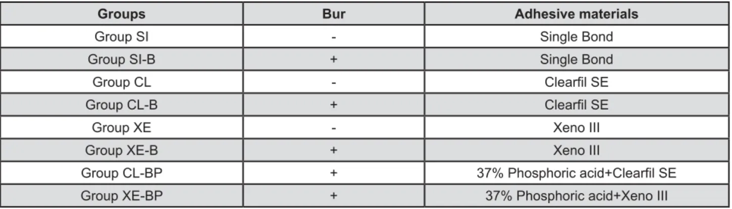
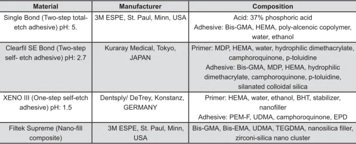
The groups are summarized in Figure 1. The adhesive systems, their manufacturers and compositions are shown in Figure 2.
Group SI: Lesion surfaces were etched for 15 s with 35% phosphoric acid gel (Vococid, Voco, Cuxhaven, Germany), rinsed with water and gently air dried. Single Bond total-etch adhesive system was applied to the etched surfaces, gently air dried and light-cured for 20 s.
Group SI-B: The outer surface of the sclerotic dentin was removed by roughening with a diamond bur (Diatech, Coltene, Whaledent AG, Switzerland) at a highspeed handpiece with water spray and Single Bond was applied in the same manner as described for Group SI.
{)+>) "# 9 applied to the lesion surfaces for 20 s and gently ) "'
Figure 1- Summary of treated groups
Groups Bur Adhesive materials
Group SI - Single Bond
Group SI-B + Single Bond
Group CL - ;!<!
Group CL-B + ;!<!
Group XE - Xeno III
Group XE-B + Xeno III
Group CL-BP + =>? H;!<!
Group XE-BP + =>? HJMMM
and light-cured for 10 s.
Group CL-B: The outer surface of the sclerotic dentin was removed by roughening with a &9 % ) " Bond system was applied in the same manner as described for Group CL.
Group XE: Xeno III Single Step Self Etching Dental Adhesive was applied to the lesion surfaces for 10 s, air-thinned and light-cured for 10 s.
Group XE-B: The outer surface of the sclerotic dentin was removed by roughening with a water-cooled high-speed diamond bur and Xeno III was applied to the roughened surfaces in the same as
explained for Group XE.
Group CL-BP: After removing the outer surface of the sclerotic dentin with a water-cooled high-speed diamond bur, the remaining dentin was etched with a 37% phosphoric acid gel for 15 s, & ) SE Bond system was applied in the same manner as described for Group CL.
Group XE-BP: After removing the outer surface of the sclerotic dentin with a water-cooled high-speed diamond bur, the remaining dentin was etched with a 37% phosphoric acid gel for 15 s, rinsed with water and gently air dried, and Xeno III
Figure 2- Composition of materials used
Material Manufacturer Composition
Single Bond (Two-step total- etch adhesive) pH: 5.
3M ESPE, St. Paul, Minn, USA N=>?
Adhesive: Bis-GMA, HEMA, poly-alcenoic copolymer, water, ethanol
;!<!O]#^ self- etch adhesive) pH: 2.7
Kuraray Medical, Tokyo, JAPAN
Primer: MDP, HEMA, water, hydrophilic dimethacrylate, camphoroquinone, p-toluidine
Adhesive: Bis-GMA, MDP, HEMA, hydrophilic dimethacrylate, camphoroquinone, p-toluidine,
silanated colloidal silica
XENO III (One-step self-etch adhesive) pH: 1.5
Dentsply/ DeTrey, Konstanz, GERMANY
Primer: HEMA, water, ethanol, BHT, stabilizer, <!!
Adhesive: PEM-F, UDMA, camphoroquinone, EPD
)!%$]}<!! composite)
3M ESPE, St. Paul, Minn, USA
O~O#~!<!! zirconi-silica nano cluster
Figure 3-$ !<($
Maxilla Mandibula Sclerosis WS SS
N An Prm Mo An Prm Mo Yes No
Group SI
30 7 8 4 4 5 2 29 1 20 10
Group SI-B
30 8 6 3 4 6 3 29 1 17 13
Group CL
41 8 6 3 7 13 4 40 1 30 11
Group CL-B
31 10 5 3 5 5 3 30 1 24 7
Group XE
30 6 5 3 6 6 4 29 1 14 16
Group XE-B
30 8 5 3 6 4 4 29 1 12 18
Group CL-BP
30 7 6 4 4 6 3 29 1 16 14
Group XE-BP
30 6 6 4 6 5 3 29 1 22 8
was then applied as explained for Group XE. Eight non-sclerotic cervical lesions were observed in only one patient. Other sclerotic lesions were randomly chosen to be roughened with a diamond bur at a high speed handpiece with water spray before phosphoric acid etching (Figure 3).
In all groups, lesions were restored with the was placed in at least two 2-mm-thick increments, which were light-cured for 40 s each using a & <|' }~ ( $ at 620 mW/cm2 maintaining the light-guide tip at
a distance of 1 mm from the composite surface. = #9{ (De Trey Dentsply, Konstanz, Germany).
The restorations were evaluated by a single investigator (not the operator) at baseline, 3 months, 1 year and 2 years after placement using the modified USPHS criteria (Figure 4). Marginal adaptation, gingival tissue response and wear were evaluated using a mirror and a probe. Gingival tissue response was evaluated according to presence of red, hypertrophic gingiva and gingival bleeding on probing around the cervical restoration5. Postoperative sensitivity was evaluated
before and after the restorative procedures with a 3-s air blast applied directly at the restoration site from a distance of 1 inch. Tooth vitality was evaluated with an electronic digital pulp tester (Parkell, Parkell Electronics Division, Farmingdale, NY, USA). Kruskal Wallis and Mann-Whitney U test were used to determine the statistical differences in clinical marginal adaptation and marginal staining data between the groups. Differences between the time intervals were analyzed using the Wilcoxon & = & 5% for all analyses.
RESULTS
Four patients with 20 lesions each, one patient with 6 lesions and one patient with 8 lesions could not be evaluated at the 3-month recall due to nonattendance (63% recall rate). All of the remaining restorations were evaluated at 3 months, 1 year and 2 years.
Retention
Number of retained restorations and retention rates of the groups are shown in Table 1. At 2 years, retention rates of the groups SI, SI-B, CL, CL-B, XE, XE-B, CL-BP, XE-BP were 70.6%, 86.7%, 78.1%, 95.5%, 70%, 85.7%, 93.3%, 93.8%, respectively. ? & & &FGGH$
Marginal staining
Number of restorations that had no marginal staining (Alpha) and comparisons of the groups are shown in Table 2. No group presented discoloration at 3 months. At 1 year, significant difference was found between group XE and other groups (p<0.05). In addition, only group XE showed & & % months and 1 year, between 3 months and 2 years) (p<0.05). Marginal staining was always seen at the gingival margin of the restorations.
Marginal adaptation
Number of restorations that had undetectable margins (Alpha) and comparisons of the groups are shown in Table 3. At all time intervals (3 months, 1 year, 2 years) when groups SI, CL and /" & % & )+ ( FGGH$
Figure 4-<&&$!! ]!!!$
Category Acceptable Unacceptable Criteria
Retention Alfa Retained
Charlie Missing
Marginal Staining Alfa None
Bravo $<!(]!!!
Charlie Deep staining (not removable, generalized)
Marginal Adaptation Alfa Undetectable margin or slight detectable
step
Detectable crevice
Bravo
Charlie Obvious crevice or fracture
Other failures (post operative sensitivity, recurrent caries, gingival response, tooth vitality,
wear)
Alpha None
Groups Baseline 3 months 1 year 2 years
Group SI = =]? > ]&?aA > ]&?aAB > ]>&?O
Group SI-B = =]? ]=&=?aA =]&>?aA =]&>?aA
Group CL ]? = =]&?aA = =]&?aA = >]>&?aA
Group CL-B = =]? ]&?aA ]&?aA ]&?aA
Group XE = =]? >]?aA ]>?aA ]>?aA
Group XE-B = =]? ]&?aA ]&?aA ]&>?aA
Group CL-BP = =]? ]?aA ]?aA ]=&=?aA
Group XE-BP = =]? ]?aA ]=&?aA ]=&?aA
Table 1- Number, and percentages of retained restorations and comparisons between the groups at 3 months, 1 year and ]&]#!& &
Q^ (< ^ ($ ]& !^ ^ (<^ ($&;! (<^ ]= ]& ! ^(<^ &
Groups Baseline 3 months 1 year 2 years
Group SI = =]? > ]&?aA > ]&?aA > ]&?aA
Group SI-B = =]? ]=&=?aA =]&>?aA ]>=&=?aA
Group CL ]? = =]&?aA = ]&?aA = ]>&?aA
Group CL-B = =]? ]&?aA ]&?aA ]&?aA
Group XE = =]? >]?aA ]?bB ]?aB
Group XE-B = =]? 21/19 (90.5)aA ]&>?aA 21/16 (76.2)aA
Group CL-BP = =]? ]?aA ]?aA =]&?aA
Group XE-BP = =]? ]?aA ]=&>?aA ]>&?aA
Q^ (< ^ ($ ]& !^ ^ (<^ ($&;! (<^ ]= ]& ! ^(<^ &
Table 2- Number and percentages of restorations that have no marginal staining (Alpha) and comparisons of the groups = ]&&]#!& &
Groups Baseline 3 months 1 year 2 years
Group SI = =]? > =]>&?aA > ]&>?aB > ]>&?aB
Group SI-B = =]? =]&>?aA ]&>?aA 15/10 (66.7)acA
Group CL ]? = ]&?abA = ]&?abB = ]=&?aB
Group CL-B = =]? ]&?aA >]>>&=?aB ]=&?aB
Group XE = =]? ]?bA >]=?bB ]?bB
Group XE-B = =]? ]>&?abcA ]&=?abB ]>&?aB
Group CL-BP = =]? ]?acA ]?cA =]&>?cA
Group XE-BP = =]? ]?acA ]=&>?cA ]=&>?cA
Q^ (< ^ ($ ]& !^ ^ (<^ ($&;! (<^ ]= ]& ! ^(<^ &
Table 3- Number and percentages of restorations that have undetectable margin (Alpha) and comparison of the groups at
% & % &(/"JGGH$?&& difference was found between the groups CL and /" FGGH$ & difference was detected at 2 years (p<0.05). At all & & &(9')+9'/"9'FGGH$ & & was observed by bur removal of dentin at 3 months FGGH$% &((9' groups CL and CL-B, and groups XE and XE-B. At @ & % group XE and group XE-B (p<0.05). Bur removal of dentin and pre-etching with phosphoric acid in the groups with self-etch adhesive systems (groups CL and CL-BP, and groups XE and XE-BP) showed a & JGGH$
When the groups were evaluated according to time, marginal adaptation did not decreased over & (9' )+9'# /"9'# FGGH$ & & % 3 months and 1 year and between 3 months and 2 years (p<0.05). While group XE-BP showed the best marginal adaptation, group XE showed the worst marginal adaptation at 2 years. Marginal deteriorations were seen especially at the gingival margin.
Remaining clinical variables
None of the restorations showed postoperative sensitivity, gingival tissue response or secondary caries and all of the retained restorations were clinically and aesthetically acceptable.
Failure analysis
When failed restorations were evaluated, 54% of totally lost restorations were in premolars, 58% in wedge-shaped lesions and 42% in saucer-shaped lesions. Failed restorations occurred almost equally in both arches, so no correlation was determined between arch and restoration failure.
DISCUSSION
Clinical trials are the ultimate test for assessment of bonding effectiveness of adhesive materials. Peumans, et al.15 (2005) reviewed clinical studies
published between 1998 and 2004 and concluded that three-step etch-and-rinse and two-step self-etch adhesive systems showed a clinically reliable % The clinical performance of two-step etch-and-rinse adhesive systems was less favorable, while one-step self-etch adhesive systems had an unacceptable clinical performance. In the present study, three adhesive systems were compared and 9 9 ) "$ more reliable marginal adaptation than the one-step
self-etch adhesive (Xeno III) in the groups without bur removal of dentin at 2 years. There was no difference between the two-step self-etch adhesive ) "$ 9 9 (Single Bond) with respect to marginal adaptation.
) "9 9 self-etching primer contains 10-methacryloxydecyl dihydrogen phosphate (10-MDP) as a functional monomer, which is dissolved in water resulting in a pH of approximately 3. In previous studies, 10-MDP has been shown to chemically react with hydroxyapatite7,26. The resulting two-fold
micromechanical and chemical bonding mechanism might have led to the better marginal adaptation ) " / ((( @ ( at 2 years, Xeno III showed the worst marginal adaptation of all three systems. This may be due to the weak bond between dentin and Xeno III adhesive resin, which may be more affected than the other adhesive systems used in this study by occlusal stresses and intraoral temperature changes in the oral cavity. Water trees in the hybrid layer may be another explanation of the lower marginal adaptation of Xeno III to dentin surfaces. Water trees represent an area where a certain volume of water is retained causing incomplete polymerization of the adhesive21. It has been speculated that this
volume of water may cause degradation of the bonded surface due to hydrolysis9. Tay, et al.22
(2003) suggested that water treeing in the hybrid layer may explain the initial problems associated %& and the underlying causes of their relative lack of durability. Tay, et al.20 (2001) noted that
one-step self-etch adhesive systems are permeable membranes that permit diffusion of water, and this residual water within the adhesive layer may lead to areas of incomplete polymerization of the adhesive. In previous nanoleakage studies, it was reported that the lowest occurrence of nanoleakage within % ) "5.
In the present study, however, the retention results were different from those of marginal & among the groups. Peumans, et al.14 (2005)
evaluated the clinical performance of the mild 9 ) "9 cervical lesions. They reported 100% retention rate after 3 years and 98% retention rate after 5 years. Burrow and Tyas4@GG $ )
between previous studies and this current study may be due to factors associated with individual patients (such as occlusal loading, dentin sclerosis).
Sugizaki, et al.19 (2007) restored class V cavities
with Xeno III and composite resin, and evaluated the outcomes at recall intervals up to 18 months. They reported that all restorations were clinically satisfactory. Similar to these results, Türkün23
(2003) reported a 96% retention rate for Xeno III & % / (((9 9 ) Protect Bond). In the present study, retention rates / ((( G$ & from those for the other groups.
The removal of the outer surface of the sclerotic dentin by roughening with a diamond bur has been recommended in the literature in order to create a better hybrid layer8. Van Dijken24 (2004) determined
that roughening of sclerotic dentin surfaces with a diamond bur did not increase the retention of restorations. In the present study, although the group of XE-B, tended to show better marginal adaptation, marginal staining and retention than the &/ & % ? & better marginal adaptation was determined as a result of the study after 2 years. On the other hand, & % % groups SI and SI-B, and groups CL and CL-B, in which the adhesive systems were applied with or without bur removal of dentin. The different results between these adhesive systems may depend on their different bonding ability to sclerotic dentin. Sclerotic dentin has diffusion barriers (obliteration of dentin tubules with sclerotic casts and presence of acid-resistant hypermineralized layer), which can inhibit the acid demineralization and compromise bonding21; & ') "/
III was completely ineffective in overcoming the diffusion barriers in sclerotic dentin and could not % % < roughening the sclerotic dentin with a diamond bur increased the bonding ability of Xeno III.
Different strategies were used in previous studies to increase the bond strength of adhesive systems to sclerotic dentin, such as bur removal of the most 9 & with phosphoric acid11,21. However, none of these
earlier studies investigated these two mechanisms together. In the present study, bur removal of dentin and additional phosphoric acid etching before self-etch primer application increased marginal adaptation of both self-etch adhesive systems, but did not change their retention rates. Although Van Landuyt, et al.25 (2006) reported that prior
&& % & of self-etch adhesive systems to sound dentin, in the current study sclerotic dentin surfaces were
used as a bonding substrate. The different dentin substrates (sclerotic versus sound dentin) may be the explanation for the different results obtained in the studies.
Several clinical co-variables that are unique to the oral environment have been described to affect the clinical performance of adhesive systems1.
With regard to location, researchers determined that the retention of cervical restorations was & & | the mandibular arch16. Differently from previous
studies, in the current study, failed restorations occurred almost equally in both arches. However, most of the failed restorations occurred in premolars H$;& "$ researchers compared tooth groups and reported that the magnitude of cervical stress was highest for premolars, followed by incisors and lowest for canines13. Powell, et al.16 (1995) reported that the
shapes of the lesions do not affect the retention of restorations. On the other hand, Eliguzeloglu, et al.6 (2011) compared the effect of cavity shape
on the stress distribution of cervical lesions which were restored with a composite using FEM analysis, and it is determined that the stress distribution of saucer-shaped non-carious cervical lesions have more advantages than wedge-shaped lesions. In the present study, 58% of the lost restorations had been placed in wedge-shaped lesions, and 42% of them in saucer-shaped lesions. These results are in good aggrement with the authors’ previous FEM study.
To be considered clinically effective, adhesive systems should keep the restoration in place and completely seal the restoration margins against & \ & Incomplete marginal seal will result in post-placement sensitivity, marginal staining and, eventually, recurrent caries, which are still the most common symptoms associated with clinical failure of adhesive restorations17. Marginal
staining is probably caused by microleakage or an accumulation of stains at a marginal defect, such as the chip fracture of a slight excess of material covering unground and/or non-treated tooth surface10. In the present study, the sealing capacity
CONCLUSIONS
In conclusion, the null hpothesis could not be accepted because;
9 9 ) SE showed better marginal adaptation than the one-step self-etch adhesive system Xeno III after 2 years;
) " & retention and marginal staining to those of the etch-and-rinse adhesive system Single Bond after 2 years;
The removal of the outer surface layer of sclerotic dentin did not increase the retention and marginal staining of the adhesive systems evaluated in this study;
The removal of the outer surface layer of dentin and additional phosphoric acid etching, increased &/ ((() " after 2 years.
REFERENCES
1- Bayne SC, Heymann HO, Sturdevant JR, Wilder AD, Sluder TB. Contributing co-variables in clinical trials. Am J Dent. 1991;4(5):247-50.
2- Browning WD, Brackett WW, Gilpatrick RO. Two-year clinical % 9% in noncarious Class V lesions. Oper Dent. 2000;25(1):46-50. 3- Brunton PA. Decision-making in operative dentistry. London: Quintessence Publishing Co. Ltd; 2002.
4- Burrow MF, Tyas MJ. Clinical evaluation of three adhesive systems for the restoration of non-carious cervical lesions. Oper Dent. 2007;32(1):11-5.
5- Duarte S Jr., Perdigao J, Lopes MM. Effect of dentin conditioning time on nanoleakage. Oper Dent. 2006;31(4):500-11.
9"&W &""<"&{' effect of cavity shape and hybrid layer on the stress distribution of cervical composite restorations. Eur J Dent. 2011;5(2):180-5. 7- Fukegawa D, Hayakawa S, Yoshida Y, Suzuki K, Osaka A, Van Meerbeek B. Chemical interaction of phosphoric acid ester with hydroxyapatite. J Dent Res. 2006;85(10):941-4.
8- Gwinnett AJ, Kanca J 3rd. Interfacial morphology of resin
composite and shiny erosion lesions. Am J Dent. 1992;5(6):315-7. 9- Hashimoto M, Ohno H, Kaga M, Endo K, Sano H, Oguchi H.
In vivo degradation of resin-dentin bonds in humans over 1 to 3 years. J Dent Res. 2000;79(6):1385-91.
10- Kubo S, Kawasaki K, Yokota H, Hayashi Y. Five-year clinical evaluation of two adhesive systems in non-carious cervical lesions. J Dent. 2006;34(2):97-105.
11- Kwong SM, Cheung GS, Kei LH, Itthagarun A, Smales RJ, Tay FR, et al. Micro-tensile bond strengths to sclerotic dentin using a self-etching and a total-etching technique. Dent Mater. 2002;18(5):359-69.
12- Lehman ML, Meyer ML. Relationship of dental caries and stress: concentrations in teeth as revealed by photoelastic tests. J Dent Res. 1966;45(6):1706-14.
13- Palamara JE, Palamara D, Messer HH, Tyas MJ. Tooth morphology and characteristics of non-carious cervical lesions. J Dent. 2006;34(3):185-94
14- Peumans M, De Munck J, Van Landuyt K, Lambrechts P, Van Meerbeek B. Three-year clinical effectiveness of two step self-etch adhesive in cervical lesions. Eur J Oral Sci. 2005;113(6):512-8. 15- Peumans M, Kanumilli P, De Munck J, Van Landuyt K, Lambrechts P, Van Meerbeek B. Clinical effectiveness of contemporary adhesives: a systematic review of current clinical trials. Dent Mater. 2005;21(9):864-81.
16- Powell LV, Johnson GH, Gordon GE. Factors associated with clinical success of cervical abrasion-erosion restorations. Oper Dent. 1995; 20(1): 7-13.
17- Qvist V, Qvist J, Mjör IA. Placement and longevity of tooth-colored restorations in Denmark. Acta Odontol Scand. 1990;48(5):305-11.
9= ?+? & surveys of periodontal disease. J Dent Res. 1956;35(3):350-9. 19- Sugizaki J, Morigami M, Uno S, Yamada T. Clinical evaluation and interfacial morphology observation of Xeno III self-etching resin bonding and restorative system. Dent Mater J. 2007;26(4):602-7.
20- Tay FR, Pashley DH. Aggressiveness of contemporary self etching systems. I: Depth of penetration beyond dentin smear layers. Dent Mater. 2001;17(4):296-308.
21- Tay FR, Pashley DH. Resin bonding to cervical sclerotic dentin: a review. J Dent. 2004;32(3):173-96.
22- Tay FR, Pashley DH. Water treeing - a potential mechanism for degradation of dentin adhesives. Am J Dent. 2003;16(1):6-12. 23- Türkün SL. Clinical evaluation of a self-etching and a one bottle adhesive system at two years. J Dent. 2003;31(8):527-34. @9~ ~% in Class V non-carious cervical lesions. Am J Dent. 2004;17(1):27-32.
25- Van Landuyt KL, Peumans M, De Munck J, Lambrechts P, Van Meerbeek B. Extension of a one-step self-etch adhesive into a multi-step adhesive. Dent Mater. 2006;22(6):533-44.
![Figure 4-<&&$!! ]!!!$](https://thumb-eu.123doks.com/thumbv2/123dok_br/14982447.511144/4.892.82.811.116.448/figure-lt-amp-amp.webp)

