Pierre Adriano Moreno NEVES(a)
Leonardo Abrantes LIMA(a)
Fernanda Cristina Nogueira RODRIGUES(b)
Tarcisio Jorge LEITÃO(c)
Cecília Cláudia Costa RIBEIRO(c)
(a) Universidade Federal do Maranhão – UFMA, School of Dentistry, Postgraduate Program in Dentistry, São Luís, MA, Brazil. (b) Universidade Federal do Maranhão – UFMA,
School of Dentistry, São Luís, MA, Brazil. (c) Universidade Federal do Maranhão –
UFMA, School of Dentistry, Department of Dentistry II, São Luís, MA, Brazil.
Clinical effect of photodynamic therapy
on primary carious dentin after partial
caries removal
Abstract: This study was conducted to assess the clinical effect of photodynamic therapy (PDT) in the decontamination of the deep dentin of deciduous molars submitted to partial removal of carious tissue. After cavity preparation, dentin samples were taken from the pulp wall of nineteen deciduous molars before and after PDT application. Remaining dentin was treated with 0.01% methylene blue dye followed by irradiation with an InGaAlP diode laser (λ – 660 nm; 40 mW; 120 J/cm2; 120 s). Dentin samples were microbiologically assessed for the enumeration of total microorganisms, Lactobacillus spp. and mutans streptococci. There was no
signiicant difference in the number of colony-forming units (CFU) for any
of the microorganisms assessed (p > 0.05). Photodynamic therapy, using 0.01% methylene blue dye at a dosimetry of 120 J/cm2 would not be a viable clinical alternative to reduce bacterial contamination in deep dentin.
Keywords: Lasers; Photochemotherapy; Tooth, Deciduous.
Introduction
The innerportion part of the carious dentin, deep dentin, is a vital tissue with a robust collagen structure that is capable of remineralization.1 Less invasive restoration techniques have been used to maintain this dentin layer. It has been shown that, after cavity restoration, there is a drastic reduction in both the genotypic and phenotypic complexity of the microbiota.2,3
However, some studies show that microorganisms, including Streptococcus mutans, may adapt to this new environment created under the restorations after the incomplete removal of the carious dentin.4
Bacteria may persist in the dentin after cavity preparation, even after “complete” removal of carious tissue,3 which is driving the search for alternative treatments aimed at reducing the number of microorganisms after interventions for removing carious tissue.5
Photodynamic Therapy (PDT) is currently being studied as an antimicrobial alternative with an effect on the microorganisms involved in caries.6 This technique is based on the application of a light source to activate a photosensitizer (PS), which in the presence of oxygen, produces singlet oxygen and free radicals that irreversibly damage cellular components and modify metabolic activities, leading to cell death.7
The antimicrobial effectiveness of PDT has been demonstrated under laboratory conditions on planktonic cultures,8 monospecies
bioilms of certain genera of streptococcus, including Streptococcus
Declaration of Interests: The authors certify that they have no commercial or associative interest that represents a conflict of interest in connection with the manuscript.
Corresponding Author: Cecília Cláudia Costa Ribeiro E-mail: cecí[email protected]
DOI: 10.1590/1807-3107BOR-2016.vol30.0047
Submitted: May 30, 2015
mutans9 and also in multi-species bioilms.10 PDT’s effects on carious dentin formed under experimental conditions through in vitro11 and in situ12 models has also been shown.
However, the response to PDT inside the oral cavity may be different from the results so far under experimental conditions.7 In fact, a study has shown that, using the same dosimetry, PDT reduced microbial levels in the in vitro bioilm and had little effect on
microbial levels in the in situ situation.13
We have found only one in vivo study that assessed the effect of PDT/ Methylene blue on contaminated dentin of permanent teeth, which showed a reduction in the number of viable bacteria (total microorganisms,
mutans streptococci, Lactobacillus spp.) on the order of
one decimal reduction after applying a low-power
laser.14 Therefore, further studies into the effectiveness of PDT in clinical situations are required, including analysis of its effects on the remaining pulp wall. Thus, the goal of this study was to evaluate the clinical effect of using photodynamic therapy (PDT) to decontaminate the deep dentin of primary molars submitted to partial removal of carious tissue.
Methodology
Sample selection
The study was approved by the Research Ethics
Committee of the Universidade Federal do Maranhão
– UFMA under no. 23115-004903/2011-24. Parents or
legal guardians received detailed information about the study and signed a free and informed consent form to permit the children to participate in the study. All children received full dental care in the Pediatric
Dentistry Clinic of the UFMA.
The sample size used in this study was based on a previous study of our group regarding the partial removal of carious dentin; there, it was determined that sixteen teeth were required to obtain a test power of
80% at a signiicance level of 5% and with a maximum error of the estimate of 0.6 colony-forming units.3
To select the samples, 418 children aged between 6 and 10 years from five public schools in the municipality of São Luís (state of Maranhão, Brazil)
were examined. Children with systemic diseases
or who had used antibiotics in the three months
preceding the study were excluded, as these factors might interfere in the etiological factors of the disease. The inclusion criteria were children with deciduous teeth with active carious cavities scoring 6 (Extensive
distinct cavity with visible dentin) in the ICDAS index (International Caries Detection and Assessment
System)15 plus inner half of the dentin conirmed by radiological examination. Teeth showing clinical or radiographic signs of irreversible damage to the pulp
such as spontaneous pain, istula or radiographic
alteration in the interradicular or periapical region were excluded.3 Thirty-four children were selected to participate in the study, but only 19 arrived at the
appointed date for the experiment (Figure 1).
Cavity preparation
The teeth of the patients were anesthetized and
isolated using a rubber dam. Next, a high-speed bur
no. 1011 (KG Sorensen, São Paulo, Brazil) was used
to gain access to the caries lesion. Cavity preparation
was limited to the total removal of carious tissue from the cavosurface angle and surrounding walls.
The infected dentin was supericially removed from the pulp wall using low-speed sterile no. 3 carbide
(KG Sorensen, São Paulo, Brazil) bur.3 Calibration of the examiner for partial caries removal was performed on 20 extracted deciduous molars which were evaluated after treatment by a second examiner
experienced in this procedure (C.C.C.R.). A kappa
value of 0.85 was obtained.
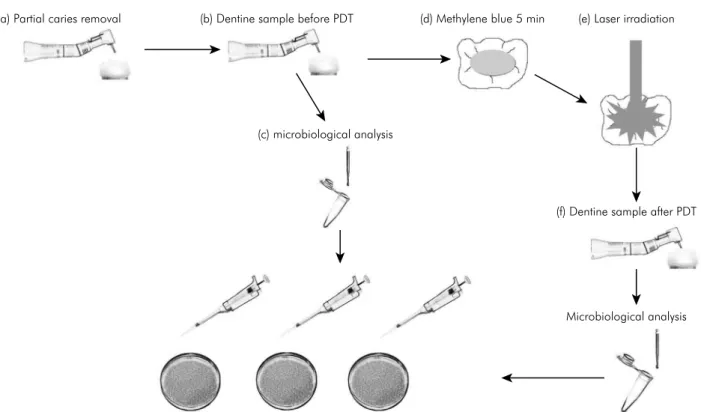
After cavity preparation, a irst sample of dentin
was collected from the remnant pulp wall using
another low-speed sterile no. 3 carbide (KG Sorensen,
São Paulo, Brazil) bur (samples before PDT) to assess the microbiological state of this tissue.16 After sampling, the bur was transferred to an Eppendorf tube containing glass beads and 1 mL 0.9% saline
(NaCl 0.9%) and kept on ice for microbiological processing within one hour (Figure 2).
Application of PDT and restorative procedure
After collecting the first sample of dentin, the photosensitizing dye methylene blue at 0.01% (Aptivalux, Belo Horizonte, Brazil) was applied to the whole
same used in permanent molarswhere PDT reduced microbial levels of caries related microorganims.14
The complex microbiota of the active caries lesion was the reason methylene blue was selected for this experiment. In vitro study have shown that methylene
blue is a cationic photosensitizer that can inactivate
both gram-positive and gram-negative bacteria after
applying PDT with a low-power red laser.17
After the 5 min with methylene blue were elapsed, every tooth was irradiated with a low power diode (a) Partial caries removal (b) Dentine sample before PDT (d) Methylene blue 5 min (e) Laser irradiation
(f) Dentine sample after PDT
Microbiological analysis (c) microbiological analysis
Total microorganisms Mutans streptococci Lactobacillus spp.
Figure 2. Experimental design of the study. Figure 1. Flow chart subject enrollment.
Excluded (n = 384)
• Not meeting inclusion criteria (n =3 59)
• Declined to participate (n = 21) • Other reasons (n = 4)
Arrived at the appointed date for the experiment (n = 19) Assessed for eligibility (n = 418 children)
Between 6 and 10 years
laser (InGaAlP – Indium Gallium Aluminum Phosphide) with dosimetry of 120 J/cm2, power density of 1 W/cm2 for 120 s, receiving energy of 4,8 J. The device parameters were: output power of 40 mW, 660 nm wavelength and spot size of 0.04 cm2. The active tip of the laser was placed perpendicular to the cavity, as close as possible to the stained dentin
(average 0.25 cm from cavity loor), being cavity’s
opening the tip approximation limit. Next, a new dentin sample (sample after PDT) was collected at a site
adjacent to that of the irst sample for microbiological processing (Figure 2).
All teeth were protected with calcium hydroxide
cement (Dentsply, Milford, USA), etched with 37%
phosphoric acid for 15 s, and restored with an adhesive and resin composite system (3M ESPE, Minneapolis,
USA) according to manufacturer instructions.
Microbiological analysis
To estimate the number of microorganisms present
in 1 mg dentin (CFU/mg), dentin weight was calculated
from the difference between the whole set – tube, glass beads, saline and bur with dentin and the previously assessed weight of the set without the dentin sample.3
After weighing, the tubes were shaken in a vortex for 30 s to disperse the sample.3 A 100 µL aliquot of the dispersed suspension was used to prepare serial dilutions (1:10, 1:100, 1:1.000, 1:10.000 e 1:100.000). Twenty microliter aliquots of each dilution were incubated in duplicate in the following media: blood agar for total microorganism count; mitis salivarius
agar supplemented with 0.2 units/mL bacitracin and 15% sucrose for mutans streptoccci count18 and Rogosa agar (RG), for Lactobacillus spp count.19 The Rogosa and blood agar plates were incubated under
anaerobic conditions for 48 h,20 whereas the MSB
plates were incubated in an atmosphere of 5% CO2
for the same period of time.18
After incubation, the CFU number was counted
in dilutions containing 6–60 colonies. Microbial counts were conducted using a stereo microscope (Quimis®, São Paulo, Brazil) by a single examiner who was blinded to the previous clinical procedure. The reproducibility of the counting method was assessed by comparing the microorganism counts for each sample’s duplicate plates: the counts were positively correlated (r = 0.92; p < 0.001).
Statistical analysis
The experimental unit used as a parameter for
analysis was the tooth, and CFU counts before and
after PDT were compared in each culture medium: blood agar, MSB agar and Rogosa agar. The data
distribution was found to be non-normal using the Shapiro-Wilk test. The Box-Cox method was used to log10-transform the data. After this transformation, CFU counts were analyzed using the Wilcoxon test.
The BioEstat 5.3 software (Instituto Mamirauá, Belém, Brazil) was used in these analyses, and a level of
signiicance of 5% was adopted.
Results
The results of this study showed no statistically
signiicant difference in the viable microorganisms
count before and after application of PDT in molars submitted to partial removal of carious tissue. The
therapy promoted an average log-reduction of 0.61
in total microorganisms, 0.44 in mutans streptococci
and 0.46 in Lactobacillus spp. (Figure 3a, 3b and
3c); however, this reduction was not significant (p > 0.05) (Table).
Table. CFU count (means ± standard deviation) before and after photodynamic therapy with a 120 J/cm2 dose.
Dose
Total microorganisms Mutans streptococci Lactobacillus spp.
before after before after before after
120 J/cm2
CFU (Log10) 3.93 ± 0.93 3.32 ± 1.68 2.17 ± 1.85 1.73 ± 1.77 1.51 ± 1.97 1.05 ± 1.67
Variation (%) 14.71 ± 41.03 34.5 ± 49.19 20.25 ± 43.71
p 0.3981 0.4265 0.4990
Discussion
In this study, PDT was not effective at reducing contamination in the deep dentin of deciduous teeth using a dosimetry of 120 J/cm2.
The susceptibility of the microorganisms involved in caries disease grown in planktonic culture to PDT, using lower energy levels than in this study, namely between 24 and 72 J/cm2, has already been reported in the literature.13,21 Additionally, the effect of PDT on microbial reduction has also been shown in experimental models of carious dentin based on induced caries lesions in bovine dentin11 and the “in situ” caries model on human dentin,12 even with intensities below the ones used in this experiment,
i.e., between 24 and 94 J/cm2, respectively.
Differences in experimental conditions may explain the discrepancies between this clinical study and studies under controlled conditions. Previous
in vitro studies used planktonic cells with reduced antimicrobial resistance,21 whereas the studies that induced the formation of in vitro and in situ bioilms
displayed less microbiological complexity than deep caries cavities.22,23 For these reasons, the response to PDT in the oral cavity may be different from the results in these more controlled studies:7 in fact, it has already been shown that the positive PDT results for planktonic culture were not repeated with the
bioilm formed “in situ” using the same dosimetry.13 The variety of protocols for applying laser or LED light and the existence of few studies that assess the properties of the available dyes and light sources
hinder the comparison of the indings on PDT.24
We have found only one clinical study on PDT in the carious dentin of permanent teeth, in which a significant reduction in the counts of total microorganisms, mutans streptococci and Lactobacillus
spp.was observed.14 It may be that the dentinal substrate contributed to the differences between the
indings of the study carried out with permanent
teeth14 and this study, which used caries lesions on deciduous teeth. However, it should be stressed that the difference in substrates may have been minimized, as that PDT was applied to caries lesions in the deep dentin in both studies.
The difference in applied dosimetry might be another explanation for why we did not observe PDT
Figure 3. a) Effect of PDT with 120 J/cm2 dose on the
total microorganism count; b) Effect of PDT with 120 J/cm2
dose on the Mutans streptococci count; c) Effect of PDT with 120 J/cm2 dose on the Lactobacillus spp. count. The data
represent the Log reduction, and the error bars represent the standard deviation. Data followed by same letters represent no significant difference (p > 0.05).
4.00 6.00
5.00
2.00
1.00
0.00 3.00
Total microorganisms
Before After
120 J/cm² A
a a
Log
10
reduction
4.00 5.00
2.00
1.00
0.00 3.00
-1.00
Mutans streptococci
120 J/cm² a
a B
Before After
Log
10
reduction
4.00 5.00
2.00
1.00
0.00 3.00
-1.00
120 J/cm² Lactobacillus spp.
a
a C
Before After
Log
10
effects in the present study, where the dosimetry used was 120 J/cm2 and, therefore, lower than in the clinical study that obtained positive effects in permanent teeth, which used 320 J/cm2.14 In an additional experiment using 180 J/cm2 in the remaining dentin of 10 primary molars, extending the time of laser application for 180 s and using the maximum power output of the device to red light (40 mW); still
found no signiicant antimicrobial effect (data not
shown). This last dosimetry is the maximum limit for the red laser of our device.
In this study, PDT caused an average log reduction of 0.46 for mutans streptococci, 0.44 for Lactobacillus
spp. and 0.61 for total microorganisms; however,
this reduction was not signiicant compared to the
controls before treatment. In the study on carious dentin of permanent teeth, the log reduction was approximately 1.38 for mutans streptococci, 0.93 for
Lactobacillus spp. and 0.91 for total microorganisms,
all of which were statistically signiicant.14 Thus,
although an effect of PDF was observed in that clinical
study with permanent teeth, its effect was low to be
considered dose-effective because it should induce a reduction of at least 2-log CFU.25
The limited penetration of the dye in the carious dentin may be another explanation for the lack of PDT effect observed in this study. It has been shown that the penetration of toluidine blue and methylene blue in healthy dentin are, respectively, 130 and 190 µm,26 and this penetration is even smaller in the carious dentin, only 52.6 µm below the surface.27 Therefore, the altered structure of the carious dentin may not allow adequate penetration of the photosensitizer28,14 and its interaction with microorganisms such as the ones in the mutans
group, which have been observed in dental tubules at depths of 200 µm or more from the dentinal surface.29 The study of new parameters such as the type and concentration of the dyes may give new perspectives for an antimicrobial effect of PDT.
In this sense, Longo et al.30 using a different
photosensitizer (aluminum-chloride-phthalocyanine – AlClPc entrapped in cationic liposomes) using
a dosimetry of 180 J/cm2 in carious dentin of primary/permanent molars (n = 12), found a
signiicant reduction in absolutes values of CFU
(82%), however no percentual CFU reduction was
observed after PDT application (p = 0.15). It would
be possible that the photosensitizer used (AlClPc
entrapped in cationic liposomes) could be more promising than Methylene blue here used to bacterial reduction in carious dentin. However that study showed no statistic difference in percentual
CFU reduction and also it was not presented an CFU-log reduction, which would impose a clinical
relevance to their results.25
However it is important to consider that residual contamination of the deep dentin, after cavity preparation, is still a cause of concern among investigators, as certain strains seem to adapt to the new environment that is created under restorations.4 Viable bacteria were found even after total removal of the carious tissue, immediately after
preparation, or re-contaminating the cavity some
months after restorations were in place.3 Therefore, even if one attempts to “sterilize” the residual bacterial contamination after cavity preparation, there seem to be other factors that can lead to the
presence of bacteria on the tooth-restoration interface
after a period of clinical intervention in the oral cavity that are not dependent on the conditions left immediately after preparation.3 Conversely, after partial removal of carious dentin, there is a drastic reduction in microbiota complexity, or even an absence of bacteria after a period of cavity sealing by restoration material.2,3
Conclusion
Our results indicate that PDT using 0.01% methylene
blue dye at a dosimetry of 120 to 180 J/cm2 would not be a viable clinical alternative to reduce bacterial
contamination in deep dentin. Other studies may be
conducted testing new photosensitizers or dosages to establish an effective protocol to PDT reduction in caries tissue with clinical relevance.
Acknowledgments
This research was inancially supported by FAPEMA
(Fundação de Amparo à Pesquisa e ao Desenvolvimento Científico e Tecnológico do Maranhão – Portuguese
1. Fusayama T, Okuse K, Hosoda H. Relationship between hardness, discoloration, and microbial invasion
in carious dentin. J Dent Res. 1966;45(4):1033-46.
doi:10.1177/00220345660450040401
2. Paddick JS, Brailsford SR, Kidd EA, Beighton D. Phenotypic and genotypic selection of microbiota surviving
under dental restorations. Appl Environ Microbiol.
2005;71(5):2467-72. doi:10.1128/AEM.71.5.2467-2472.2005
3. Lula EC, Monteiro-Neto V, Alves CM, Ribeiro CC. Microbiological analysis after complete or partial removal of carious dentin primary teeth: a randomized clinical
trial. Caries Res. 2009;43(5):354-8. doi:10.1159/000231572
4. Duque C, Negrini TC, Sacono NT, Boriollo MFG, Höfling
JF, Hebling J, et al. Genetic polymorphism of Streptococcus
mutans strains associated with incomplete caries removal.
Braz J Oral Sci. 2009;8(1):2-8.
5. Williams JA, Pearson GJ, Colles MJ, Wilson M. The
photo-activated antibacterial action of toluidine blue O in a collagen matrix and in carious dentine. Caries Res. 2004;38(6):530-6. doi:10.1159/000080582
6. Santin GC, Oliveira DSB, Galo R, Borsatto MC,
Corona SAM. Antimicrobial photodynamic therapy
and dental plaque: a systematic review of the literature. ScientificWorldJournal. 2014;2014:824538. doi:10.1155/2014/824538
7. Paschoal MAB, Duarte S, Santos-Pinto LAM.
Photodynamic antimicrobial chemotherapy for prevention
and treatment of dental caries: a critical review. OA Dentistry. 2013;1(1):1-5. doi:10.13172/2054-2526-1-1-946
8. Hakimiha N, Khoei F, Bahador A, Fekrazad R. The susceptibility of Streptococcus mutans to antibacterial photodynamic therapy: a comparison of two different
photosensitizers and light sources. J Appl Oral Sci. 2014;22(2):80-4. doi:10.1590/1678-775720130038
9. Pereira CA, Costa AC, Carreira CM, Junqueira JC, Jorge
AO. Photodynamic inactivation of Streptococcus mutans
and Streptococcus sanguinis biofilms in vitro. Lasers Med
Sci. 2013;28(3):859-64. doi:10.1007/s10103-012-1175-3
10. O’Neill JF, Hope CK, Wilson M. Oral bacteria in multi-species biofilms can be killed by red light in the presence
of toluidine blue. Lasers Surg Med. 2002;31(2):86-90.
doi:10.1002/lsm.10087
11. Giusti JS, Santos-Pinto L, Pizzolito AC, Helmerson K,
Carvalho-Filho E, Kurachi C, et al. Antimicrobial photodynamic action on dentin using a light-emitting diode light source. Photomed Laser Surg. 2008;26(4):281-7.
doi:10.1089/pho.2007.2149
12. Lima JPM, Melo MAS, Borges FMC, Teixeira AH,
Steiner-Oliveira C, Santos MN, et al. Evaluation of the
antimicrobial effect of photodynamic antimicrobial
therapy in an in situ model of dentine caries. Eur J Oral Sci. 2009;117(5):568-74. doi: 10.1111/j.1600-0722.2009.00662.x
13. Teixeira AH, Pereira ES, Rodrigues LK, Saxena D,
Duarte S, Zanin IC. Effect of photodynamic antimicrobial chemotherapy on in vitro and in situ biofilms. Caries Res. 2012;46(6):549-54. doi: 10.1159/000341190
14. Guglielmi CA, Simionato MR, Ramalho KM, Imparato JC,
Pinheiro SL, Luz MA. Clinical use of photodynamic
antimicrobial chemotherapy for the treatment of
deep carious lesions. J Biomed Opt. 2011;16(8):088003.
doi:10.1117/1.3611009
15. Ekstrand KR, Martignon S, Ricketts DJ, Qvist V. Detection and activity assessment of primary coronal caries lesions:
a methodologic study. Oper Dent. 2007;32(3):225-35. doi:10.2341/06-63
16. Kidd EAM, Joyston-Bechal S, Beighton D. Microbiological validation of assessments of caries activity during
cavity preparation. Caries Res. 1993;27(5):402-8.
doi:10.1159/000261571
17. Müller P, Guggenheim B, Schmidlin PR. Efficacy of gasiform ozone and photodynamic therapy on
a multispecies oral biofilm in vitro. Eur J Oral Sci. 2007;115(1):77-80. doi:10.1111/j.1600-0722.2007.00418.x
18. Gold OG, Jordan HV, Van Houte J. A selective medium for
Streptococcus mutans. Arch Oral Biol. 1973;18(11):1357-64. doi:10.1016/0003-9969(73)90109-X
19. Rogosa M, Mitchell JA, Wiseman RF. A selective medium for the isolation and enumeration of oral lactobacilli. J Dent
Res. 1951;30(5):682-9. doi:10.1177/00220345510300051201
20. Ersin NK, Eronat N, Cogulu D, Uzel A, Aksit S. Association of
maternal-child characteristics as a factor in early childhood caries and salivary bacterial counts. J Dent Child (Chic). 2006;73(2):105-11.
21. Paschoal MAB, Lin M, Santos-Pinto L, Duarte S. Photodynamic antimicrobial chemotherapy on Streptococcus mutans using curcumin and toluidine blue activated by a novel LED device. Lasers Med Sci.
2015;30(2):885-90. doi:10.1007/s10103-013-1492-1
22. Marsh PD. Dental plaque as a microbial biofilm. Caries
Res. 2004;38(3):204-11. doi:10.1159/000077756
23. Marsh PD. The role of microbiology in models of dental
caries. Adv Dent Res. 1995;9(3):244-54.
24. Nagata JY, Hioka N, Kimura E, Batistela VR, Terada RS,
Graciano AC, et al. Antibacterial photodynamic therapy
for dental caries: evaluation of the photosensitizers used and light source properties. Photodiagn Photodyn Ther.
2012;9(2):122-31. doi:10.1016/j.pdpdt.2011.11.006
25. Concannon SP, Crowe TD, Abercrombie JJ, Molina
CM, Hou P, Sukumaran DK, et al. Susceptibility of oral
bacteria to an antimicrobial decapeptide. J Med Microbiol.
2003;52(12):1083-93. doi:10.1099/jmm.0.05286-0
26. Nogueira AC, Graciano AX, Nagata JY, Fujimaki
M, Terada RS, Bento AC, et al. Photosensitizer and
light diffusion through dentin in photodynamic
therapy. J Biomed Opt. 2013;18(5):055004. doi:10.1117/1.JBO.18.5.055004
27. Melo MAS, Rolim JPML, Zanin ICJ, Silva JJA, Paschoal AR, Ayala AP, et al. A comparative study of photosensitizer penetration into artificial caries lesion in dentin measured by the confocal Raman microscopy. Photochem Photobiol.
2013;90(1):183-8. doi:10.1111/php.12186
28. Kouchi, Y, Ninomiya J, Yasuda H, Fukui K, Moriyama
T, Okamoto H. Location of Streptococcus mutans in the
dentinal tubules of open infected root canals. J Dent Res.
1980;59(12):2038-46. doi:10.1177/00220345800590120301
29. Love RM, McMillan MD, Jenkinson HF. Invasion of dentinal tubules by oral streptococci is associated with collagen recognition mediated by the antigen I/II family
of polypeptides. Infect Immun. 1997;65(12):5157-64.
30. Longo JPF, Leal SC, Simione AR, Almeida-Santos
MFM, Tedesco AC, Azevedo RB. Photodynamic
therapy disinfection of carious tissue mediated by
aluminum-chloride-phthalocyanine entrapped in cationic
liposomes: an in vitro and clinical study. Lasers Med