Rev Bras Anestesiol. 2013;63(1):99-106
Offi cial Publication of the Brazilian Society of Anesthesiology www.sba.com.br/rba/index.asp
REVISTA
BRASILEIRA DE
ANESTESIOLOGIA
Abstract
Background and objectives: The aim of this paper is to report a case in which the damage control resuscitation (DCR) approach was successfully used to promote hemostatic resuscitation in a polytraumatized patient with severe hemorrhagic shock.
Case report:Female patient, 32 years of age, with severe hemorrhagic shock due to polytrauma with hip fracture, who developed acidosis, coagulopathy, and hypothermia. During fl uid resuscitation, the patient received blood products transfusion of fresh frozen plasma/packed red blood cells/platelet concentrate at a ratio of 1:1:1 and evolved intraoperatively with improvement in perfusion parameterswithout requiring vasoactive drugs. At the end of the operation, the patient was taken to the intensive care unit and discharged on the seventh postoperative day. Conclusion:The ideal management of traumatic hemorrhagic shock is not yet established, but the rapid control of bleeding and perfusion recovery and well-defi ned therapeutic protocols are fundamental to prevent progression of coagulopathy and refractory shock.
© 2013 Sociedade Brasileira de Anestesiologia. Published by Elsevier Editora Ltda. All rights reserved.
Hemostatic Resuscitation in Traumatic
Hemorrhagic Shock: Case Report
José Osvaldo Barbosa Neto*
1, Marcos Fernando Breda de Moraes
2,
Ricardo Souza Nani
3, Joel Avancini Rocha Filho
4, Maria José Carvalho Carmona
51. Anesthesiologist at Department of Anesthesia, Hospital das Clínicas da Faculdade de Medicina da Universidade de São Paulo (FMUSP).
2. Resident Physician at Department of Anesthesia, Hospital das Clínicas, FMUSP; Specialization in Anesthesiology, Centro de Ensino e Treinamento, Sociedade Brasileira de Anestesiologia (SBA).
3. TSA; Anestheiologist, at Department of Anesthesia, Hospital das Clínicas, FMUSP
4. TSA; Anestheiologist, at Department of Anesthesia, Hospital das Clínicas, FMUSP; PHD in Medical Sciences, FMUSP 5. TSA; Associate Professor, FMUSP
Received from Hospital das Clínicas da Faculdade de Medicina da Universidade de São Paulo, Brazil.
Submitted on June 29, 2011. Approved on March 5, 2012.
Keywords:
Blood Component; Multiple trauma; Shock,
Hemorrhagic.
CLINICAL INFORMATION
ISSN/$ - see front metter © 2013 Sociedade Brasileira de Anestesiologia. Published by Elsevier Editora Ltda. All rights reserved. * Corresponding author: Avenida Angélica, 1071/101 Santa Cecília
01227-100 – São Paulo, SP, Brazil. E-mail: [email protected]
Introduction
100 J. O. Barbosa Neto et al.
obtaining large-bore venous access (venoclysis with a 14G catheter), fl uid resuscitation and invasive vascular monitor-ing with mean arterial pressure (MAP) and central venous pressure (CVP) was initiated.
Initial phase of resuscitation was made with the rapid infusion system of warm fl uids (Level 1 infuser®) for transfu-sion and adjusted to maintain MAP > 70 mm Hg, obtained with adrenergic pharmacological support (noradrenaline).
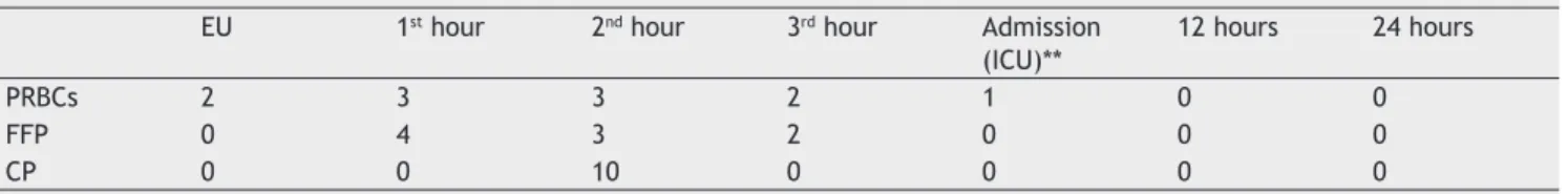
After the fi fth PRBC transfusion, the fl uid transfusion protocol was triggered and the transfusion strategy fol-lowed the FFP/PRBC/PC ratio of 1:1:1, along with Ringer’s Lactate infusion of 15 mL.kg-1.h-1 and correction of metabolic disorders. Table 2 presents data regarding the transfusion of blood products.
At the end stage of initial resuscitation, the patient had received PRBC (10U), FFB (9U), and PC (10U); the pelvic frac-ture had been surgically fi xed, with surgical bleeding under control; there was evolution with improved perfusion rates, progressive reduction of the pharmacological hemodynamic support, and temperature recovery.
Laboratory tests confi rmed the clinical improvement and successful recovery of perfusion, hemodynamics, and hemostasis. The massive transfusion protocol and infusion of norepinephrine were then discontinued (Table 3).
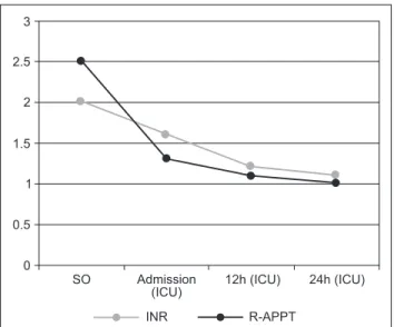
Surgical time was approximately 120 minutes; 30 minutes after the procedure the patient developed a skin rash and mild angioedema, which resolved spontaneously 2 hours later. Figure 1 shows the evolution of coagulopathy during patient’s care.
The patient was taken to the ICU where she received packed red blood cells, without requiring other blood products. Postoperatively, she was hemodynamically stable without requiring norepinephrine and was kept under seda-tion and serial neurological evaluaseda-tion due to the blood pres-ence in the right ventricle, which evolved without surgical indication.
On the third postoperative day, sedation was suspended, resulting in extubation on the seventh day after surgery and discharged from the ICU to a secondary support unit on the eighth day. The patient was discharged from the hospital on the 63rd postoperative day.
Table 1 Initial Data Monitoring in the Operating Room. Mean blood pressure 40 mm Hg
Heart rate 144 ppm
Temperature 34.6ºC
Central venous pressure 3 mm Hg
ETCO2* 25 mm Hg
*ETCO2: end tidal CO2.
Table 2 Blood Products transfused within 24 hours.
EU 1st hour 2nd hour 3rd hour Admission (ICU)**
12 hours 24 hours
PRBCs 2 3 3 2 1 0 0
FFP 0 4 3 2 0 0 0
CP 0 0 10 0 0 0 0
EU: emergency unit; ICU: intensive care unit; PRBCs: packed red blood cells, FFP: fresh frozen plasma, CP: platelet concentrate.
Fluid resuscitation in traumatic hemorrhagic shock is still a matter of debate. The classical approach (infusion of large volumes of fl uid), type of fl uid, and the goals to be achieved have been controversial, as there is evidence that conventional strategies may exacerbate coagulopathy, bleeding, and mortality 2,3.
The concept of hemostatic resuscitation was developed from the experience of emergency medicine in the military, especially during wars in Vietnam, Iraq, and Afghanistan. Hemostatic resuscitation involves early administration of blood products in order to restore both perfusion and coagu-lation and minimize the use of large volumes of crystalloid and their dilutive effect on coagulation 4,5.
In this context, resuscitation is the early use of whole blood or packed red blood cells (PRBC), fresh frozen plasma (FFP), and platelet concentrate (PC) high administration with fi xed ratio between products 5-7.
The objective of this case report is to discuss a case in which the hemostatic resuscitation has been used success-fully in a patient victim of polytrauma with severe hemor-rhagic shock.
Case report
Female patient, aged 32 years, with polytrauma result-ing from car crash against truck, who was brought to the emergency department of the Hospital das Clínicas of the Faculdade de Medicina da Universidade de São Paulo by the Emergency Care Rescue Time (Grupo de Resgate e Atendimento a Urgências – GRAU), air medical service, in the city of São Paulo.
Upon admission, the patient was unconscious, using a cervical immobilization collar, intubated, and on ventilatory support. During clinical examination, she presented with tachycardia; hypotension; anisocoria; Glasgow coma scale 3; and pelvic, humerus, and left jaw fractures; in addition to active bleeding in the right frontoparietal region and large hematoma in the pelvic and hypogastric region.
Additional tests, such as ultrasound, showed no free fl uid in the abdominal cavity, chest X-ray without changes, and brain computed tomography with evidence of bleeding into the right ventricle, but without signs of intracranial hypertension.
The initial fl uid resuscitation was performed with two lit-ers of Ringer’s Lactate and three PRBCs. After closed external immobilization of the pelvis, the patient was immediately taken to the operating room (OR) for hip fracture surgical treatment. The time interval between the patient’s arrival to hospital and OR admission was approximately 60 minutes.
101 Hemostatic Resuscitation in Traumatic Hemorrhagic Shock: Case Report
Discussion
This case report shows that an early, aggressive approach for hemorrhagic shock management, using the damage control resuscitation (DCR) strategy that focuses on hypotensive resuscitation and hemostatic transfusion to control surgical bleeding, plays an important role towards a successful treat-ment. Current approaches prioritize the rapid identifi cation and correction of circulatory collapse, the major event responsible for mortality in these patients.
Although the administration of crystalloids is an estab-lished practice in cases of class I-II hemorrhage, the fl uid resuscitation strategy in trauma with hemorrhagic shock (class III-IV) is up for debate 8,9. In this scenario, the DCR strategy has received greater attention.
In traumatic hemorrhagic shock, the DCR strategy is one of the major therapeutic advances and its application is based on three pillars: abbreviated surgery, reversal of coagulopathy (hemostatic resuscitation), and hypoperfusion (permissive hy-potension). This therapy is indicated in class III-IV hemorrhage and hemorrhagic shock and its goal is focused on fi ghting the lethal triad to abort the vicious cycle of bleeding and avoid the picture irreversibility.
The identifi cation of viable candidates to therapy is the critical point of DCR. The triggers accepted for instituting the hemostatic resuscitation therapy include coagulopathy; blood transfusion > 10 U or > 4 U.h-1; metabolic acidosis with base defi cit > 5; temperature < 35ºC; and hemodynamic instability with insuffi cient response to resuscitation 3,10-13.
Reversing coagulopathy is the main goal for treating severe bleeding. The predominant mechanism of acute trauma coagulopathy (ATC) depends on the degree of mi-croperfusion dysfunction, nature and severity of trauma, and deleterious effects of subsequent medical therapies. ATC has complex and multifactorial pathophysiology, which compromises hemostasis in its entire cascade. In addition, tissue hypoperfusion, metabolic acidosis, hypothermia, and hemodilution are other factors that trigger and maintain this disorder 14,15.
The metabolic markers of hypoperfusion in acute trauma have strong correlation with the incidence of ATC. In 20% of cases, frequently admitted patients with base defi cit over six showed an incidence of coagulopathy, whereas patients without base defi cit showed no change in laboratory markers of coagulation 14,16.
On the other hand, in a rapid attempt to restore tissue perfusion and oxygen delivery, polytrauma patients usually receive crystalloids and packed red cells that have no clotting factors, which causes strong dilutional effect on coagulation factors. Moreover, the clinical condition of these patients is worsened with the usual fi nding of metabolic acidosis (pH < 7.1) and hypothermia (temperature < 34°C) – factors that solely affect hemostasis.
The causes of hypothermia are multifactorial and inter-dependent, including changed central thermoregulation; reduced endogenous heat production, due to tissue hypop-erfusion in hemorrhagic shock; exposure to low temperatures in the operating room; and infusion of crystalloid solution and blood products inadequately warmed 17.
Because the hemostasis process consists of an enzyme cascade dependent on a body temperature of about 37°C to occur normally, in case of hypothermia there is impairment of thrombin generation, platelet aggregation, and fi brin throm-bi, which occurs simultaneously with hyperfi brinolysis 17-20.
Confi rming the deleterious effects of hypothermia on coagulation, acidosis appears as a complicating factor, as it substantially reduces the rate of thrombus formation, assessed by thromboelastography, and platelet aggrega-tion 5. The high level of lactate (lactate > 90 mg.dL-1) alone is proven to contribute to impaired hemostasis via in vitro thromboelastography 21.
Table 3 First 24 Hours Laboratory Evolution.
1st hour 2nd hour 3rd hour Admission (ICU)** 12 hours 24 hours
INR 2 1.6 1.2 1.1
APTT-R 2.5 1.3 1.1 1
Temperature (ºC) 34.6 34.8 34.7 35 35.8 35.8
pH 6.8 7.2 7.2 7.2 7.3 7.4
Base excess (mEq.L-1) -12 -3.1 -5.1 -7.3 -4.3 -1.8
Lactate (mg.dL-1) 58 39 35
Hemoglobin (g.dL-1) 3.3 3.7 9.9 6.7 8.6 9.3
Hematocrit (%) 10.1 11.6 28.7 18.2 25.8 27.9
INR: institutional normalized ratio; R-APTT: ratio of activated partial thromboplastin time.
Figure 1 Behavior of INR and R-APTT over 24 Hours.
2.5
SO Admission
(ICU) 12h (ICU) 24h (ICU)
3
2
1.5
1
0.5
0
102 J. O. Barbosa Neto et al.
The acidosis effect on coagulation is not reversed by simple pharmacological correction of acidosis with bicarbo-nate. Several investigators agree that pH must be over 7.2 before implementing circumstantial therapies for coagula-tion disorders, which reinforces the mandatory recovery of perfusion in coagulopathytreatment 19,20.
In the case reported, the patient was admitted with se-vere anemia (Hb = 3.3 g.dL-1), acidosis (pH = 6.8-1), hyperlac-tatemia (lactate = 58 mg.dL-1), and moderate hypothermia. Given the gravity of the case, transfusion was initiated during surgery to maintain a ratio of 1:1:1 between blood products, with the intention of correcting anemia without increasing hemostatic disorder.
In a cohort study, Sperry et al. 6 found that when mas-sive transfusion is performed, maintaining a high ratio of fresh frozen plasma and packed red blood cells, there is an overall reduction in transfusion requirements in the fi rst 24 hours, despite the greater amount of plasma transfused. In our case, in the fi rst 12 hours the patient received 11 units of PRBCs, 9 units of fresh frozen plasma, and 10 units of platelet concentrate.
There were some limitations in this case, such as the time to perform laboratory tests and the lack of platelet count. Thus, therapy during intraoperative period was given empirically based on surgical bleeding, clot formation in the fi eld, and presence of microvascular bleeding.
Although the optimal therapeutic approach to trau-matic hemorrhagic shock is not yet fully established, the rapid control of bleeding and recovery of perfusion, in addition to well-defi ned therapeutic protocols are fundamental to prevent progression of coagulopathy and refractory shock.
References
1. Sauaia A, Moore FA, Moore EE et al. – Epidemiology of trauma deaths: a reassessment. J Trauma,1995;38:185-193.
2. Nunez TC, Voskresensky IV, Dossett LA, Shinall R, Dutton WD, Cotton BA – Early prediction of massive transfusion in trauma: simple as ABC (assessment of blood consumption)? J Trauma, 2009;66:346-352.
3. Malone DL, Hess JR, Fingerhut A – Massive transfusion practices around the globe and a suggestion for a common massive transfusion protocol. J Trauma,2006;60:S91-96.
4. Miller RD, Robbins TO, Tong MJ, Barton SL – Coagulation defects associated with massive blood transfusions. Ann Surg,1971;174:794-801.
5. Beekley AC – Damage control resuscitation: a sensible approach to the exsanguinating surgical patient. Crit Care Med, 2008;36:S267-274.
6. Sperry JL, Ochoa JB, Gunn SR et al. – An FFP:PRBC transfusion ratio >/=1:1.5 is associated with a lower risk of mortality after massive transfusion. J Trauma, 2008;65:986-993.
7. Dente CJ, Shaz BH, Nicholas JM et al. – Early predictors of massive transfusion in patients sustaining torso gunshot wounds in a civilian level I trauma center. J Trauma, 2010;68:298-304.
8. Krausz MM. Fluid resuscitation strategies in the Israeli army. J Trauma, 2003;54:S39-42.
9. Bickell WH, Wall MJ, Jr., Pepe PE et al. – Immediate versus delayed fluid resuscitation for hypotensive patients with penetrating torso injuries. N Engl J Med,1994;331:1105-1109. 10. Parr MJ, Alabdi T – Damage control surgery and intensive care.
Injury,2004;35:713-722.
11. Blackbourne LH – Combat damage control surgery. Crit Care Med,2008;36:S304-310.
12. Bormanis J – Development of a massive transfusion protocol. Transfus Apher Sci, 2008;38:57-63.
13. Hardy JF, De Moerloose P, Samama M – Massive transfusion and coagulopathy: pathophysiology and implications for clinical management. Can J Anaesth, 2004;51:293-310.
14. Brohi K, Cohen MJ, Davenport RA – Acute coagulopathy of trauma: mechanism, identifi cation and effect. Curr Opin Crit Care,2007;13:680-685.
15. Scalea TM, Bochicchio KM, Lumpkins K et al. – Early aggressive use of fresh frozen plasma does not improve outcome in critically injured trauma patients. Ann Surg, 2008;248:578-584.
16. Brohi K, Cohen MJ, Ganter MT, Matthay MA, Mackersie RC, Pittet JF – Acute traumatic coagulopathy: initiated by hypoperfusion: modulated through the protein C pathway? Ann Surg, 2007;245:812-818.
17. Spahn DR, Rossaint R – Coagulopathy and blood component transfusion in trauma. Br J Anaesth, 2005;95:130-139. 18. Cosgriff N, Moore EE, Sauaia A, Kenny-Moynihan M, Burch JM,
Galloway B – Predicting life-threatening coagulopathy in the massively transfused trauma patient: hypothermia and acidoses revisited. J Trauma,1997;42:857-861.
19. Lier H, Krep H, Schroeder S, Stuber F – Preconditions of hemostasis in trauma: a review. The infl uence of acidosis, hypocalcemia, anemia, and hypothermia on functional hemostasis in trauma. J Trauma, 2008;65:951-960.
20. Martini WZ, Pusateri AE, Uscilowicz JM, Delgado AV, Holcomb JB – Independent contributions of hypothermia and acidosis to coagulopathy in swine. J Trauma, 2005;58:1002-1009. 21. Engstrom M, Schott U, Romner B, Reinstrup P. Acidosis impairs