ABSTRACT
INTRODUCTION
Increased life expectancies, improvements interventions have all contributed towards a longer retention of natural teeth in old age. Exposed root surfaces are common in adults and older populations, generally due to gingival recession, which is associated with anatomical factors,
gels on root dentin
Fernanda Tavares BORGES1, Wagner Reis da Costa CAMPOS2, Lais Sant’ana MUNARI3, Allyson Nogueira
MOREIRA4, Saul Martins PAIVA5 MAGALHÃES6
1- DDS, Graduate student, Department of Restorative Dentistry, School of Dentistry, Federal University of Minas Gerais, Belo Horizonte, MG, Brazil. 2- Metallurgist Engineer, PhD, Center of Nuclear Technology Development, Belo Horizonte, MG, Brazil.
3- Graduate student, Department of Restorative Dentistry, School of Dentistry, Federal University of Minas Gerais, Belo Horizonte, MG, Brazil.
4- DDS, PhD, Associate Professor, Department of Restorative Dentistry, School of Dentistry, Federal University of Minas Gerais, Belo Horizonte, MG, Brazil. 5- DDS, MS, PhD, Associate Professor, Department of Pediatric Dentistry and Orthodontics, School of Dentistry, Federal University of Minas Gerais, Belo Horizonte, MG, Brazil.
6- DDS, MS, PhD, Associate Professor, Department of Restorative Dentistry, School of Dentistry, Federal University of Minas Gerais, Belo Horizonte, MG, Brazil.
Corresponding address: Claudia Silami Magalhães - Rua Ludgero Dolabela, 139 / 301 - Bairro Gutierrez - 30-430130 - Belo Horizonte - MG - Phone / Fax: 31 33321784 / 34092430 - e-mail:[email protected]
!
S
econdary caries is still the main cause of restoration replacement, especially on the root surface Objective: This in vitroamong them at different distances from restoration margins. Standardized cavities were prepared on 240 bovine root specimens and randomly assigned to 15 groups of treatments (n=16). Cavities were ! " #$%&'*&+;<$%&'*&+;> ?* & Bond NT (Dentsply); Charisma/Gluma One Bond (Heraeus Kulzer) and the control, Z250/Single Bond (3M-ESPE). The specimens were subjected to a pH-cycling model designed to simulate >QV%W VXW or deionized/distilled water (control) was applied to the specimens for 4 min. The surface Knoop microhardness test was performed before (KHNi) and after (KHNf) the pH cycles at 100, 200 and 300
P > !
values (KHNi - KHNf). Data were analyzed by Friedman’s and Wilcoxon’s tests, ANOVA and Tukey’s
test (D=5%)]^ !
$_XQXV+ > " ! highest cariostatic effect. Vitremer presented a moderate effect, while Dyract and Charisma did not `VqX^ {{ ! ! | ^ demonstrated.
Key words: Fluorides. Dental restoration. Dental caries.
microbially induced periodontal diseases, surgical periodontal therapies, orthodontic movement of the teeth and various forms of trauma14,16. Exposed
dentin on root surfaces becomes more susceptible to caries than enamel because of its low mineral content and its higher critical pH for dissolution8,14.
lesions and reinforcing prophylactic programs must be always the choice for stopping caries
progression8,14. However, when the dental
anatomy and function are involved, is necessary to institute restorative treatments using materials with adequate strength, esthetics, adhesive and cariostatic effects8.
Secondary caries has been shown in studies worldwide to be the most common reason for the replacement of all types of restorations, regardless of the material used18,19. Secondary caries occur
mainly at the cervical margin of restoration, and restorative techniques are sensitive and complicated by the lack of an excellent adhesion to dentin19.
Conventional glass ionomer cements (GICs), ! releasing composite resins could be indicated for the restoration of root caries cavities8,30. Previous
studies have suggested that the release of prevent the formation of wall lesions and prevent caries at the margins of restorations10,12,24,28. The
mechanisms involved in the anticariogenic effects " the enhancement of remineralization5,6,15, the
interference of pellicle and plaque formation and the inhibition of microbial growth and metabolism21,30.
In vitro studies have demonstrated the synergic effect of restorative materials in association with caries adjacent to the restorations11,24. Professional
# $ %
based on
brushing has been suggested, especially for the treatment of caries-active patients. This could # abilities, to the low cost and short operational time of self-applied procedures6,15. Therefore, we
hypothesized that there may be an additional effect restorative materials. This in vitro study compared the cariostatic effects of 5 restorative materials –
$& investigated whether there was any difference containing restorative materials, associated with from the restoration margins.
MATERIAL AND METHODS
Experimental Design
A randomized complete block design was used
containing restorative materials associated with $'* strategy used to control the variability of the dentin substrate. By using this design, the specimens formed a more homogeneous experimental unit on which to compare the treatments20. “Randomized
complete” indicates that each block contains all of the treatments, applied according the randomly determined order.
The factors studied were: restorative material at 5 levels (Figure 1), topical treatments at 3 levels (Figure 1) and distance from the restoration
margins at 3 levels (100, 200 and 300 mm).
The experimental units were 240 bovine root specimens randomly assigned to 15 groups of treatments (n=16). Each block contained each one of all possible combinations of 5 (restorative !+/! (Figure 2). The control group of restorative system 7;<>/ ESPE Dental Products, Sumaré, SP, Brazil). Control group of topical treatments was distilled/deionized $% and water were applied to the restoration and adjacent dentin to test the associated effect of restorative materials and gels.
The restorative materials and topical treatments were applied according to the order randomly
determined by Microsoft Excel® software. The
dependent variable was the Knoop microhardness (KHN) loss of root dentine around the restorations.
Preparation of specimens
Two hundred and forty incisors were extracted from bovine jaws immediately after butchering. The teeth were thoroughly scaled, polished with pumice/water slurry, and washed with distilled water to clean them from debris, and were kept in a 1% chloramine-T solution for 1 week.
The roots were separated from crows and axially hemi-sectioned in a mesiodistal direction. Sections (5.0 mm x 5.0 mm x 2.0 mm) were obtained from the buccal or the lingual root surfaces at 3.0 mm from the cementonamel junction using a double-faced diamond disc (KG Sorensen Ind. e Com. Ltda, Barueri, SP, Brazil) in a low-speed handpiece under water spray coolant. Each section was embedded + mechanical grinder (Polipan-U, Panambra, São Paulo, SP, Brazil) with #400- and #600-grit Al2O3 abrasive papers (Norton Abrasivos, Guarulhos, SP, Brazil).
Cavity preparation and restorative procedures
""!"#$ Manufacturer %&
Ketac-Fil
Glass-ionomer cement
3M ESPE Dental Products (Sumaré, SP, Brazil)
Powder: 142152 Liquid: 135132
Vitremer
]!
3M ESPE Dental Products (Sumaré, SP, Brazil)
Powder: 20011019 Liquid: 20011025 Primer: 20010928 Glaze: 20011031
Dyract-Prime & Bond NT
* !
Dentsply Caulk (Milford, DE, USA)
Dyract: 0108000442 Prime & Bond NT: 3708
Charisma-GLUMA One Bond Fluoride composite resin
Heraeus Kulzer - GmbH (Gonsennheumer, Mainz, Germany)
Charisma: 060044 GLUMA One Bond: 195001
Filtek Z250-Single Bond
Non-Fluoride composite resin (control)
3M ESPE Dental Products (Sumaré, SP, Brazil)
Z250 2XM Single Bond 1105
1.23% Acidulated Phosphate Fluoride gel Vigodent
(Rio de Janeiro, RJ, Brazil)
-2% Neutral Sodium Fluoride gel Vigodent
(Rio de Janeiro, RJ, Brazil)
-Deionized/distilled water (control) -
-Figure 1- The restorative materials and topical treatments applied to the specimens
Groups ""$$"
1 Ketac-Fil + 1.23% Acidulated Phosphate Fluoride gel
2 Ketac-Fil + 2% Neutral Sodium Fluoride gel
3 Ketac-Fil + Deionized/distilled water
4 Vitremer + 1.23% Acidulated Phosphate Fluoride gel
5 Vitremer + 2% Neutral Sodium Fluoride gel
6 Vitremer + Deionized/distilled water
7 Dyract + 1.23% Acidulated Phosphate Fluoride gel
8 Dyract + 2% Neutral Sodium Fluoride gel
9 Dyract + Deionized/distilled water
10 Charisma + 1.23% Acidulated Phosphate Fluoride gel
11 Charisma + 2% Neutral Sodium Fluoride gel
12 Charisma + Deionized/distilled water
13 Filtek Z250 + 1.23% Acidulated Phosphate Fluoride gel
14 Filtek Z250 + 2% Neutral Sodium Fluoride gel
15 Filtek Z250 + Deionized/distilled water
handpiece under a constant water-spray coolant. The prepared cavities were treated in the following manner, according to each restorative material:
Ketac-Fil (3M ESPE): 10% polyacrylic acid was spread over the cavity for 10 s, rinsed for 30 s, and gently air-dried for 5 s, avoiding desiccation. Ketac-Fil powder and Ketac-Ketac-Fil liquid were dispensed at 3.2:1 by weight, hand-mixed within 60 s, loaded into a syringe (Centrix, DFL Ind. e Com. SA, Rio de Janeiro, RJ, Brazil), and injected into the preparation. Then, the surface was covered with a polyester strip and a glass slab and pressed with a weight of 1,000 g for 30 s to extrude any excess. Seven minutes after the beginning of the mixture, the strip was removed and a surface protector (Ketac Glaze, 3M ESPE) was immediately applied and light-cured for 20 s.
Vitremer (3M ESPE): Vitremer primer was applied to the cavity preparation for 30 s, air-dried for 15 s and light-cured for 20 s. Vitremer powder and Vitremer liquid were dispensed at 2.5:1 by weight, hand-mixed within 45 s, loaded into a Centrix syringe, and injected into the preparation. Then, the surface was covered with a polyester strip and a glass slab and pressed with a weight of 1,000 g for 30 s to extrude any excess. The restoration was light cured for 40 s, the strip was W and light-cured for 20 s.
Dyract/Prime & Bond NT (Dentsply Caulk, Milford, DE, USA): 35% phosphoric acid was applied to the cavity for 15 s and rinsed for 30 s. Excess water was blotted using absorbent paper. One coat of Prime & Bond NT was applied to the cavity, left undisturbed for 30 s, blown with air for 5 s, and light-cured for 10 s. Dyract was injected into the cavity preparation, covered with a polyester strip and a glass slab, pressed with a weight of 1,000 g
for 30 s to extrude any excess and light cured for 40 s.
Charisma/GLUMA One Bond (Heraeus Kulzer, Gonsennheumer, Mainz, Germany): 20% phosphoric acid was applied to the cavity for 20 s and rinsed for 40 s. Excess water was blotted using absorbent paper. Two consecutive coats of adhesive were applied, gently air-dried for 5 s and light-cured for 20 s. The bonded cavity was
bulk-and a glass slab bulk-and pressed with a weight of 1,000
g for 30 s to extrude any excess. After removal of the slab, the restoration was light-cured for 20 s.
Z250/Single Bond (3M ESPE): 35% phosphoric acid was applied to the cavity for 15 s and rinsed for 10 s. Excess water was blotted using absorbent paper. Immediately after blotting, 2 consecutive coats of adhesive were applied for 15 s with gentle agitation using a fully saturated applicator. After removal of the solvent by brief air-drying for 5 s, the adhesive was light-cured for 10 s. The bonded #* 7;<> polyester strip and a glass slab and pressed with a
weight of 1,000 g for 30 s to extrude any excess. After removal of the slab, the restoration was light-cured for 20 s.
The materials were light-cured using a halogen light-curing unit (XL300, 3M Dental Products, St. Paul, MN, USA) with an irradiance of 500 mW/cm2.
The specimens were stored in 100% relative humidity for 24 h at 37±1°C. Then, excess restorative material was removed using a water-cooled mechanical grinder (Polipan-U) with #1000-grit Al2O3 abrasive paper (Norton Abrasivos). The restored fragments were cleaned with deionized/ distilled water in an ultrasonic bath for 6 min in order to remove remnants of polishing debris.
pH-Cycling model
In order to standardize the dentin area exposed
to the pH-cycling, the specimens were entirely
covered with dental wax (Herpo Produtos Dentários Ltda, Rio de Janeiro, RJ, Brazil), leaving exposed only the restorations and 2.0 mm around their margins.
Each specimen was individually subjected to a demineralization/remineralization dynamic model, similar to that proposed by Featherstone, et al.9
]^_`!# { 26 (1992),
simulating in vivo high caries activity conditions. Initially, each specimen was immersed in 5 mL remineralizing solution (Ca+2 1.5 mmol/L, PO
4-3 0.9
mmol/L, KCl 150 mmol/L, Tris[hydroxyme2sthyl] aminomethane 2.0 mmol/L at pH 7.0) for 24 h. After, the specimens were washed in deionized/ distilled water and immersed individually, in 5 mL demineralizing solution (Ca+2 2.0 mmol/L, PO
4 -3
2.0 mmol/L in a buffer solution of 0.075 mmol/L of CH3COO- at pH 4.3) for 1 h. After rinsing with
deionized/distilled water, 1.23% acidulated >$>/!;$> >$>/!">$>/ ml) was applied to the restoration and 2.0 mm around its margins for 4 min. Then, the specimens were immersed in 5 mL of remineralizing solution, completing the 24-h cycle. Each pH-cycle totaled 24 h (demineralization: 1 h; topical treatment: 4 min; remineralization: about 23 h). Three pH-cycling runs were completed during 3 consecutive days. The demineralizing and remineralizing solutions were renewed daily11.
Surface Knoop microhardness measurements
Dentin surface microhardness measurements were obtained using a microhardness tester (Durimet, Ernst Leitz GmbH, Wetzlar, Germany) with a Knoop diamond and a 5 g-static-load applied for 30 s1. The surface microhardness values were
measured before and after the demineralization/ remineralization cycles. Nine initial indentations (KHNi) were made at three positions: 0°, 60° and
];>^f) were made at
restoration. In each position, parallel indentations were made 100, 200 and 300 mm from the margin of the restoration11. The KHN values were calculated
using the following equation: KHN = (14229.P) / L2
where P is the applied load (g) and L is the indentation length (mm).
Statistical analysis
For each specimen, the mean of the nine initial
indentations (KHNi!
indentations (KHNf) were considered for statistical analysis. The difference between the initial and
i - KHNf) was
on root dentin. The statistical analysis considered # microhardness values for each association of material and treatment at 100, 200 and 300 mm distances.
The effect of distance on root dentin microhardness loss was evaluated by Friedman’s and Wilcoxon’s tests (D=0.05). An analysis of variance (ANOVA) tested the effects of blocking, the restorative systems, the topical treatments and their interactions for each level of the factor
distance. A multiple-comparison Tukey’s test
(D>$><! #
means of microhardness values. The software SPSS 8.0 (SPSS Inc., Chicago, IL, USA) was used for the analysis.
RESULTS
AFriedman’
differences among the three levels of the factor >$>>!$ & difference between the loss of dentin microhardness at 100 and 300 mm. (Table 1).
%W% factors blocking (p=0.000), the restorative system (p=0.000) and topical treatment (p=0.000) on the surface microhardness loss at 100, 200 and 300 mm. The interaction of particular interest (restorative system x topical treatment) was not ]>> >$]>;! 200 (p=0.849) and 300 mm (p=0.580). As this analyzed separately.
There were differences among
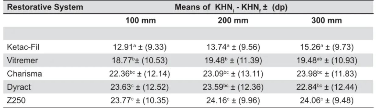
the restorative systems at 100 (p=0.000), 200 (p=0.000) and 300 (p=0.000) mm (Table 2). At a distance of 100 mm, microhardness loss around restorative systems (Vitremer, Charisma, Dyract and Z250). The highest microhardness loss was # (Z250), which was similar to Charisma and Dyract. Vitremer showed a cariostatic effect lower than # Z250. At the distance of 200 mm, the effect of the restorative system was similar to that shown at 100 #W
'"*"+9< KHNi - KHNf9#=<
100 19.39a ± (-12.77/53.30)
200 19.53ab ± (-13.09/54.93) 300 19.56b ± (-11.58/57.93)
Table 1- ! $"i - KHNf), minimum and maximum
values, considering all of the restorative systems and treatments at 100, 200 and 300 mm distances
Statistical differences are expressed by different superscript letters (p< 0.05)
" !""*>?@i - KHNf ± (dp)
D
Ketac-Fil 12.91a ± (9.33) 13.74a ± (9.56) 15.26a ± (9.73)
Vitremer 18.77b± (10.53) 19.48b ± (11.39) 19.48ab ± (10.93)
Charisma 22.36bc ± (12.14) 23.09bc ± (13.11) 23.98bc ± (11.83)
Dyract 23.63c ± (12.52) 23.59bc ± (12.36) 22.84bc ± (12.44)
Z250 23.77c ± (10.35) 24.16c ± (9.96) 24.06c ± (9.48)
Means followed by distinct letters are different when compared in columns (p<0.05) Tukey dms (100 mm) = 4.41; Tukey dms (200 mm) = 4.61; Tukey dms (300 mm) = 4.41
Table 2- ! $"i - KHNf) for the restorative systems
Charisma and Dyract. At the distance of 300 mm, effect than for all other tested materials except for Vitremer, from which it did not differ. Vitremer showed a cariostatic effect higher than Z250, but similar to Charisma and Dyract.
than the control group for all the restorative systems and distances evaluated (p=0.000) (Table 3).
DISCUSSION
& supported by the literature. Fluorides affect caries formation through a variety of mechanisms involving the reduction of demineralization, the enhancement of remineralization, the interference of pellicle and plaque formation and the inhibition of microbial growth and metabolism5,6,15,21. Fluoride
release from restorative materials is related to content and depends on the environmental conditions3,4,7,29,30.
%]>>! # *
material is formed. Depending on the pH of the
2 layer, which has a higher
# + # calcium and phosphate ions.The products of these
# 2
incorporated on tooth surface. This reservoir of ##" and enhance remineralization5,15.
In this study, the differences on cariostatic effect could be attributed to the amount of evaluated by dentin microhardness loss at various distances from restoration margins. Although it was not able to completely inhibit microhardness loss adjacent to the restoration, Ketac-Fil exhibited properties. GIC success in reducing the production of caries-like lesions in the adjacent root surface
corroborates the results obtained by other in
vitro studies10,11,12,24,28. Typical GICs are materials
powder and an aqueous solution of a poly(acrylic acid-itaconic acid) copolymer containing tartaric acid22. It is well known that the setting of these
cements involves neutralization of the polyacid by the basic glass and results in the formation of an ionically cross-linked polyacrylate matrix. Initially, # into the aqueous acid phase and become trapped in the hardening gel matrix. Once the cement has + is loosely bound and free to move7. Moreover, the
erosion of GIC in an acidic environment increases of an acid-resistant, radiopaque, hypermineralized layer around the restorations27.
RMGICs consist substantially of GIC components (water-soluble polymeric acid, ion-leachable glass and water) together with methacrylate monomers and their associated initiation systems. These hybrid materials set partially by means of an acid-base reaction and partially by means of a polymerization reaction that is initiated by photochemical and/or chemical generation of free radicals. The action of 2-hydroxyethyl methacrylate (HEMA) as a cosolvent for organic and aqueous components affects the GIC setting reaction because ions are reluctant to form in the organic medium. Additionally, organic * of the polyacrylic acid molecule22. RMGICs have the
to conventional cements, but this capacity may be affected not only by the formation of complex polyacrylic acid, but also by the type and amount of resin used for the photochemical polymerization reaction30.
In this study, under high cariogenic challenge, # W # development was observed, as dentin microhardness loss was statistically lower in this group than in the control along all distances. However, Vitremer did not inhibit caries development as effectively as
Ketac-$$"*9>?@i - KHNf) ± (dp)
D
Neutral 17.73a±(11.14) 18.53a±(11.56) 19.23a±(11.33)
Acidulate 19.16a±(10.28) 18.81a±(10.63) 18.99a±(10.10)
Control 23.99b±(11.51) 25.08b±(11.64) 24.86b±(11.20)
Means followed by distinct letters are different when compared in columns (p<0.05) Tukey dms (100 mm) = 2.94; Tukey dms (200 mm) = 3.06; Tukey dms (300 mm) = 2.94
Table 3- ! $"i - KHNf) for the topical treatments
Fil at 100 and 200 mm. The extent of the cariostatic effect provided by Ketac-Fil dropped at 300 mm and became equivalent to Vitremer. Moreover, at 200 mm and 300 mm, Vitremer behaved in the same release is expected to be low compared to RMGICs. Hara, et al.13 (2002) demonstrated that the extent
of cariostatic effects provided by conventional GICs and RMGICs was estimated to be about 300 and 150 mm, respectively, in root dentin.
& ! ! were shown to be ineffective in the inhibition of # the control group (Z250) at all the distances.
These results are in agreement with other in
vitro studies11,13,28$
# RMGICs, some cariostatic effect was expected for Dyract. This material does not contain water in its formulation. With time, water must diffuse from the environment toward the material before the
can begin17$ &
released by the ion-exchange mechanism and the short-term de/remineralization regimen used in this study could explain why these materials were not able to inhibit caries lesion progression around the restorations.
Obviously, in Charisma, a TEGDMA-Bis-GMA '% there is no acid-base reaction. The only source resulting in a slow diffusive release. Moreover, the polymer composition of Charisma does not contain HEMA, which would increase the hydrophilicity of the matrix and facilitate the transport of water
4$
levels much lower than the levels released from conventional GICs or RMGICs and also somewhat
3,30.
Under the in vitro simulation of high cariogenic
restorative materials did not completely inhibit dentin microhardness loss around the restorations. % drop to very low levels after a higher initial release
of ions3,4,30
gels could be an interesting strategy to maintain high risk of developing caries. Some guidelines * are available. In the present study, an intensive therapeutic program was simulated with daily 6,15,25. This could
be applied at home using a toothbrush or a custom-made tray, or it could be professionally supervised for noncompliant home users.
The interaction of particular interest (restorative +!
any of the distances studied. Hence, the analysis the materials tested. Neutral 2% NaF and 1.23% APF gels promoted a similar protection against surfaces. This effect is explained by the capacity of * onto tooth surfaces, which serves as a reservoir for
30.
However, in vitro studies have also shown
that 1.23% APF gel is more reactive than 2% NaF gel. CaF2 formation increases in acidulate media microhardness and acid-resistance of dental structures23. Additionally, the ppmF concentration
of APF gel [12,300 ppm] is higher than that of 2% NaF gel [9,040 ppm], with a higher formation of *$& similar cariostatic effects of neutral and acidulated +$ # that the acidic pH of APF gel promoted an initial microhardness loss in root dentin in spite of the *$
The ability to carry out experiments under highly controlled conditions represents the major advantage of in vitro experimentation27. However,
caries lesions does not take into account the effects of loss of the dentin organic phase during root caries, the effects of vital dentin reactions in vivo
or the activity of salivary enzymes, thus limiting its clinical relevance. Nevertheless, the ability to quantify changes in mineral content in response to of value in assessing their potential to inhibit root caries2,23,28.
In vitro models for caries induction on root complicate microhardness analysis. Hara, et al.11
(2002) suggested reducing the number of de/ remineralization cycles and the time of immersion in the demineralizing solution in order to avoid $ the model proposed by Featherstone, et al.9 (1986)
# { 26 (1992) was
adjusted to make it suitable for the microhardness test. Hence, the specimens were immersed in remineralizing solution for 24 h prior to submission to three cycles of de/remineralization.
The Knoop microhardness value evaluates mineral gain and loss over the course of the de/ remineralization process, taking into account the organic properties of the root dentin. This is an advantage over other methods like microradiography that quantify the mineral content + matrix11-13. However, dentin is more porous and less
measures. Hence, to minimize the effects of the dentin substrate on surface microhardness, a Knoop diamond with a 5 g static load was applied
for 30 s1^^
on a representative circular area of dentin around the restoration.
CONCLUSIONS
Under the conditions of this in vitro study, it # application inhibit the progression of caries-like lesions adjacent to restorations, but are not able to prevent the formation of the lesion. Conventional GICs and RMGICs reduced the microhardness # recommended for root caries control, primarily in high-risk patients. The use of neutral or acidulate # the effects associated with restorative materials have not been demonstrated.
ACKNOWLEDGEMENTS
This research study was based on a thesis submitted to the Federal University of Minas for granting a MS degree, and was supported by a % (Grant #CDS79/2003) and PRPq/UFMG.
REFERENCES
1- American Society for Testing and Materials (ASTM). Annual book of ASTM standards. Norm E384-99: Standard test method for microindentation hardness of materials. Philadelphia: ASTM; 1999.
2- Arends J, Ruben JL, Christoffersen J, Jongebloed WL, Zuidgeest TGM. Remineralization of human dentine in vitro. Caries Res. 1990;24:432-5.
3- Can-Karabulut DC, Batmaz I, Solak H, Tastekin M. Linear various restorative materials. Dent Mater. 2007;23:1057-65. 4- Chan WD, Yang L, Wan W, Rizkalla AS. Fluoride release from dental cements and composites: a mechanistic study. Dent Mater. 2006;22:366-73.
5- Cury JA, Tenuta LMA. Enamel remineralization: controlling the caries disease or treating early caries lesions? Braz Oral Res. 2009;23(Sp. Issue 1):23-30.
6- Delbem ACB, Brighenti FL, Vieira AEM, Cury JA. In vitro
comparison of the cariostatic effect between topical application $ % {$ 2004;12:121-6.
7- Dhondt CL, De Maeyer EA, Verbeek RM. Fluoride release $$ 2001;80:1402-6.
8- Donovan T. Critical appraisal: Protocol for the prevention and management of root caries. J Esthet Restor Dentistry. 2008;20:405-11.
9- Featherstone JDB, O’Reilly MM, Shariati M, Brugler S. Enhancement of remineralization in vitro and in vivo. In: Leach SA. Factors relating to demineralization and remineralization of the teeth. Oxford: IRL Press; 1986. p. 23-4.
10- Gonzalez Ede H, Yap AU, Hsu SC. Demineralization inhibition of direct tooth-colored restorative materials. Oper Dent. 2004;29:578-85.
11- Hara AT, Magalhães CS, Serra MC, Rodrigues AL Jr. Cariostatic
dentifrices on root dentin. J Dent. 2002;30:205-12.
12- Hara AT, Queiroz CS, Freitas PM, Giannini M, Serra MC, Cury JA. Fluoride release and secondary caries inhibition by adhesive systems on root dentin. Eur J Oral Sci. 2005;113:245-50. 13- Hara AT, Turssi CP, Serra MC, Nogueira MCS. Extent of the # restorative materials. Oper Dent. 2002;27:480-7.
14- Heijnsbroek M, Paraskevas S, Van der Weijden GA. Fluoride interventions for root caries: a review. Oral Health Prev Dent. 2007;2:145-52
]<%"$% in situ study. J Dent. 2008;36:396-401.
16- Kassab MM, Cohen RE. The etiology and prevalence of gingival recession. J Am Dent Assoc. 2003;134;220-5.
17- Meyer JM, Cattani-Lorente MA, Dupuis V. Compomers: between glass-ionomer cements and composites. Biomaterials.1998;19:529-39.
18- Mjör IA. Clinical diagnosis of recurrent caries. J Am Dent Assoc. 2005;136:1426-33.
19- Mjör IA. Secondary caries: a literature review with case reports. Quintessence Int. 2000;31:165-79.
20- Montgomery DC. Design and analysis of experiments. 3rd ed. New York: Wiley; 1991.
21- Nakajo K, Imazato S, Takahashi Y, Kiba W, Ebisu S, Takahashi N. Fluoride released from glass-ionomer cement is responsible to inhibit the acid production of caries-related oral streptococci. Dent Mater. 2009;25:703-8.
22- Nicholson JW. Chemistry of glass-ionomer cements: a review. Biomaterials. 1998;19:485-94.
23- Nyvad B, Ten Cate JM, Fejerskov O. Arrest of root surface caries in situ. J Dent Res. 1997;76:1845-53.
24- Okuyama K, Nakata T, Pereira PN, Kawamoto C, Komatsu H, { $ # $ $ 2006;31:135-42.
25- Pedrini D, Delbem ACB, França JGM, Machado Tde M. Fluoride release by restorative materials before and after a topical $'$;>>/]]/]$ 26- Serra MC, Cury JA. The in vitro effects of glass-ionomer cement restoration on enamel subjected to a demineralization and remineralization model. Quintessence Int. 1992;23:143-7. 27- Ten Cate JM, Buijs MJ, Damen JJM. The effects of GIC restorations on enamel and dentin demineralization and remineralization. Adv Dent Res. 1995;9:384-8.
28- Torii Y, Itota T, Okamoto M, Nakabo S, Nagamine M, Inoue $ # # releasing restorative materials. Oper Dent. 2001;26:36-43. 29- Weidlich P, Miranda LA, Maltz M, Samuel SM. Fluoride release and uptake from glass-ionomer cements and composite resins. Braz Dent J. 2000;11:89-96.