Martinez CAR, Fabris FM, Silva CMG, Rodrigues MR, Sato DT, Ribeiro ML, Pereira JA. Oxidative stress and changes in the content and pattern of tissue expression of β-catenin protein in diversion colitis. J Coloproctol, 2012;32(4): 343-358.
ABSTRACT: Objective: The aim of this study is to verify if oxidative stress is related to changes in content and pattern of β-catenin protein expression in an experimental model of diversion colitis. Methods: Sixty Wistar rats were submitted to intestinal bypass. The animals were di-vided into three groups according to the sacriice to take place in six, 12 and 18 weeks. For each group, ive animals only underwent laparotomy (control). The presence of colitis was diagnosed by histological study, and its severity, by inlammation grading scale. Cellular oxidative stress was measured by comet assay. Tissue expression of β-catenin protein was analyzed by the immunohistochemistry and quantiication of its tissue content by computerized morphometry. Statistical analysis was performed with the Student’s t-test, median, Mann-Whitney, ANOVA and Kruskal-Wallis, adopting a signiicance level of 5% (p <0.05). Results: Colon segments without fecal stream developed colitis, which worsened with time of exclusion. Segments without fecal stream suffer higher levels of oxidative stress when compared to those with stream, and it worsens with time of exclusion. The levels of cellular oxidative stress are directly related to the degree of inlammation. The total con-tent of β-catenin in segments without fecal stream reduces after six weeks, and does not vary thereafter. The content of β-catenin in the apical portion of the colon crypts decreases with time, whereas in the basal region, it increases. The total content of β-catenin is inversely related to the degree of inlammation and levels of tissue oxidative stress levels. Conclusion: There are changes in tissue content of E-cadherin and increased expression of β-catenin in proliferative regions of colonic crypts, related with oxidative tissue stress.
Keywords: colon; colitis; oxidative stress; adherens junctions; cell adhesion molecules; catenins; comet assay; immunohistochemistry; fatty acids, volatile; rats.
ReSuMO: Objetivo: O objetivo do presente estudo é avaliar a relação entre estresse oxidativo e conteúdo tecidual de β-catenina em modelo experimental de colite de exclusão. Métodos: Sessenta ratos Wistar foram submetidos à derivação intestinal e divididos em três grupos experimentais segundo o sacrifício ser realizado em 6, 12 e 18 semanas. Para cada grupo, cinco animais foram submetidos apenas a laparotomia (controle). A colite foi diagnosticada por estudo histológico, enquanto sua intensidade por escala de graduação inlamatória. Os níveis de estresse oxidativo foram mensurados pelo ensaio cometa, enquanto a expressão e o conteúdo tecidual de β-catenina por imunoistoquímica e morfometria computadorizada, respectivamente. Os resultados foram analisados pelos testes t de Student, Mann Whitney, ANOVA e Kruskal-Wallis, estabelecendo-se nível de signiicância de 5% (p<0,05). Resultados: Nos segmen-tos sem trânsito fecal ocorre desenvolvimento de colite que piora com o tempo de exclusão. Segmensegmen-tos sem trânsito sofrem maiores
Oxidative stress and changes in the content and pattern of tissue
expression of β-catenin protein in diversion colitis
Carlos Augusto Real Martinez1, Fabiano Marcelo de Fabris2, Camila Morais Gonçalves da Silva3, Murilo Rocha Rodrigues4, Daniela Tiemi Sato4, Marcelo Lima Ribeiro5, José Aires Pereira6
1Adjunct Professor of the Postgraduate Program of Health Sciences at Universidade São Francisco (USF) – Bragança Paulista (SP), Brazil. 2Master’s Postgraduate Program of Health Sciences at USF – Bragança Paulista (SP), Brazil. 3Master’s degree in Health Sciences in the Postgraduate Program of Health Sciences at USF – Bragança Paulista (SP), Brazil. 4Medical student at USF – Bragança Paulista (SP), Brazil. 5Doctor in Pharmacology at Universidade Estadual de Campinas (UNICAMP); Assistant Professor of the Postgraduate Program of Health Sciences at USF – Bragança Paulista (SP), Brazil. 6Master’s degree in Pharmacology at USF; Assistant Professor of Pathology at the Medical School of USF –
Bragança Paulista (SP), Brazil.
Study carried out at the Postgraduate Program of Health Sciences at Universidade São Francisco – Bragança Paulista (SP), Brazil. Financing source: São Paulo Research Foundation (FAPESP) – Project number: 2010/12492-7
Conlict of interest: nothing to declare.
INTRODuCTION
The colonic mucosa is one of the most perfect functional barriers of the human body1. It is formed
by a single cell layer, separating the intestinal content, which is rich in bacteria, from the internal sterile intes
-tinal wall. The maintenance of this eficient
function-al barrier is determined by a series of defense mecha
-nisms. When they act together, they protect the internal
intestinal wall from bacterial invasion1. The main
com-ponents of this defense system are represented by the mucus that covers the mucosal surface, the colonocyte apical and basolateral membranes, the adhesion sys -tems formed by intercellular junctions, desmosomes,
hemidesmosomes and, inally, the basal membrane1,2.
From the defense systems, the intercellular junc
-tions (ICJ) represent one of the most eficient
mecha-nisms1,3. They are formed by three types of junctions:
occluding (OJ), adherens (AJ) and communication
(CJ). The OJs are located in the apical portion of the
intercellular space and seal the space between neigh -boring cells, thus preventing the migration of small molecules3-6. The AJs, located right below the OJs,
connect the cytoskeleton of a cell to its neighbor, and
they also play a relevant role in the mechanisms of cellular signalling7. They are formed by a
transmem-brane protein called E-cadherin and by cytoplasmatic
proteins of the catenin family (α, β, γ)8. Finally, the CJs control the transmission of electrical and chemical
signals from one cell to the other, making a complex cell communication network9-11.
For the development of colitis, it is necessary that epithelial barrier mechanisms be compromised12-15. However, the rupture of these defense systems should
be the irst stage, which comes before the bacterial in-vasion of the submucosa and the subsequent
inlam-matory response1. Among the ICJs, AJs are the most
compromised ones in patients with inlammatory bowel diseases (IBD)16. Important changes in
con-tent and chemical structure of proteins that form JA were described in patients with ulcerative rectoco -litis (URC)5,8,17,18.
Studies demonstrated that the tissue content and lo
-cation of the proteins that constitute AJs are modiied in the tissue that is chronically inlammed in IBD, URC,
colorectal cancer (CRC), CRC associated with URC, and in models of experimental colitis13,14,19-23. A strong rela-tion was found between the reduced tissue expression of
β-catenin protein and the worsening of URC24. The
im-portance of β-catenin protein in the early stages of the development of colitis is more evident when it is dem
-onstrated that knockout mice (Min-/-), for the genes that translate the AJ proteins, develop severe forms of colitis at early stages25. These indings present the existing relation between β-catenin protein tissue changes and the worsened colitis24,26,27. However, the molecular
mechanisms that determine the ruptured ICJs in pa-tients with URC are not yet enlightened. Among the
possibilities, it has been recently shown that oxygen free radicals (OFR) can be the molecules responsible for the initial damage to the mucosal barrier1. Since
they are toxic radicals, its excessive production is able
to damage the ICJs, allowing the migration of bacteria
through the intercellular space1. Experimental studies
conirmed the harmful role of OFR by demonstrating
that the exposure of colonic mucosa to hydrogen per -oxide (H2O2), which is a strong OFR producer, enables the appearance of colitis28,29. When demonstrated that OFR can cause damage to the epithelial barrier, it is reasonable to suppose that they can also damage the AJs in the early stages of colitis1,30,31. However, most
experimental models of colitis do not allow the analy
-níveis de estresse oxidativo quando comparados àqueles com trânsito, piorando com o tempo de exclusão. Os -níveis de estresse oxida-tivo encontram-se diretamente relacionados a piora da inlamação. O conteúdo total de β-catenina no cólon sem trânsito reduz após seis semanas de exclusão. O conteúdo de β-catenina no ápice das criptas cólicas diminui com o tempo, enquanto na região basal, aumenta. O conteúdo total da β-catenina encontra-se inversamente relacionado ao grau de inflamação e aos níveis de estresse oxidativo. Conclusão: Existe redução no conteúdo de β-catenina, principalmente no ápice das glândulas cólicas e aumento nas regiões basais, relacionadas à piora do estresse oxidativo.
sis of this possibility, since it causes damage to the colonic barrier due to the exposure of the mucosa to harmful substances, such as trinitrobenzene sulfonic
acid (TNBS), acetic acid or dextran sulfate sodium
(DSS)26,30,32. Actually, these models do not reproduce
the initial molecular mechanisms that cause the muco
-sal barrier to rupture in the different forms of colitis. However, they conirm that the integrity of defense mechanisms is indispensable to stop bacterial inil-tration, thus keeping the local immune response at a quiescent state33. Therefore, the ideal experimental
model to conirm the initial stages of colitis should
cause damage in the defense systems, and among
them, in ICJs, just with changes in the metabolism
of epithelial cells, without damaging the functional
barrier artiicially32.
Glotzer et al.34, in 1981, described the
develop-ment of an inlammation in the colonic mucosa
with-out fecal stream, similar to what happens with URC,
and this condition is called diversion colitis (DC). DC appears due to the interruption in the supply of
short-chain fatty acids (SCFA), main energetic substrate for the oxidative metabolism of epithelial cells in the co -lonic mucosa32,35,36. It has been demonstrated that cells from the colonic epithelium without fecal stream pro -duce increasing amounts of OFR with time of exclu -sion, and that the resulting oxidative stress is related to the epithelial lesion31-33,37-40. It is possible that the lesion in the colonic mucosa, which triggers DC, is
related to the ruptured ICJs, and especially AJs, which
is a result of the increased production of OFR by the
changes in the oxidative cellular metabolism.
Although it has been demonstrated, in models of chemically induced colitis, that there are changes in the expression of β-catenin protein in the sore mu-cosa, the evaluation of content and changes in the pat -tern of the protein expression has not been studied in
experimental models of DC. Thus, the objective of this study is to check if there is a relation between the
oxidative stress and the changes in content and pat -tern of β-catenin protein expression in an
experimen-tal model of DC.
MeTHOD
This study obeys the Federal Law 11,794,
from October 8, 2008, and the guidelines of Colégio
Brasileiro de Experimentação Animal (COBEA). This
project was approved by the Animal Research Ethics Committee of Universidade São Francisco, Bragança
Paulista (SP).
Animals used for experimentation and experimental groups
Sixty SPC male Wistar rats, whose weight var
-ied from 300 to 350 g and with mean age of 4 months were used. Three experimental groups with 20
ani-mals were randomly constituted and divided accord
-ing to the sacriice to take place in 6, 12 and 18 weeks after surgical intervention. Each group was then
di-vided into two subgroups called experiment and con
-trol. In the experiment group, with 15 animals, bowel
stream was bypassed in the left colon, while in the
control group, composed of ive rats, there was only laparotomy, without stream bypass. Animals were kept in individual cages during the experiment, in a
climatized environment, with temperature, light, hu
-midity and noise control. For surgery, they were anes-thetized with 2% xylazine hydrochloride (Anasedan®)
and ketamine chloride (Dopalen®), 0.1 mL/100 g,
ad-ministered via intramuscular in the left back paw.
Surgical technique
After the anesthesia, a trichotomy of the anterior abdominal region was performed, followed by a medi -al longitudin-al 3 cm long incision and posterior open
-ing of the abdominal wall by layers. The left colon was identiied and cut 4cm above the Peyer’s patch,
and the proximal segment was exteriorized as termi
-nal colostomy in the left hypocondrium, ixated to the skin with separate stitches made with absorbable monoilament thread. The caudal segment of the large
intestine was catheterized and irrigated with 40 mL of
0.9% physiological solution at 37ºC, until the efluent drained by the anus did not present with stool. After the irrigation, the distal colon was exteriorized, like a distal mucus istula in the left iliac fossa. Afterwards,
the abdominal wall was closed with two suture lay
-ers (aponeurosis and skin). After the surgery, the rats were kept warm in an incubator, and after anesthesia
recovery, they were placed in individual cages previ
after they regained consciousness. They remained in individual cages until the day of sacriice, and no addi-tional care was taken in relation to the surgical wound and stomas. No antibiotics or analgesic were adminis-tered, and during postoperative follow-up it was nec-essary to sacriice one animal, which was replaced, due to bowel obstruction caused by internal hernia.
Material collection
The day before the date scheduled for material collection, the animals were fasting for 24 hours, ex
-cept for water. For the removal of colonic fragments to
be analyzed, they were under anesthesia with the same
technique previously described. After the cavity was
reopened, the whole colon with stream was removed, including colostomy, and the caudal segment, with
-out fecal stream, involving the anus. In the animals
of the control subgroup, the whole large intestine,
including the anus, was resected. Immediately after
removal, the colonic segments were opened longitu -dinally by the anti mesocolic border and washed care -fully with warm physiological serum to remove fecal
residue. Two fragments were taken from each colonic
segment, and each of them were 20 mm long, being one of them sent to histological and immunohisto -chemical analysis, and the other one to determine the tissue levels of cellular oxidative stress by the comet
assay. In the animals of the control group, the same
number of fragments was collected from the left co
-lon, also 20 mm long, 10 mm above the Peyer’s patch.
For those addressed to measuring the levels of cellular oxidative stress, the mucosa was separated from the
other layers of the wall by microdissection. The
re-moved part was immediately stored in eppendorf, with
freezing solution and at -80ºC.
Histological analysis
For the histological analysis, the fragments of colons with and without stream of each animal in the experiment group, and of the left colon of animals in
the control group, were ixated onto a lat cork surface with the mucus side facing up. After identiication, they were stored in a 10% buffered formaldehyde so-lution, and remained there for 72 hours. After this
pe-riod, they were washed in running and distilled water, and then dehydrated in successive increasing concen
-trations of alcohol and clariied in xylene. Afterwards,
they were included in parafin blocks and submitted to two longitudinal cuts, 5 μ thick, for the histologi-cal and immunohistochemihistologi-cal studies. The slides des-tined to diagnose colitis and to grade the inlammation score were stained with hematoxylin-eosin (HE). For
the evaluation of colitis severity, the grading system
for inlammation was used, being previously proposed
and validated, which considers the presence of ero -sions and ulcers on the colonic mucosal surface and
the intensity of the inlammatory cell iniltrate41.
Immunohistochemical technique
For the research of tissue β-catenin protein ex-pression, histological cuts obtained from all the sam -ples were analyzed (colons with and without stream and left colon of the control group) in the three peri
-ods of exclusion proposed. After being deparafinized,
the cuts were rehydrated in alcohol at decreasing con
-centrations and washed in distilled water. Afterwards, they were submersed in PBS (0.05 M, pH 7.2) for 10 minutes and the dry slides at ambient temperature. The endogenous peroxidase activity was blocked with 3% H2O2 at ambient temperature for 10 minutes, fol
-lowed by another wash with PBS for more 10 minutes.
Afterwards, the antigen recovery with 10 mM sodi
-um citrate was performed (pH 6.0) in bath water at 95ºC for 45 minutes. For the study of tissue β-catenin expression, the anti-β-catenin primary antibody was used (Dako — Denmark A/S, Glostrup, DE — Ref. M3539, Lot 10025022) diluted at 1:50 in bovine al-bumin (1%). The slides were covered with 100 µL
of the solution containing the primary antibody and
stored at 4ºC for 24 hours. After the conclusion of this stage, they were washed with PBS, incubated with
the secondary antibody and submitted to the complex
biotin-streptavidin peroxidase staining for 45 min-utes, prepared with 1:100 dilution in PBS. The slides
were developed with a solution of diaminobenzidine
tetrahydrochloride (DAB, 10 mg in 10 mL of PBS
with 3 mL H2O2), which was dropped over the blades
and incubated for 3 minutes. Afterwards, they were
washed and counterstained with methyl green, and
again washed in distilled water. After the
counter-staining, there was dehydration by immersion in in
-creasing solutions of ethanol and xylene. Finally,
they were set, labeled and stored in the horizontal
Measuring the content and evaluating the pattern of β-catenin expression
The presence of β-catenin was considered as
pos-itive when the brownish coloration was diffusely pres-ent, with variable intensity regions and i ne granular
distribution in the apical and basolateral membrane,
cytoplasm and cell nucleus. According to the
recom-mendation of the manufacturer, the negative control
of immunocoloration was performed without the
addi-tion of the primary antibody, and the positive one used the neoplastic colonic tissue, which is positive for the
protein. The analysis of the β-catenin expression was performed with a common optical microscope, Nikon Eclipse DS50 (Nikon Inc., Osaka, Nippon), with i -nal 200x magnii cation, by an experienced pathologist in immunohistochemical techniques. He did not know the origin of the material and objectives of the study.
The photographic documentation was obtained
with a video capture camera DS-Fi-50 (Nikon Inc., Osaka, Nippon), previously attached to the
micro-scope, and the images were digitized, identified
and filed in a computer.
The pattern of expression of β-catenin protein
was evaluated according to the place of greater ex-pression along the colonic crypts (apex or base),
clas-sifying the intensity of immunocoloration in each of
the sites into crosses: + mild expression; ++ moder-ate expression; and +++ intense expression. The pat-tern of i nal tissue expression for each slide was the median found after reading three different i elds with at least three full and contiguous crypts. The grading
intensity expressed into crosses was performed by
two independent observers, and the conl icting results were analyzed afterwards.
The total tissue content of β-catenin was
mea-sured by computer assisted image analysis (comput-erized morphometry) in three i elds, which showed, in a longitudinal cut, three contiguous and full crypts. The selected image was captured by the camera,
pro-cessed and analyzed by the software NIS-Elements
(Nikon Inc., Osaka, Nippon). For the quantii cation
of β-catenin content in each chosen Field, in 100
mi-crometers rgb wavelength i lter was selected contain-ing the brown color (which identii ed the tissue
immu-noexpression of β-catenin). Afterwards, the software transformed the captured content into a binary image in which the white color represented the presence of
protein, and black represented the rest of the i eld.
The values found for the total content of β-catenin
were expressed as percentage per i eld. The i nal
num-ber adopted for the animals of subgroups control and experiment (segments with and without intestinal stream) was always represented by median, with the
respective standard deviation.
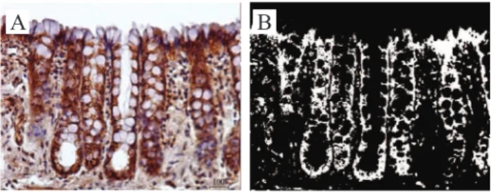
Figure 1A exemplii es the expression of β-catenin
protein in the mucosal layer of the colon with fecal
stream after 18 weeks of bypass, while Figure 1B shows the quantii cation of protein in the same i eld shown by Figure 1A, in binary image, using the com-puter-assisted image processing.
evaluation of oxidative stress levels
The quantii cation of oxidative stress levels by
gel electrophoresis of isolated cells (comet assay) was
performed according to the previously described tech-nique42. Briel y, all the samples from animals in the
control and experiment groups, with colon with or
without intestinal stream, underwent triplicate analy-sis. The specimens were incubated in 3 mL of Hank’s buffered solution (Invitrogen, Carlsbad, CA, USA), with 5.5 mg of proteinase K (Sigma Chemical, CO, St. Louis, MO, USA) and 3 mg collagenase for 45 minutes at 37ºC for the isolation of colonic mucosal cells. Parts were removed and the cellular viability was assessed. Finally, the l uorescein diacetate (FDA) / ethidium bro-mide (EtBr) method (Sigma-Aldrich, St. Louis, MO, USA) was used. The solution of cell coloration was
prepared immediately before its use, and it contained
30 mL of FDA in acetone (5 mg/mL), 200 mL of EtBr in phosphate buffered solution (PBS; 200 mg/mL), and 4.8 mL of PBS.
Figure 1. (A) Pattern of β-catenin expression in the mucosa of the colonic segment with fecal steram after 18 weeks of intestinal
bypass (immunohistochemistry 400x). (B) Tissue β-catenin content
(white color) by binary analysis of computer assisted image in the
same i eld described in Figure 1A (400x).
The suspension containing isolated cells was
isolated, and then mixed to 25 mL of stain solution,
placed over the slide, covered with glass slides and
read in luorescence microscope. The nuclei of viable
cells were stained green, and those of unviable cells
were stained red. After the analysis of the slides, only tissue samples that presented more than 75% of the viable cells were selected. For the alkaline version of the comet assay in the viable samples, 15 ml of
the previously obtained cell suspension were mixed
to the 0.5% low melting point agarose placed onto a blade and covered with a glass slide. Finally, they were immersed in cold lysis solution (2.5 M NaCl, 100 mM EDTA, 10 mM Tris, 1º SDS, pH10 with 1% Triton X-100 and 10% DMSO) and remained at 4ºC for 12 hours. Subsequently, they were exposed to an alkaline buffer (1 mM EDTA and 300 mM NaOH, pH 13.4) for 40 minutes at 4ºC. Electrophoresis was per-formed in this buffer, inside the refrigerator, at 4ºC, for 30 minutes at 25 V and 300 mA. After electropho-resis, the slides were neutralized (0.4 M Tris, pH 7.5),
stained with Sybr Safe (Invitrogen, Carlsbad, CA,
USA) and analyzed at the luorescence microscope. The whole material was processed and veriied at the same time to avoid technical variations. Two hundred
cells were randomly selected (100 of each intestinal segment, with and without stream and of animals in the control group), which were analyzed with the
Komet 5.5 software (Kinetic Imaging, NY, USA). So,
the value of tail moment (TM) was obtained, and the
means were determined. According to the manufac-turer’s manual, TM is deined as the product between the fragments of tail DNA and the mean distance of migration of the comet tail, which relects the exten-sion of the rupture of DNA helix (oxidative stress). The value can be quantiied by the intensiication of image and computational analysis. For each animal,
the mean obtained from the reading of 100 cells of each colonic segment performed by the same techni
-cian, was used. The technician did not know about the origin of the material and the objectives of the study.
Statistical method
The results were described by mean with the re
-spective standard deviation. The Student’s t-test was
used to assess the total content of oxidative stress, and
the Mann-Whitney test analyzed the total content of
β-catenin, comparing animals from the control and ex-periment groups. The median test was used to analyze
the intensity of expression of β-catenin protein in the apical and basal regions of the colonic glands in con
-trol and experiment groups. The inlammation grading
scale in the different times of exclusion was assessed
by the Main-Whitney test. We applied the ANOVA
test for the analysis of variance in relation with time
of exclusion of stream and total tissue protein content. Kruskal-Wallis was used to evaluate the variation in
the protein expression in apical and basal regions of
the colonic glands in relation to the time of experiment. The established signiicance level was 5% (p<0.05) for all the tests. Statistical analysis was per-formed with the software SPSS (SPSS Inc., Chicago, USA version 13.0).
ReSuLTS
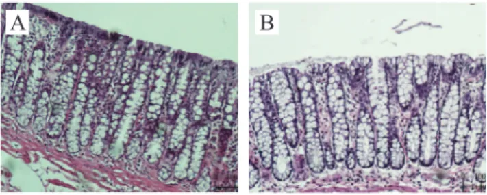
Figure 2A shows the epithelial surface of the
colonic mucosa with fecal stream, while Figure 2B shows the colon without stream after 18 weeks of ex-clusion. It was possible to observe that in segments
without stream the colonic glands were dilated, with
a great amount of mucus in the lumen. The caliciform
cells are dilated and replace the cells with absorptive function in the apical surface, which no longer pres -ent the same juxtaposition by the edema in the stroma,
thus coniguring an aspect similar to a “brush border”.
Figure 3 indicates, by mean and the respective
standard deviation, the values found for the
inlamma-tion score, comparing animals in the control and ex -periment subgroups (segments with and without fecal
stream) in the different proposed periods of exclusion.
We found higher score for those without stream, re
-gardless of considered time of exclusion. The inlam-mation score in the colon without stream after 6 weeks
was 3±0.40, while after 12 and 18 weeks of bowel
ex-clusion these values were 8±0.37, presenting statistical signiicance when compared to the colon with stream and to the animals in the control group (p<0.01). The colon without stream presented increased inlamma-tion score in sacriiced animals in 12 and 18 weeks in relation to those sacriiced after 6 weeks (p<0.05).
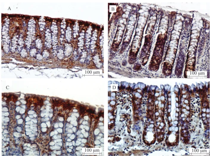
Figures 4A and 4B show, respectively, show the
tissue expression of β-catenin protein in the colonic
of fecal exclusion, while Fgures 4C and D present the protein expression in segments with and without fecal
stream after 18 weeks of exclusion. It is observed that
in the colon with stream (Figures 4A and C), the greater protein expression is concentrated in cells of the apical
surface of colonic glands, while the cells of deeper re-gions present with lower expression. On the contrary, in segments without fecal stream (Figures 4B and D), the
expression of β-catenin protein is more intense in
the deep portions of the Lieberkühn glands, exactly in the proliferative regions of colonic glands.
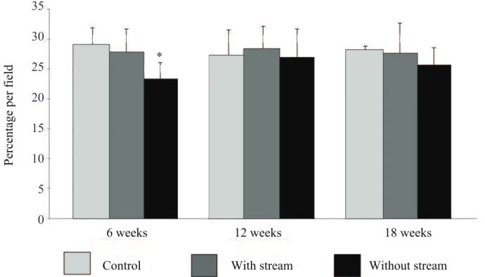
Figure 5 indicates, in average, with the
respec-tive standard deviation, the total tissue content of
β-catenin, comparing the control and experiment sub-groups (with and without fecal stream) in the
differ-ent periods of fecal stream exclusion, measured by
computerized morphometry. In colonic glands, after six weeks of intestinal exclusion, the mean percentage
of β-catenin tissue content was 23.30±3.00%, while
in animals submitted to bypass from 12 to 18 weeks it was 26.79±4.85% and 25.52±3.08%, respectively.
When the total β-catenin tissue content was
consid-ered in the glands of the colonic epithelium, a reduc-tion in the segments without fecal stream was
ob-served in comparison to the colon with stream and the
animals of the control group after only six weeks of fecal exclusion. The total protein content in the colon without stream did not range with the time of exclu-sion (Table 1).
Figure 2. (A) Colonic mucosa with stream after 18 weeks of fecal stream diversion (hematoxylin-eosin 200x); (B) Colonic mucosa without fecal stream after 18 weeks of intestinal diversion (hematoxylin-eosin 200x).
Figure 3. Inl ammation score comparing the control and experiment subgroups (colons with and without stream) in the different periods of exclusion.
Infl
am
m
at
ion gra
de
6 weeks escore
12 weeks 18 weeks
Control With stream Without stream
9 8 7 6 5 4 3 2 1 0
** **
** Signii cant (without stream X control and with stream) (p<0.01). Mann-Whitney test.
Figure 6 shows, in median, the intensity of the variation of β-catenin protein expression in the api-cal regions of the colonic mucosal crypts in animals of control and experiment groups (segments with and
without fecal stream) in different times of exclusion. It was observed that, regardless of the time of
exclu-sion, there was a signii cant reduction in the content
of β-catenin in the apical regions of colonic crypts,
es-pecially after 12 and 18 weeks of exclusion (p<0.05).
The protein content in the apical portions of the colonic glands in the segments without stream did not show
variation according to the time of exclusion.
Figure 4. (A) β-catenin expression in the mucosa of the colonic segment with fecal stream after 12 weeks of intestinal bypass (immunohistochemistry 200x); (B) Protein expression in the mucosa of the colonic segment without fecal stream after 12 weeks of intestinal
bypass (immunohistochemistry 400x); (C) β-catenin protein expression in the mucosa of the colonic segment with fecal stream 18 weeks
after surgery (immunohistochemistry 400x); (D) Expression in the colonic mucosa without fecal stream 18 weeks after intestinal bypass (immunohistochemistry 400x).
Colon without stream Mean±SD.
6 weeks 12 weeks 18 weeks p-value Oxidative stress (TM) 3.24±0.44 3.74±0.40 4.37±0.32 0.0007*
β-catenin 23.13±3.02 26.79±4.95 25.52±3.08 0.49
Table 1. Variation of cellular oxidative stress levels and β-catenin content in the colon without fecal stream in relation to the different times of exclusion.
Figure 5. Total β-catenin tissue content measured by computerized morphometry comparing animals from the control and experiment subgroups (colons with and without stream) in the different periods of intestinal stream exclusion.
P
erc
ent
age
pe
r fi
el
d
6 weeks
12 weeks
18 weeks
Control
With stream
Without stream
35
30
25
20
15
10
5
0
*
*without stream<Control and Proximal. (p<0.05). Mann-Whitney test.
Figure 6. Variation of β-catenin tissue content in the apical region of the crypts, comparing the animals in control and experiment subgroups (colons with and without stream) in the different periods of intestinal stream exclusion.
6 weeks
E
sc
ore
12 weeks
18 weeks
Control
With stream
Without stream
3.5
3
2.5
2
1.5
1
0.5
0
*
*
*
*
On the other hand, Figure 7 shows, in median, the variation of intensity in the β-catenin protein ex-pression in the basal regions of the colonic mucosa glands in animals from the control and experiment groups (with and without fecal stream) in the dif
-ferent considered times of exclusion. The increased β-catenin contentin the deeper regions of colonic
glands was observed, regardless of time of exclusion,
and such increase was even more signiicant after 12 and 18 weeks of exclusion. The increased protein
content in the basal regions of the crypts was also ob
-served after six weeks of exclusion, when these val-ues started to stabilize.
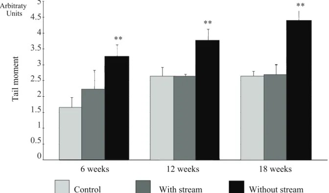
Figure 8 shows, in mean, with the respective
standard deviation, the values found for the levels of tissue oxidative stress, comparing the animals in the control and experiment subgroups (with and without fecal stream) in the different times of fecal exclu
-sion. The levels of oxidative stress were similar in
the subgroup control and in segments with stream, re
-gardless of the considered time of exclusion. In the colon without stream for 6 weeks, these values were 3.24±0.44 TM, while, after 12 and 18 weeks of exclu-sion, they were 3.74±0.40 TM and 4.37±0.32 TM, re-spectively. The results showed that the levels of tissue
oxidative stress were higher in the segments without stream when compared to those with stream and to the subgroup control, and time of exclusion was not
relevant (p=0.0001). Table 1 indicates that levels of
oxidative stress in the colon without stream increase
with time of exclusion (p=0.0007).
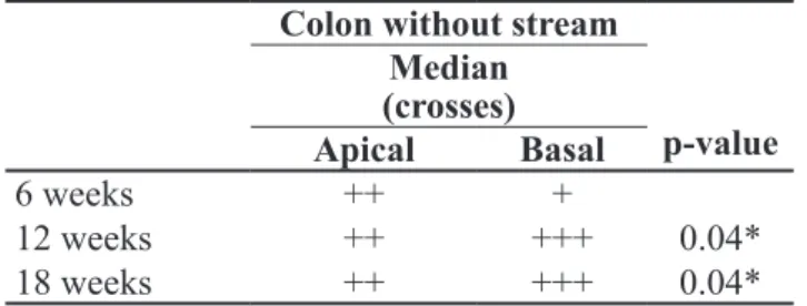
Table 2 presents the variation of β-catenin content between the apical and basal regions of the colonic crypts in segments without fecal stream, in relation to time of
exclusion. The variation in the pattern of expression
of the tissue protein in the basal region of colonic glands was observed, in comparison to colons without stream
for 6 weeks and those for 12 and 18 weeks (p=0.04 and p=0.04, respectively).
DISCuSSION
Cell adhesion is a primary characteristic of the
architecture of most of the tissues in the human body.
The epithelium that covers the digestive tube is formed by an isolated layer of specialized cells, with absorp -tion and secre-tion func-tions, intimately adhered with
each other and to the basal blade11,43. These cells are
gathered to one another by systems of cell-cell
adhe-sion, which support most of the mechanical stress and
also work as a functional barrier. The specialized ICJs
are located in places where there is no contact between two cells, or between one cell and the extracellular
matrix. In order for the cells to function in an
inte-grated manner in a compact set, the already mentioned
specialized ICJs, formed by group systems, are
need-ed43. There are three functional groups of ICJ: The OJs
or zonula occludens, the AJs or the zonula adherens,
and the communicating or electrotonic ones. There are
also specialized junctions in the adhesion of the cell with the extracellular matrix or basal membrane, rep
-resented by hemidesmosomes. All of these ICJs are
composed by different types of proteins, which have
speciic roles inside the complex that form them. The AJs connect the internal cytoskeleton of a
cell to its neighbor, by means of a protein complex formed by the proteins in the cadherin, catenin, vin -culin and actin families11. The AJs are also related to
the proteins of the intracellular signaling pathway, enabling them to participate in the communication mechanisms that are present inside the neighboring cells, and the cadherins are the main proteins that form AJs11. These are proteins dependent on calcium that
play an important role in intercellular adhesion, tis
-sue differentiation, polarization and epithelial
stratii-cation44. The cytoplasmic domain of E-cadherin joins
one or more intracellular anchor protein, represented especially by β-catenin, while the extracellular do-main interacts with the dodo-main of another homolo -gous molecule, from the neighbor cell11,43.
In order for the occurrence of the anchor between the E-cadherin and the actin protein, which is the most important component of the cellular cytoskeleton,
pro-teins from the catenin family (α-catenin, β-catenin e
γ-catenin) are essential11. β-catenin is translated from
the transcription of the CTNNB1 gene, located on the
chromosome 8q32, with molecular weight of 88 kDa.
The β-catenin isoform presents double cell function, be-cause besides participating in the mechanisms of adhe -sion between two neighboring cells, it is important for the Wnt signaling11. When the protein system that form
the AJs in the intercellular space breaks, there is the
Figure 7. Variation of β-catenin tissue content in the deep region of the crypts, comparing the animals in control and experiment subgroups (colons with and without stream) in the different periods of intestinal stream exclusion.
6 weeks
E
sc
ore
12 weeks
18 weeks
Control
With stream
Without stream
3.5
3
2.5
2
1.5
1
0.5
0
*
*
* *
* *
*Without stream 6 weeks > with stream 12 weeks (p<0.05); **Without stream 12 and 18 weeks > with stream 12 and 18 weeks (p<0.01). Median test
Figure 8. Levels of cell oxidative stress comparing the control and experiment subgroups (colons with and without stream) in the different times of exclusion.
6 weeks
T
ai
l m
om
ent
12 weeks
18 weeks
Control
With stream
Without stream
5
4.5
4
3.5
3
2.5
2
1.5
1
0.5
0
**
**
**
Arbitraty Units
cytosol, the ubiquitin system is unable to degrade the β-catenin in the proteasome, and this way the excess
protein migrates inside the nucleus and joins the gene transcription factors, thus activating the transcription of genes related to cell proliferation, such as C-MYC and
cyclin-17,8,11,13. Therefore, β-catenin, besides being
es-sential to AJs, actively participates in the Wnt signal -ing, one of the main mechanisms responsible for the
induction of cell division. This double functional
do-main is important for the renewal of cells in the colonic
epithelium, in constant replacement.
It has been shown that, in patients with URC, who suffers from constant apoptosis of the supericial
cells of the colonic epithelium, the β-catenin protein presents more expression in the deep regions of colon -ic glands, where the germinative zone is located, and is the main responsible for the process of cell prolif -eration13,45. On the contrary, the substantial reduction of β-catenin expression in cells of the mucosal surface of subjects with URC was shown, and in this situation there is constant cellular death13,22,24. The comparison
between normal and sore tissues in subjects with URC
showed signiicant reduction of the tissue content of E-cadherin and β-catenin proteins just in locations in
which the disease is active, but not in normal tisses26.
These results conirm the strong relation between the
rupture of AJs and the development of colitis24.
Many mechanisms are able to damage the AJs
in the colonic epithelium. Among them, the oxidative
stress stands out20,21,32,45. Studies show that the colonic mucosa exposed to high concentrations of OFR oxi -dizes the Ca++ ions, which keep the E-cadherin
mol-ecules together in the intercellular space20. The rupture
of Ca++ molecules degrading the intercellular bridges
of E-cadherin causes the accumulation of β-catenin
cytoplasmic content, thus stimulating cellular divi -sion1,11,20,21,46. The OFRs can also dissociate the
junc-tions between E-cadherin and β-catenin in the cyto-sol by a dependent tyrosine-kinase mechanism, which
leads to the cytoplasmic accumulation of β-catenin44.
It was experimentally shown that the migration of β-catenin from its membranous cytoplasmic domain to the inner nuclear cell is considered as the irst event
in the animal models of chemically induced colitis,
which comes even before the neutrophilic
iniltra-tion19,47,48. However, it is dificult to demonstrate this possibility in the models of chemical colitis, since the rupture of AJs could result from the action of harmful
agents, such as TNBS and DSS. With the DV
mod-el proposed in this study, it is possible to observe the role of OFR in the rupture of AJs30. Recent evidence
shows that in experimental models of DC, despite the non existence of intestinal epithelium exposure to any toxic substance, there is increased production of OFR, which determines the appearance of colitis by the rup -ture of different lines of defense of the colonic muco -sal barrier29-31,37-40. Even though the relations between the changes in content and pattern of tissue expres
-sion of E-cadherin and β-catenin in sick patients and
experimental models of URC have been subjectively demonstrated, this possibility had not been assessed in experimental models of DC19,23.
It is also worth mentioning that the evaluation of
tissue content of proteins that compose AJs in models of colitis is usually subjective, which depends on the
pathologist’s experience. The possibility to use
meth-ods of image analysis with the assistance of computers leads to more accuracy, uniformity and reliability in
relation to the results, since it quantiies the tissue con-tent of the protein analyzed objectively. The analysis of assisted image, also known as
computer-ized morphometry, presents additional advantages in relation to conventional methods, such as the fast and
low cost quantitative evaluation of microscopic
struc-tures37-40. In this study, with the use of computerized morphometry, it was possible to determine the tissue content of the β-catenin protein objectively, which enabled more precise comparisons between normal and sore tissues38. Computerized morphometry had
not been previously used to quantify tissue levels of β-catenin in models of colitis.
Colon without stream Median (crosses)
Apical Basal p-value
6 weeks ++ +
12 weeks ++ +++ 0.04*
18 weeks ++ +++ 0.04*
Table 2. Variation of β-catenin content values in the apical and basal regions of colonic glands in the segments without fecal stream in relation to the different times of exclusion.
The evaluation of tissue oxidative stress levels
can also be performed by different techniques. Among
the most used ones are the malondialdehyde tissue dosimetry, which is a product of the cell membrane phospholipid peroxidation, the plasmatic and urinary
dose of 8-hydroxyguanosine levels, and the advanced
oxidation protein products29,49. However, these are
sensitive biochemical techniques, subject to
varia-tions, and they need a reasonable amount of tissue to be executed42. With the advent of the single cell gel
electrophoresis (comet assay), it became possible to
quantify the oxidative stress levels in the whole
tis-sue50. The technique enables the comparison of oxi-dative damage levels in cells of the normal colonic mucosa and the sore epithelium50.
The comet assay is one of the most sensitive methods to assess levels of oxidative stress, present -ing greater accuracy when compared to other tech
-niques50. Because of its high sensitivity, allied to its low cost, it has been more and more used30,31,40,42,50. The comet assay had not been employed to assess the relation between tissue oxidative stress and changes in tissue content and expression of the β-catenin protein
in DC models.
At irst, with the objective to conirm if animals
used for experimentation developed colitis in seg -ments without stream, we evaluated the histological
changes in the diverted colon. We observed the
pres-ence of colitis in this segment for all rats, regardless of diversion time, when compared to segments with preserved stream30,31. In the control group and in
co-lonic segments with fecal stream in the animals from
the experiment group, even though we did not ind the formation of epithelial ulcers, we identiied some de-gree of inlammatory iniltrate, especially those con-stituted of neutrophils. Differently, in the colon
with-out stream there was the formation of more epithelial ulcers, sometimes deep, destroying the whole epithe
-lial surface, and larger tissue inlammatory iniltrate, regardless of considered time. It is worth mentioning that in segments without stream, after 12 and 18 weeks of intestinal exclusion, the inlammation grading scale
increased in relation to animals submitted to bypass
for 6 weeks, thus suggesting that the worsened epithe-lial aggression could be related to the deicient supply
of SCFA, modifying the cellular energetic metabolism
for a longer period of time. Conirming this
possibil-ity, we found higher levels of oxidative stress in these
segments, which increased with the experiment. It is
possible that the increased production of OFR be re -sponsible for the larger number of ulcers, and, conse
-quently, for the higher inlammation score observed in segments without stream after 12 and 18 weeks. When
we measure the tissue levels of oxidative stress in the segments without stream we observe that they were
directly related to the worst inlammation score. The high levels of oxidative stress after six weeks of
di-version may be related to the increased production of OFR, both for the presence of neutrophils – cells that produce OFR – and for the changes in cell metabolism
resulting from SCFA deiciency.
When we measure the tissue content of β-catenin
in animals submitted to bypass for six weeks, we ind
reduction in the segments without fecal stream in re -lation to those in the control group and the segments
with preserved stream. When we study the β-catenin
content separating the apical and basal regions of the colonic glands, we notice the reduction of content in the apical region, and, on the other hand, its increase
in the basal region. These indings suggest that the
re-duction of β-catenin expression in cells of the epithe-lial surface may be related to higher levels of damage to the cells in this region, probably due to greater lo
-cal oxidative stress. The worsened inlammation in the
segments without stream could lead to the degradation of β-catenin, and consequently to the greater
produc-tion of OFR. It is possible that cytokines and proteases
produced by activated neutrophils, which are present at this stage of the experiment, could also be respon -sible for the greater degradation of β-catenin, and also
that the rupture of E-cadherin/β-catenin bridges in the
cells of the epithelial surface could lead to the migra -tion of β-catenin free of cytosol to the inner part of the nucleus in cells of the germinative zone, with the objective to increase the transcription of genes related
to cellular division. It is also possible that the greater β-catenin content found in deep cells of the colonic
glands can be explained by the greater need for cell proliferation, with the goal to replace the dead cells
from the apical surface, destroyed by tissue stress.
However, only the analysis of cell division genes, such as C-myc or Ciclyn-1, and the β-catenin expres-sion, comparing apical and basal cells from the co
In animals submitted to intestinal bypass for 12 weeks, we found worsened inlammation score in
the colonic segments without fecal stream, when com
-pared to those with bypass for six weeks. This is mainly
due to the greater presence of epithelial ulcers, once we
found lower neutrophilic iniltrate. The levels of
oxida-tive stress in these segments were higher when compared
to those found in animals with bypass for six weeks, and were directly related to the worst inlammation score.
The worsened levels of tissue oxidative stress in the colon without stream in animals with bypass for
12 weeks could also be a result of the presence of neu-trophils. However, it is likely that for these animals,
the greater formation of OFRs because of the SCFA
deiciency is more relevant for tissue damage when compared to animals with bypass for six weeks.
The total β-catenin content in segments without
stream after 12 weeks was similar to that of animals in the control groups and segments with fecal stream. In the diverted colon, there was no variation in protein
content when compared to that found in animals with
bypass for six weeks. When analyzing the β-catenin
expression separately in the apical and basal regions of colonic glands, we noticed the reduction of con -tent in the apical region, while the basal one presented
with increase. We found signiicant variation in
pro-tein content between the apical and basal regions of
colonic glands in relation to time of exclusion. These indings reinforce our suspicion that the proportional
increase of β-catenin concentration in the proliferative zones of the crypts is related to the greater need for cell division in the proliferative zones of crypts, with the goal to replace the cells from an increasingly dam
-aged epithelial surface.
The inlammation score of the colon without stream in animals with bypass for 18 weeks did not increase in relation to those with bypass for 12 weeks. However, in the diverted segments for 18 weeks,
de-spite the major presence of epithelial ulcers, the neu
-trophilic iniltrate was insigniicant. These indings
suggest that the worsened epithelial lesion cannot be
related to the higher presence of neutrophils. In these
segments, we found higher levels of oxidative stress
than for animals with bypass for 12 weeks, suggesting that after 18 weeks, the greater tissue oxidative stress caused by energy deiciency can be the main respon-sible for the worsened epithelial lesion.
The total β-catenin tissue content in segments
without fecal stream, diverted for 18 weeks, was similar to that of animals with bypass for 6 and 12 weeks. The
intensity of β-catenin expression in the apical regions of colonic glands was similar to that of animals submit
-ted to bypass for 6 and 12 weeks. However, in the basal
region, the intensity of the expression remained high, with values similar to those found in the colon without
stream for 12 weeks, thus suggesting the greater need
for gene activation related to cell division by β-catenin.
As what happened with diverted colons for 12 weeks,
the intensity of β-catenin expression in segments
with-out stream presented signiicant variation when com-paring apical and basal regions. The greater β-catenin
expression in the proliferative zone of colonic glands
after 18 weeks reafirms the need to maintain the cell division process in the cells by the germinative zone.
Studies have shown the important role of SCFA to maintain the proper tropism of epithelial cells from the colonic mucosa35-38. They are important substracts to pre-serve the integrity of barrier mechanisms, since they in -duce the expression of genes that form the proteins that are responsible for the selective permeability of AJs and avoid oxidative stress, which cause lesions in these de -fense systems35-38. The inhibition of SCFA metabolization leads to the appearance of colitis36, while the establishment
of fecal stream and the administration of SCFA, mixed
nutritional solutions and omega-3 and omega-6 rich
poly-unsaturated fatty acids, improve histological alterations, probably by diminishing tissue oxidative stress30.
The results found in this study add new evidence
to support the theory of colitis induction by OFR. In
this paper, it was possible to demonstrate that cells from the colonic mucosa without regular SCFA sup -ply suffer from more oxidative stress, especially in the
late stages of fecal diversion. It was possible to show
that colonic segments without stream present histo -logical changes that are indistinguishable from those found in human DC, and similar to those described in experimental models of chemically induced colitis37-40.
The results also showed that SCFA deiciency, despite practically keeping the total β-catenin tissue content,
drastically changes the place of protein expression, re -ducing its content in the damaged epithelial surface
and increasing it in the proliferative region. These
favoring the mechanisms of cellular adhesion to the epithelial surface and leading to cellular proliferation
in germinative zones of colonic glands. Finally, the re-sults in this study show, for the irst time in literature,
that the content and place of β-catenin expression is
changed in DC, as it happened with URC. From the
practical point of view, the indings suggest that the
reestablishment of SCFA supply to the diverted colon, be it by the reconstitution of fecal stream or by the ad -ministration of nutritional solutions rich in SCFA, or even the use of antioxidant substances, can be consid
-ered as a valid strategy to prevent and treat DC.
ReFeReNCeS
1. Pravda J. Radical induction theory of ulcerative colitis. World J Gastroenterol 2005;11(16):2371-84.
2. Gaudier E, Hoebler C. Physiological role of mucins in the colonic barrier integrity. Gastroenterol Clin Biol 2006;30(8-9): 965-74.
3. Laukoetter MG, Nava P, Nusrat A. Role of the intestinal barrier in inlammatory bowel disease. World J Gastroenterol 2008;14(3):401-7.
4. Berkes J, Viswananthan VK, Savkovic SD, Hecht G. Intestinal epithelial responses to enteric pathogens: effects on the tight junction barrier, iron transport, and inlammation. Gut 2003;52(3):439-51.
5. Clayburgh DR, Shen L, Turner JR. A porous defense: the leaky epithelial barrier in intestinal disease. Lab Invest 2004;84(3):282-91.
6. Usami Y, Chiba H, Nakayama F, Ueda J, Matsuda Y, Sawada N, et al. Reduced expression of claudin-7 correlates with invasion and metastasis in squamous cell carcinoma of the esophagus. Hum Pathol 2006;37(5):569-77.
7. Gumbiner B, Stevenson B, Grimaldi A. The role of the cell adhesion molecule uvomorulin in the formation and maintenance of the epithelial junctional complex. J Cell Biol 1988;107(4):1575-87.
8. Gumbiner BM, McCrea PD. Catenins as mediators of the cytoplasmic functions of cadherins. J Cell Sci Suppl 1993;17:155-8.
9. Yeager M, Unger VM, Falk MM. Synthesis, assembly and structure of gap junction intercellular channels. Curr Opin Struct Biol 1998;8(6):810-1.
10. Hynes RO, Zhao Q. The evolution of cell adhesion. J Cell Biol 2000;150(2):F89-96.
11. Kypta R, Bernield M, Burridge K, Geiger B, Goodenough D, Humphries M, Hynes R, Reichardt L, Rosenbaum J, Rucislahti E, Sanes J, Springer T, Yurchenco P. Junções celulares, adesão celular e matriz extracelular. In: Alberts B, Johnson A, Lewis J, Raff M, Roberts K, Walter P. (eds.). Biologia Molecular da Célula. Porto Alegre: ARTMED; 2006. p. 1065-1125.
12. Demetter P, De Vos M, Van Damme N, Baeten D, Elewaut D, Vermeulen S, et al. Focal up-regulation of E-cadherin-catenin complex in inlamed bowel mucosa but reduced expression in ulcer-associated cell lineage. Am J Clin Pathol 2000;114(3):364-70.
13. Aust DE, Terdiman JP, Willenbucher RF, Chew K, Ferrell L, Florendo C, et al. Altered distribution of β-catenin, and its binding proteins E-cadherin and APC, in ulcerative colitis-related colorectal cancers. Mod Pathol 2001;14(1):29-39. 14. Kucharzik T, Walsh SV, Chen J, Parkos CA, Nusrat A. Neutrophil
transmigration in inlammatory bowel disease is associated with differential expression of epithelial intercellular junction proteins. Am J Pathol 2001;159(6):2001-9.
15. Laukoetter MG, Nava P, Nusrat A. Role of the intestinal barrier in inlammatory bowel disease. World J Gastroenterol 2008;14(3):401-7.
16. Gassler N, Rohr C, Schneider A, Kartenbeck J, Bach A, Obermüller N, et al. Inlammatory bowel disease is associated with changes of enterocytic junctions. Am J Physiol Gastrointest Liver Physiol 2001;281(1):G216-28. 17. Ozawa M, Ringwald M, Kemler R. Uvomorulin-catenin
complex formation is regulated by a speciic domain in the cytoplasmic region of the cell adhesion molecule. Proc Natl Acad Sci USA 1990;87(11):4246-50.
18. Schmitz H, Barmeyer C, Fromm M, Runkel N, Foss HD, Bentzel CJ, et al. Altered tight junction structure contributes to the impaired epithelial barrier function in ulcerative colitis. Gastroenterology 1999;116(2):301-9.
19. Takahashi M, Fukuda K, Sugimura T, Wakabayashi K. Beta-catenin is frequently mutated and demonstrates altered cellular localization in azoxymethane-induced rat colon tumors. Cancer Res 1998;58(1):42-6.
20. Parrish AR, Catania JM, Orozco J, Gandoli AJ. Chemically induced oxidative stress disrupts the E-cadherin/catenin cell adhesion complex. Toxicol Sci 1999;51(1):80-6.
21. Meyer TN, Schwesinger C, Ye J, Denker BM, Nigam SK. Reassembly of the tight junction after oxidative stress depends on tyrosine kinase activity. J Biol Chem 2001;276(25):22048-55.
22. Dorudi S, Shefield JP, Poulsom R, Northover JM, Hart IR. E-cadherin expression in colorectal cancer. An immunocytochemical and in situ hybridization study. Am J Pathol 1998;142(4):981-6.
23. Chen J, Huang XF. The signal pathways in azoxymethane-induced colon cancer and preventive implications. Cancer Biol Ther 2009;8(14):1313-7.
25. Hermiston ML, Gordon JI. Inlammatory bowel disease and adenomas in mice expressing a dominant negative N-cadherin. Science 1995;270(5239):1203-7.
26. Karayiannakis AJ, Syrigos KN, Efstathiou J, Valizadeh A, Noda M, Playford RJ, et al. Expression of catenins and E-cadherin during epithelial restitution in inlammatory bowel disease. J Pathol 1998;185(4):413-8.
27. Nollet F, Berx G, van Roy F. The role of the E-cadherin/ catenin adhesion complex in the development and progression of cancer. Mol Cell Biol Res Commun 1999;2(2):77-85. 28. Sheehan JF, Brynjolfsson G. Ulcerative colitis following
hydrogen peroxide enema: case report and experimental production with transient emphysema of colonic wall and gas embolism. Lab Invest 1960;9:150-68.
29. Marques LHS, Silva CMG, Lameiro TMM, Almeida MG, Cunha FL, Pereira JA, et al. Avaliação dos níveis de peroxidação lipídica em células da mucosa cólica após aplicação de enemas com peróxido de hidrogênio: estudo experimental em ratos. Rev bras colo-proctol 2010;30(3):272-80.
30. Martinez CA, Ribeiro ML, Gambero A, Miranda DD, Pereira JA, Nadal SR. The importance of oxygen free radicals in the etiopathogenesis of diversion colitis in rats. Acta Cir Bras 2010;25(5):387-95.
31. Longatti TS, Acedo SC, de Oliveira CC, Miranda DD, Priolli DG, Ribeiro ML, et al. Inlammatory alterations in excluded colon in rats: a comparison with chemically induced colitis. Scand J Gastroenterol 2010;45(3):315-24.
32. Damiani CR, Benetton CA, Stoffel C, Bardini KC, Cardoso VH, Di Giunta G, et al. Oxidative stress and metabolism in animal model of colitis induced by dextran sulfate sodium. J Gastroenterol Hepatol 2007;22(11):1846-51.
33. Liu Q, Shimoyama T, Suzuki K, Umeda T, Nakaji S, Sugawara K. Effect of sodium butyrate on reactive oxygen species generation by human neutrophils. Scand J Gastroenterol 2001;36(7):744-50.
34. Glotzer DJ, Glick ME, Goldman H. Proctitis and colitis following diversion of the fecal stream. Gastroenterology 1981;80(3):438-41.
35. Agarwal VP, Schimmel EM. Diversion colitis: a nutritional deiciency syndrome? Nutr Rev 1989;47(9):257-61.
36. Butzner JD, Parmar R, Bell CJ, Dalal V. Butyrate enema therapy stimulates mucosal repair in experimental colitis in the rat. Gut 1996;38(4):568-73.
37. Nonose R, Spadari AP, Priolli DG, Máximo FR, Pereira JA, Martinez CA. Tissue quantiication of neutral and acid mucins in the mucosa of the colon with and without fecal stream in rats. Acta Cir Bras 2009;24(4):267-75.
38. Martinez CA, Nonose R, Spadari AP, Máximo FR, Priolli DG, Pereira JA, et al. Quantiication by computerized morphometry of tissue levels of sulfomucins and sialomucins in diversion colitis in rats. Acta Cir Bras 2010;25(3):231-40. 39. Sousa MV, Priolli DG, Portes AV, Cardinalli IA, Pereira JA,
Martinez CA. Evaluation by computerized morphometry of histopathological alterations of the colon wall in segments
with and without intestinal transit in rats. Acta Cir Bras 2008;23(5):417-24
40. Caltabiano C, Máximo FR, Spadari AP, Miranda DDC, Serra MM, Ribeiro ML, et al. 5-aminosalicylic (5-ASA) can reduce the levels of oxidative DNA damage in cells of colonic mucosa with and without fecal stream. Dig Dis Sci 2011;56(4):1037-46.
41. Gupta RB, Harpaz N, Itzkowitz S, Hossain S, Matula S, Kornbluth A, et al.. Histologic inlammation is a risk factor for progression to colorectal neoplasia in ulcerative colitis: a cohort study. Gastroenterology 2007;133(4):1099-105. 42. Ribeiro ML, Priolli DG, Miranda DD, Arçari DP, Pedrazzoli
J Jr. Martinez CA. Analysis of oxidative DNA damage in patients with colorectal cancer. Clin Colorectal Cancer 2008;7(4):267-72.
43. Lodish H, Berk A, Zipursky SL, Matsudaira P, Baltimore D, Darnell J. A integração das células nos tecidos. In: Lodish H, Berk A, Zipursky SL, Matsudaira P, Baltimore D, Darnell J. (eds.). Biologia celular e molecular. Rio de Janeiro: Revinter; 2004. p.968-1002.
44. Duband JL, Thiery JP. Spatio-temporal distribution of the adherens junction-associated molecules vinculin and talin in early avian embryo. Cell Differ Dev 1990;30(1):55-76. 45. Rao RK, Basuroy S, Rao VU, Karnaky Jr KJ, Gupta A.
Tyrosine phosphorylation and dissociation of occludin-ZO-1 and E-cadherin-b-catenin complexes from the cytoskeleton by oxidative stress. Biochem J 2002;368(Pt 2):471-81. 46. Schmehl K, Florian S, Jacobasch G, Salomon A, Körber
J. Deiciency of epithelial basement membrane laminin in ulcerative colitis affected human colonic mucosa. Int J Colorectal Dis 2000;15(1):39-48.
47. Cooper HS, Murthy S, Kido K, Yoshitake H, Flanigan A. Dysplasia and cancer in the dextran sulfate sodium mouse colitis model. Relevance to colitis-associated neoplasia in the human: a study of histopathology, B-catenin and p53 expression and the role of inlammation. Carcinogenesis 2000;21(4):757-68. 48. Fodde R, Tomlinson I. Nuclear beta-catenin expression
and Wnt signalling: in defence of the dogma. J Pathol 2010;221(3):239-41.
49. Baskol M, Baskol G, Koçer D, Ozbakir O, Yucesoy M. Advanced oxidation protein products: a novel marker of oxidative stress in ulcerative colitis. J Clin Gastroenterol 2008;42(6):687-91
50. Glei M, Hovhannisyan G, Pool-Zobel BL. Use of Comet-ish in the study of DNA damage and repair: review. Mutat Res 2009;681(1):33-43.
Correspondence to:
Carlos Augusto Real Martinez Rua José Raposo de Medeiros