Non-HFE hemochromatosis
Introduction
Hereditary hemochromatosis (HH) is a disorder characterized by enhanced intestinal absorption of dietary iron. Without therapeutic intervention, iron overload leads to multiple organ damage such as liver cirrhosis, cardiomyopathy, diabetes, arthritis, hypogonadism and skin pigmentation. The most common intervention is therapeutic phlebotomy, which consists of regular blood withdrawal (usually 400-500 mL per session) until serum ferritin is controlled(1-5).
HFE mutations are, by far, the most common genetic abnormality involved in HH, especially the genotypes: homozygosity for p.Cys282Tyr or the p.Cys282Tyr/p.His63Asp compound heterozygosity. However, since the causal association between HFE mutations and HH was discovered in 1996, it became evident that there are cases of HH that cannot be explained by HFE gene mutations. As a consequence, cases of HH that are not associated with
HFE mutations are collectively referred to as non-HFE hemochromatosis; these comprise mutations in the genes that encode hemojuvelin (HJV), hepcidin (HAMP), transferrin receptor 2 (TFR2) and ferroportin (SLC40A1)(6-8). Cases of HH due to HJV or HAMP mutations are denominated type 2 HH; those related to TFR2 mutations are named type 3 HH; and cases associated with SLC40A1 mutations, which can be signiicantly different from classic cases of
HH, receive the denomination of type 4 HH or “ferroportin disease”.
Considering that the group of non-HFE hemochromatosis has many peculiarities, the aim of this review is to explore molecular, clinical and management aspects of non-HFE
hemochromatosis.
Juvenile Hemochromatosis or Type 2 hereditary hemochromatosis
Juvenile hemochromatosis (JH), also classiied as type 2, is a rare autosomal recessive disorder of iron overload that leads to organ damage before the age of 30. JH is characterized by severe iron overload usually associated with liver damage, cardiomyopathy and/or hypogonadotrophic hypogonadism. Hypogonadism is the main symptom at disease presentation, and the course of symptoms is more rapid and severe than classic HFE
hemochromatosis (type 1)(9). Men and women are equally affected. Typically, patients with JH die prematurely of cardiovascular causes before reaching their fourth decade of life. JH is subdivided in types 2A (OMIM 602390) and 2B (OMIM 613313), which are caused by mutations in the HJV and HAMP genes, respectively(10-12).
Both types 2A and 2B HH are associated, in their inal pathophysiology, with hepcidin regulation. Hepcidin is a hormone produced by hepatocytes, which plays an important role in iron homeostasis by regulating its absorption and release in the enterocytes and macrophages(13).
The HJV (OMIM 608374) gene is constituted by 4 exons located in chromosome 1. It was identiied in 2004 and encodes a protein called hemojuvelin(10). This protein is critical for iron homeostasis regulation and for hepcidin expression in response to iron. In this scope, patients with type 2A JH and HJV knockout mice models demonstrate low hepcidin levels suggesting that hemojuvelin is involved in hepcidin synthesis(14). Several HJV mutations associated with JH Paulo Caleb Júnior de Lima Santos1
Carla Luana Dinardo1 Rodolfo Delini Cançado2
Isolmar Tadeu Schettert1
José Eduardo Krieger1
Alexandre Costa Pereira1
Conlict-of-interest disclosure:
The authors declare no competing inancial
interest
Submitted: 6/28/2012 Accepted: 7/10/2012
Corresponding author:
Paulo Caleb Júnior de Lima Santos Laboratory of Genetics and Molecular Cardiology, Heart Institute (InCor), Faculdade
de Medicina da Universidade de São Paulo -
USP
Av. Dr. Enéas de Carvalho Aguiar, 44 Cerqueira César
05403-000 São Paulo, SP, Brazil Phone: 55 11 2661-5329
www.rbhh.org or www.scielo.br/rbhh
DOI: 10.5581/1516-8484.20120079 1 Faculdade de Medicina da Universidade de São Paulo - USP, São Paulo, SP, Brazil 2 Faculdade de Ciências Médicas da Santa Casa de São Paulo – FCMSCSP, São Paulo, SP, Brazil
Hereditary hemochromatosis (HH) is an autosomal recessive disorder classically related to HFE mutations. However, since 1996, it is known that HFE mutations explain about 80% of HH cases, with the remaining around 20% denominated non-HFE hemochromatosis. Nowadays, four main genes are implicated in the pathophysiology of clinical syndromes classiied as non-HFE hemochromatosis: hemojuvelin (HJV, type 2A juvenile HH), hepcidin (HAMP, type 2B juvenile HH), transferrin receptor 2 (TFR2, type 3 HH) and ferroportin (SLC40A1, type 4 HH). The aim of this review is to explore molecular, clinical and management aspects of non-HFE hemochromatosis.
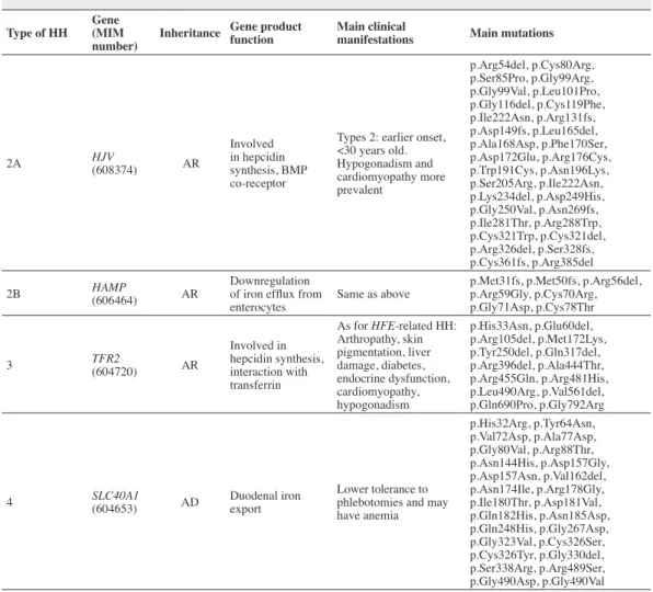
Table 1 - Characteristics according to non-HFE hemochromatosis(19, 59-61)
Type of HH Gene (MIM number)
Inheritance Gene product function
Main clinical
manifestations Main mutations
2A HJV
(608374) AR
Involved in hepcidin synthesis, BMP co-receptor
Types 2: earlier onset, <30 years old. Hypogonadism and cardiomyopathy more prevalent
p.Arg54del, p.Cys80Arg, p.Ser85Pro, p.Gly99Arg, p.Gly99Val, p.Leu101Pro, p.Gly116del, p.Cys119Phe, p.Ile222Asn, p.Arg131fs, p.Asp149fs, p.Leu165del, p.Ala168Asp, p.Phe170Ser, p.Asp172Glu, p.Arg176Cys, p.Trp191Cys, p.Asn196Lys, p.Ser205Arg, p.Ile222Asn, p.Lys234del, p.Asp249His, p.Gly250Val, p.Asn269fs, p.Ile281Thr, p.Arg288Trp, p.Cys321Trp, p.Cys321del, p.Arg326del, p.Ser328fs, p.Cys361fs, p.Arg385del
2B HAMP
(606464) AR
Downregulation of iron eflux from enterocytes
Same as above
p.Met31fs, p.Met50fs, p.Arg56del, p.Arg59Gly, p.Cys70Arg, p.Gly71Asp, p.Cys78Thr
3 TFR2
(604720) AR
Involved in hepcidin synthesis, interaction with transferrin
As for HFE-related HH: Arthropathy, skin pigmentation, liver damage, diabetes, endocrine dysfunction, cardiomyopathy, hypogonadism
p.His33Asn, p.Glu60del, p.Arg105del, p.Met172Lys, p.Tyr250del, p.Gln317del, p.Arg396del, p.Ala444Thr, p.Arg455Gln, p.Arg481His, p.Leu490Arg, p.Val561del, p.Gln690Pro, p.Gly792Arg
4 SLC40A1
(604653) AD
Duodenal iron export
Lower tolerance to phlebotomies and may have anemia
p.His32Arg, p.Tyr64Asn, p.Val72Asp, p.Ala77Asp, p.Gly80Val, p.Arg88Thr, p.Asn144His, p.Asp157Gly, p.Asp157Asn, p.Val162del, p.Asn174Ile, p.Arg178Gly, p.Ile180Thr, p.Asp181Val, p.Gln182His, p.Asn185Asp, p.Gln248His, p.Gly267Asp, p.Gly323Val, p.Cys326Ser, p.Cys326Tyr, p.Gly330del, p.Ser338Arg, p.Arg489Ser, p.Gly490Asp, p.Gly490Val
MIM = Mendelian inheritance in man; HJV= encodes hemojuvelin; HAMP= encodes hepcidin; TFR2= encodes transferrin receptor 2; SLC40A1= encodes ferroportin; BMP = bone morphogenetic protein, AR = autosomal recessive; AD = autosomal dominant have been described in the literature (Table 1). However, HJVp.
Gly320Val is the most important mutation and has been reported in JH patients in several different populations around the world(10,15-19).
The HAMP (OMIM 606464) gene directly encodes
hepcidin which is produced by hepatocytes and plays a role in iron absorption related to ferroportin degradation of the enterocytes(12,20). Mutations in the HAMP gene, which is constituted by 3 exons and located in chromosome 19, are a very rare cause of JH. Since its description, some mutations have already been described (Table 1)(12,19,21,22).
Type 3 hereditary hemochromatosis
Type 3 HH (OMIM 604250) is an autosomal recessive disease caused by mutations in the TFR2 gene. The irst
description of this disease, in two Sicilian families, dates back to 2000 and is the irst diagnosis of hemochromatosis attributed to a gene mutation other than HFE(23).
Type 3 HH leads to an iron overload similar to HFE
hemochromatosis, and, consequently, may present with abnormal
liver function, diabetes, hypogonadism, cardiomyopathy and arthritis(24). The typical onset is during adulthood, but inheritance of both TFR2 and HFE mutations are known to lead to an earlier onset of the disease(25).
TFR2 gene (OMIM 604720) is constituted by 18 exons and encodes the transferrin receptor 2 protein (TFR2). Different to TFR1, TFR2 expression is restricted almost entirely to the liver(26). Rather than only being involved with the uptake of transferrin-bound iron by hepatocytes, TFR2 is a sensor of iron levels and is also involved in hepcidin synthesis(23,27-29).
Type 3 HH is a rare condition and usually presents with decreased hepcidin levels. Known TFR2 mutations are shown in Table 1(19,23,30-34).
Type 4 hereditary hemochromatosis or ferroportin disease
a protein named ferroportin, which is a transmembrane iron transporter expressed in macrophages, enterocytes, hepatocytes and syncytiotrophoblasts(35,41). Ferroportin is responsible for iron transportation across the enterocyte surface and for iron recycling in the reticuloendothelial system(26). It is known that hepcidin binds to ferroportin, promoting its internalization and degradation leading to a decrease in iron absorption and, consequently, to a reduction in serum iron. Even ferroportin expression on the cell surface can be regulated by hepcidin(20).
Patients with ferroportin disease, differently from HFE
HH, typically present with low to normal transferrin saturation (TS) and iron overload within macrophages, mainly from the liver, spleen and bone marrow. In these cases, a mild iron-deicient anemia may be present at the initial stage leading to a reduced tolerance to therapeutic phlebotomy(40-42). However, it is also known that some cases of ferroportin disease may present phenotypically very similar to HFE HH with high TS and an iron overload predominantly in hepatocytes(43,44). Regarding these two possible phenotypes of the disease, it has recently been shown that this difference may be due to the patterns of SLC40A1
mutations. While the most common phenotype is related to a loss of iron exporting activity of ferroportin, the latter (more similar to HFE HH), may be associated with mutations that lead to a hepcidin-resistant ferroportin(41).
Diagnosis of non-HFE hemochromatosis
Similarly to HFE HH, initial suspicions of non-HFE HH are related to abnormalities in iron biochemical assays. Typically, patients present with increased levels of TS (≥ 45%), which is the earliest phenotypic biochemical indication of HH, and raised serum ferritin. It is important to point out that ferritin is an acute phase reactant and, as a consequence, can be elevated in many situations other than HH; other possible causes must be discarded before proceeding with the HH investigation process(45-47). However, hyperferritinemia remains one of the most common signs and is identiied from either a systematic biochemical workout or the diagnostic procedure with a large number of opening symptoms such as fatigue, joint pain, jaundice, skin pigmentation, neurological signs, impotence, diabetes, heart disease and even anemia.
A four-step strategy can be proposed to progressively narrow the ield of putative causes of hyperferritinemia(48,49).
Step 1) Rule out an acquired cause of hyperferritinemia unrelated to signiicant iron overload (IOL). Non-hereditary causes of hyperferritinemia are numerous and much more prevalent than hereditary abnormalities of iron metabolism. Thus, neglecting this step usually results in unnecessary genetic testing. The main causes of non-IOL related hyperferritinemia are: inlammatory syndrome, cell necrosis, chronic alcohol consumption and metabolic syndrome. Personal and family history, clinical examination including biometric evaluation (body mass index, waist circumference and blood pressure), iron parameters (serum ferritin and TS) and some simple biochemical tests (C-reactive protein, hemoglobin, alanine aminotransferase and aspartate aminotransferase) in most cases, allow the diagnosis of acquired hyperferritinemia.
Step 2) Conirm IOL and rule out acquired causes. The determination of TS is necessary at an early stage in the diagnostic algorithm. However, due to the test variability throughout a day and depending on technical procedures, any increase in TS must be veriied. Repeatedly high TS levels usually denote IOL. It should be noted that a high TS is a particularly valuable indicator for the presence of a HFE mutation. The main causes of acquired IOL are: chronic anemia (thalassemia major, myelodysplastic syndrome, sideroblastic anemia, chronic hemolysis), excessive iron supplementation (oral or parenteral iron, transfusions), porphyria cutanea tarda, chronic liver disease (alcoholic, viral or metabolic), end stage chronic liver disease.
Step 3) As serum ferritin may be increased due to a variety of causes unrelated to IOL, the third step is to assess hepatic iron stores directly. Magnetic resonance imaging is then necessary to authenticate high hepatic iron content. Liver biopsy is indicated if it can supply information that imaging or blood tests cannot and that will help patient management.
Step 4) Conirm the hereditary character of IOL and the precise gene(s) involved. The term of hereditary IOL is restricted to IOL conditions related to primary (genetic) abnormalities of iron metabolism. However in clinical practice, once hepatic IOL has been proven, phlebotomy must be initiated rapidly without waiting for sequencing results. Quantiication of the total iron removed by phlebotomies may serve as an additional argument for retrospective evaluation of the extent of iron accumulation.
Molecular assaying of HFE mutations should be performed only in cases with increased biochemical values and in those with familial history of HFE HH(50). Initially, the two main HFE mutations (p.Cys282Tyr and p.His63Asp) should be tested and, in their absence, non-HFE HH should be suspected. Hence, when there is iron overload in an under 30-year-old patient with cardiac or endocrine manifestations, a diagnosis of type 2 HH needs to be considered. Thus, the evaluation of the p.Gly320Val mutation in the HJV gene must be the molecular test of choice(5,51). If the result is negative, sequencing should be considered to evaluate the HJV and HAMP genes (Figures 1 & 2).
In addition, mutations in the TFR2 and SLC40A1 genes are rare, but they have been reported in child, adolescent, and adult cases. Considering the current advances in sequencing, it is recommended that, ideally, these genes should be evaluated to investigate non-HFE HH in patients with negative results for the
HFE mutation but with clinical manifestations of primary iron overload (Figures 1 & 2). Considering that direct sequencing is yet not widely available, usually this last approach is reserved for scientiic studies and in the investigation of refractory cases(5,7,52-54).
Therapeutic management
µg/L (through weekly or two-weekly phlebotomy sessions with withdrawal of 400-500 mL of total blood), maintenance sessions should be initiated, aiming to keep ferritin levels between 50 and 100 µg/L. All patients should be advised against the abusive use of alcohol and vitamin C during the treatment.
Patients with ferroportin disease are usually intolerant of conventional phlebotomies. In these cases, a less aggressive phlebotomy regimen should be attempted, and adjunctive therapy with erythropoietin may be beneicial(11). Also, in cases of intense side effects with phlebotomy, such as in patients with anemia or heart failure, the use of oral iron chelators such as deferasirox may be a safe therapeutic option(1-5).
Erythrocytapheresis has also been mentioned as a possible therapeutic option for patients with HH, but its use is rarely seen in clinical practice(50).
Conclusions
Advances in the understanding of non-HFE HH have been obtained over the years including: association of HJV and HAMP
mutations with the juvenile form, several pathogenic mutations associated with non-HFE HH, hepcidin as an iron hormone and its relationship with HFE protein, comprehension of the molecules involved in iron homeostasis, new techniques for the laboratorial evaluation, and increased knowledge about HH therapeutic management. Nonetheless, there are still unclear points to be explored in the non-HFE HH context, such as the better approach to the molecular investigation and therapeutic management.
In this scope, considering the rapid development of molecular techniques, which are becoming faster, more precise and
HJV p.Gly320Val
Increased TS and/or SF *
< 30 years old and cardiac or endocrine manifestations
Negative results for the HFE gene, specially for the p.Cys282Tyr and p.His63Asp mutations
HJV sequencing
+
+
-Type 2A JH HAMP
sequencing
Type 2B JH Types 3 and 4 HH
TFR2 and SLC40A1
sequencing**
+
Hereditary hemochromatosis
ADULT TS
> 30 years < 30 years
HFE TFR2 HJV HAMP SLC40A1
JUVENILE
Autosomal recessive
Autosomal domina
t
FERREPORTIN DISEASE to normal TS
Figure 1 - Representation of molecular investigation strategy for non-HFE hereditary hemochromatosis.
* Recommendations report TS > 45%, SF > 200 µg/L in females and > 300 µg/L in males; or in advanced stages: TS > 50% in females and TS > 60% in males,
in the absence of secondary causes(50,62)
** Some patients with primary iron overload may not present mutations during this genetic approach. Very rare mutations in other genes may be involved(7,63)
TS = transferrin saturation; SF = serum ferritin; JH = juvenile hemochromatosis. + means positive result and - means negative result
Figure 2 - Representation of the hereditary hemochromatosis types according to involved genes and phenotypes.
and diabetes are irreversible irrespective of treatment(52). The beneits of phlebotomy for HH have been demonstrated in cohort studies, but not in clinical randomized trials. However, it is known that survival of HH patients subjected to phlebotomies without diabetes and cirrhosis is similar to that of the general population(56,57).
economically viable, it is possible to consider that the diagnosis of non-HFE HH, or even the identiication of combinations of
mutations in the HJV, HAMP, TFR2, SLC40A1 and HFE genes, may become more common in the clinical practice.
Excluding HFE mutations and secondary iron overload are crucial steps before considering the diagnosis of non-HFE
HH. Thus, genetic testing can lead to more adequate and faster therapeutic management.
Acknowledgments
Paulo Caleb Júnior de Lima Santos is recipient of a fellowship from FAPESP, Proc. 2010-17465-8, Brazil. This work was inanced by FAPESP, Proc. 2011-18702-6.
References
1. Alexander J, Kowdley KV. HFE-associated hereditary hemochromatosis. Genet Med. 2009;11(5):307-13.
2. Bacon BR. Hemochromatosis: diagnosis and management. Gastroenterology. 2001;120(3):718-25.
3. Moyer TP, Highsmith WE, Smyrk TC, Gross JB Jr. Hereditary hemochromatosis: laboratory evaluation. Clin Chim Acta. 2011;412(17-18):1485-92.
4. Phatak P, Brissot P, Wurster M, Adams PC, Bonkovsky HL, Gross J, et al. A phase 1/2, dose-escalation trial of deferasirox for the treatment of iron overload in HFE-related hereditary hemochromatosis. Hepatology. 2010;52(5):1671-779.
5. Santos PC, Cancado RD, Pereira AC, Chiattone CS, Krieger JE, Guerra-Shinohara EM. HJV hemochromatosis, iron overload, and hypogonadism in a Brazilian man: treatment with phlebotomy and deferasirox. Acta Haematol. 2010;124(4):204-5.
6. Lok CY, Merryweather-Clarke AT, Viprakasit V, Chinthammitr Y, Srichairatanakool S, Limwongse C, et al. Iron overload in the Asian community. Blood. 2009;114(1):20-5. Comment in: Blood. 2009;114(1):1-2.
7. Santos PC, Cancado RD, Pereira AC, Schettert IT, Soares RA, Pagliusi RA, et al. Hereditary hemochromatosis: mutations in genes involved in iron homeostasis in Brazilian patients. Blood Cells Mol Dis. 2010;46(4):302-7.
8. Swinkels DW, Janssen MC, Bergmans J, Marx JJ. Hereditary hemochromatosis: genetic complexity and new diagnostic approaches. Clin Chem. 2006;52(6):950-68.
9. Lamon JM, Marynick SP, Roseblatt R, Donnelly S. Idiopathic hemochromatosis in a young female. A case study and review of the syndrome in young people. Gastroenterology. 1979;76(1):178-83.
10. Papanikolaou G, Samuels ME, Ludwig EH, MacDonald ML, Franchini PL, Dube MP, et al. Mutations in HFE2 cause iron overload in chromosome
1q-linked juvenile hemochromatosis. Nat Genet. 2004;36(1):77-82. 11. Pietrangelo A. Non-HFE hemochromatosis. Semin Liver Dis.
2005;25(4):450-60.
12. Roetto A, Papanikolaou G, Politou M, Alberti F, Girelli D, Christakis J, et al. Mutant antimicrobial peptide hepcidin is associated with severe
juvenile hemochromatosis. Nat Genet. 2003;33(1):21-2.
13. Fleming MD. The regulation of hepcidin and its effects on systemic and cellular iron metabolism. Hematology Am Soc Hematol Educ Program. 2008:151-8.
14. Lin L, Goldberg YP, Ganz T. Competitive regulation of hepcidin mRNA
by soluble and cell-associated hemojuvelin. Blood. 2005;106(8):2884-9.
15. Aguilar-Martinez P, Lok CY, Cunat S, Cadet E, Robson K, Rochette J. Juvenile hemochromatosis caused by a novel combination of hemojuvelin G320V/R176C mutations in a 5-year old girl. Haematologica. 2007;92(3):421-2.
16. de Lima Santos PC, Pereira AC, Cancado RD, Schettert IT, Hirata RD, Hirata MH, et al. Hemojuvelin and hepcidin genes sequencing in Brazilian patients with primary iron overload. Genet Test Mol Biomarkers. 2010;14(6):803-6.
17. Lanzara C, Roetto A, Daraio F, Rivard S, Ficarella R, Simard H, et al. Spectrum of hemojuvelin gene mutations in 1q-linked juvenile hemochromatosis. Blood. 2004;103(11):4317-21.
18. Lee PL, Beutler E, Rao SV, Barton JC. Genetic abnormalities and juvenile hemochromatosis: mutations of the HJV gene encoding hemojuvelin. Blood. 2004;103(12):4669-71.
19. Lee PL, Beutler E. Regulation of hepcidin and iron-overload disease. Annu Rev Pathol. 2009;4:489-515.
20. Nemeth E, Tuttle MS, Powelson J, Vaughn MB, Donovan A, Ward DM, et al. Hepcidin regulates cellular iron eflux by binding to ferroportin and
inducing its internalization. Science. 2004;306(5704):2090-3. Comment in: Science. 2004;306(5704):2051-3.
21. Jacolot S, Le Gac G, Scotet V, Quere I, Mura C, Ferec C. HAMP as
a modiier gene that increases the phenotypic expression of the HFE
pC282Y homozygous genotype. Blood. 2004;103(7):2835-40.
22. Porto G, Roetto A, Daraio F, Pinto JP, Almeida S, Bacelar C, et al. A Portuguese patient homozygous for the -25G>A mutation of the HAMP promoter shows evidence of steady-state transcription but fails to up-regulate hepcidin levels by iron. Blood. 2005;106(8):2922-3.
23. Camaschella C, Roetto A, Cali A, De Gobbi M, Garozzo G, Carella M, et al. The gene TFR2 is mutated in a new type of haemochromatosis
mapping to 7q22. Nat Genet. 2000;25(1):14-5.
24. Girelli D, Bozzini C, Roetto A, Alberti F, Daraio F, Colombari R, et
al. Clinical and pathologic indings in hemochromatosis type 3 due
to a novel mutation in transferrin receptor 2 gene. Gastroenterology. 2002;122(5):1295-302.
25. Pietrangelo A, Calefi A, Henrion J, Ferrara F, Corradini E, Kulaksiz H,
et al. Juvenile hemochromatosis associated with pathogenic mutations of adult hemochromatosis genes. Gastroenterology. 2005;128(2):470-9.
26. Wallace DF, Subramaniam VN. Non-HFE haemochromatosis. World J
Gastroenterol. 2007;13(35):4690-8.
27. Kawabata H, Yang R, Hirama T, Vuong PT, Kawano S, Gombart AF, et al. Molecular cloning of transferrin receptor 2. A new member of the transferrin receptor-like family. J Biol Chem. 1999;274(30):20826-32.
28. Nemeth E, Roetto A, Garozzo G, Ganz T, Camaschella C. Hepcidin is
decreased in TFR2 hemochromatosis. Blood. 2005;105(4):1803-6.
29. Wallace DF, Summerville L, Lusby PE, Subramaniam VN. First
phenotypic description of transferrin receptor 2 knockout mouse, and the role of hepcidin. Gut. 2005;54(7):980-6.
30. Biasiotto G, Roetto A, Daraio F, Polotti A, Gerardi GM, Girelli D, et al.
Identiication of new mutations of hepcidin and hemojuvelin in patients
with HFE C282Y allele. Blood Cells Mol Dis. 2004;33(3):338-43.
31. Fleming RE, Ahmann JR, Migas MC, Waheed A, Koefler HP,
Kawabata H, et al. Targeted mutagenesis of the murine transferrin
receptor-2 gene produces hemochromatosis. Proc Natl Acad Sci U S A.
2002;99(16):10653-8.
32. Koyama C, Wakusawa S, Hayashi H, Ueno T, Suzuki R, Yano M, et al.
A Japanese family with ferroportin disease caused by a novel mutation of SLC40A1 gene: hyperferritinemia associated with a relatively low transferrin saturation of iron. Intern Med. 2005;44(9):990-3.
with type 3 hereditary hemochromatosis and early onset iron overload. Haematologica. 2006;91(8 Suppl):ECR33.
34. Mattman A, Huntsman D, Lockitch G, Langlois S, Buskard N, Ralston D,
et al. Transferrin receptor 2 (TfR2) and HFE mutational analysis in
non-C282Y iron overload: identiication of a novel TfR2 mutation. Blood.
2002;100(3):1075-7.
35. Abboud S, Haile DJ. A novel mammalian iron-regulated protein involved in intracellular iron metabolism. J Biol Chem. 2000;275(26):19906-12.
36. Beutler E. Hemochromatosis: genetics and pathophysiology. Annu Rev Med. 2006;57:331-47.
37. Cremonesi L, Forni GL, Soriani N, Lamagna M, Fermo I, Daraio F, et al.
Genetic and clinical heterogeneity of ferroportin disease. Br J Haematol. 2005;131(5):663-70.
38. Hetet G, Devaux I, Souir N, Grandchamp B, Beaumont C. Molecular
analyses of patients with hyperferritinemia and normal serum iron values reveal both L ferritin IRE and 3 new ferroportin (slc11A3) mutations. Blood. 2003;102(5):1904-10.
39. Kasvosve I, Gomo ZA, Nathoo KJ, Matibe P, Mudenge B, Loyevsky M,
et al. Effect of ferroportin Q248H polymorphism on iron status in African
children. Am J Clin Nutr. 2005;82(5):1102-6.
40. Schimanski LM, Drakesmith H, Merryweather-Clarke AT, Viprakasit V, Edwards JP, Sweetland E, et al. In vitro functional analysis of human
ferroportin (FPN) and hemochromatosis-associated FPN mutations.
Blood. 2005;105(10):4096-102.
41. Mayr R, Janecke AR, Schranz M, Grifiths WJ, Vogel W, Pietrangelo
A, et al. Ferroportin disease: a systematic meta-analysis of clinical and
molecular indings J Hepatol. 2010;53(5):941-9.
42. Camaschella C, Poggiali E. Rare types of genetic hemochromatosis. Acta Haematol. 2009;122(2-3):140-5.
43. Liu XB, Yang F, Haile DJ. Functional consequences of ferroportin 1 mutations. Blood Cells Mol Dis. 2005;35(1):33-46.
44. Sham RL, Phatak PD, West C, Lee P, Andrews C, Beutler E. Autosomal dominant hereditary hemochromatosis associated with a novel ferroportin mutation and unique clinical features. Blood Cells Mol Dis. 2005;34(2):157-61.
45. Bacon BR. Screening for hemochromatosis. Arch Intern Med. 2006;166(3):269-70.
46. McLaren CE, McLachlan GJ, Halliday JW, Webb SI, Leggett BA, Jazwinska EC, et al. Distribution of transferrin saturation in an Australian population: relevance to the early diagnosis of hemochromatosis. Gastroenterology. 1998;114(3):543-9.
47. Olynyk JK, Cullen DJ, Aquilia S, Rossi E, Summerville L, Powell LW. A population-based study of the clinical expression of the hemochromatosis
gene. N Engl J Med. 1999;341(10):718-24. Comment in N Engl J Med. 1999;341(10):755-7.
48. Aguilar-Martinez P, Schved JF, Brissot P. The evaluation of hyperferritinemia: an updated strategy based on advances in detecting genetic abnormalities. Am J Gastroenterol. 2005;100(5):1185-94.
49. Brissot P, Troadec MB, Bardou-Jacquet E, Le Lan C, Jouanolle AM, Deugnier Y, et al. Current approach to hemochromatosis. Blood Rev. 2008;22(4):195-210.
50. European Association For The Study Of The Liver. EASL clinical practice guidelines for HFE hemochromatosis. J Hepatol. 2010;53(1):3-22.
51. Gehrke SG, Pietrangelo A, Kascak M, Braner A, Eisold M, Kulaksiz H, et al. HJV gene mutations in European patients with juvenile hemochromatosis. Clin Genet. 2005;67(5):425-8.
52. Gan EK, Powell LW, Olynyk JK. Natural history and management of
HFE-hemochromatosis. Semin Liver Dis. 2011;31(3):293-301.
53. Santos PC, Krieger JE, Pereira AC. Molecular diagnostic and pathogenesis of hereditary hemochromatosis. Int J Mol Sci. 2012;13(2):1497-511.
54. Santos PC, Pereira AC, Cancado RD, Schettert IT, Sobreira TJ, Oliveira PS, et al. HFE gene mutations in patients with primary iron overload: is
there a signiicant improvement in molecular diagnosis yield with HFE
sequencing? Blood Cells Mol Dis. 2010;45(4):302-7.
55. Niederau C, Fischer R, Purschel A, Stremmel W, Haussinger D,
Strohmeyer G. Long-term survival in patients with hereditary hemochromatosis. Gastroenterology. 1996;110(4):1107-19. Comment in: Gastroenterology. 1996;110(4):1304-7.
56. Adams PC, Speechley M, Kertesz AE. Long-term survival analysis in hereditary hemochromatosis. Gastroenterology. 1991;101(2):368-72.
57. Niederau C, Fischer R, Sonnenberg A, Stremmel W, Trampisch
HJ, Strohmeyer G. Survival and causes of death in cirrhotic and in
noncirrhotic patients with primary hemochromatosis. N Engl J Med.
1985;313(20):1256-62.
58. Milman N, Pedersen P, a Steig T, Byg KE, Graudal N, Fenger K.
Clinically overt hereditary hemochromatosis in Denmark 1948-1985:
epidemiology, factors of signiicance for long-term survival, and causes
of death in 179 patients. Ann Hematol. 2001;80(12):737-44.
59. Kaplan J, Ward DM, De Domenico I. The molecular basis of iron overload disorders and iron-linked anemias. Int J Hematol. 2011;93(1):14-20.
60. Pietrangelo A. Hereditary hemochromatosis: pathogenesis, diagnosis, and treatment. Gastroenterology. 2010;139(2):393-408, e1-2.
61. Pietrangelo A. Hepcidin in human iron disorders: therapeutic implications. J Hepatol. 2011;54(1):173-81.
62. Brissot P, de Bels F. Current approaches to the management of hemochromatosis. Hematology Am Soc Hematol Educ Program. 2006:36-41.
63. van Bokhoven MA, van Deursen CT, Swinkels DW. Diagnosis and management of hereditary haemochromatosis. BMJ. 2011;342:c7251.