Genetic Polymorphisms of
Interleukin-16
and
Risk of Knee Osteoarthritis
Shi-Xing Luo1,2☯‡, Shan Li3☯‡, Xue-Hui Zhang4,5, Jun-Jing Zhang2, Guang-Hua Long2, Gui-Fu Dong2, Wei Su1, Yan Deng3, Yanqiong Liu3, Jin-Min Zhao1*, Xue Qin3*
1Department of Orthopedic Trauma and Hand Surgery, The First Affiliated Hospital of Guangxi Medical University, Nanning, Guangxi, China,2Department of Trauma Orthopedics, Ninth Affiliated Hospital of Guangxi Medical University, Beihai, Guangxi, China,3Department of Clinical Laboratory, First Affiliated Hospital of Guangxi Medical University, Nanning, Guangxi, China,4Department of Nuclear medicine, Ninth Affiliated Hospital of Guangxi Medical University, Beihai, Guangxi, China,5Graduate school of Guangxi Medical University, Nanning, Guangxi, China
☯These authors contributed equally to this work. ‡These authors share first authorship on this work.
*[email protected](JMZ)[email protected](XQ)
Abstract
Background
Interleukin-16 (IL-16), a pleiotropic cytokine, plays a fundamental role in inflammatory dis-eases. This study investigates the association betweenIL-16polymorphisms and the risk of
knee osteoarthritis (OA) in a Chinese population.
Methods
TheIL-16 rs11556218,rs4072111, andrs4778889polymorphisms were determined in 150
knee OA cases and 147 healthy controls through polymerase chain reaction-restriction frag-ment length polymorphism.
Results
The results suggested that the variants inIL-16geners11556218site were associated with a decreased knee OA risk after adjusting for age, sex, BMI, and smoking and drinking status (TG vs. TT: OR, 0.69; 95% CI, 0.53–0.89;P= 0.006; GG vs. TT: OR, 0.64; 95% CI, 0.45–
0.90;P= 0.042; dominant model: OR, 0.68; 95% CI, 0.29–0.87;P= 0.002; G vs. T allele: OR, 0.77; 95% CI, 0.66–0.90;P= 0.003). Similarly, subjects bearing thers4072111variant genotypes and alleles also had a lower susceptibility to knee OA compared with those bear-ing the wild-type (CT vs. CC: OR, 0.66; 95% CI, 0.53–0.83;P= 0.002; TT vs. CC: OR, 0.57; 95% CI, 0.40–0.82;P= 0.027; dominant model: OR, 0.65; 95%, CI 0.52–0.80;P<0.001; T vs. C allele: OR, 0.69; 95% CI, 0.58–0.81;P<0.001). Further, the C allele and the combined genotype (CC+CT) ofrs4778889were associated with a slightly decreased risk of knee OA. In addition, we found two high-risk haplotypes: TTT (OR, 3.70) and GCC (OR, 6.22). Fi-nally, serum IL-16 levels of knee OA patients were significantly higher than those of controls (P= 0.001).
OPEN ACCESS
Citation:Luo S-X, Li S, Zhang X-H, Zhang J-J, Long G-H, Dong G-F, et al. (2015) Genetic Polymorphisms ofInterleukin-16and Risk of Knee Osteoarthritis. PLoS ONE 10(5): e0123442. doi:10.1371/journal. pone.0123442
Academic Editor:Tuan Van Nguyen, Garvan Institute of Medical Research, AUSTRALIA
Received:August 19, 2014
Accepted:March 3, 2015
Published:May 8, 2015
Copyright:© 2015 Luo et al. This is an open access article distributed under the terms of theCreative Commons Attribution License, which permits unrestricted use, distribution, and reproduction in any medium, provided the original author and source are credited.
Data Availability Statement:All relevant data are within the paper.
Funding:This research was supported by National Natural Science Foundation of China (No. 81460345); National Natural Science Youth Foundation of Guangxi (2012GXNSFBA053124); Health Department Self-raised Foundation of Guangxi (Z2011064). The funders had no role in the study design, data collection and analysis, decision to publish or preparation of the manuscript.
Conclusions
Despite the small sample size, this is the first study suggestingIL-16gene polymorphisms to be associated with the risk of knee OA.
Introduction
Osteoarthritis (OA) of the knee, which affects about 10% of adults over 55 years old, is a com-mon but complex disease characterized by the degradation of articular cartilage, often resulting in joint disability [1]. Although many risk factors have been associated with OA, including age, previous injury, obesity, diet, hormone therapy, and smoking habits [2–4], the pathogenesis of OA remains largely unknown and needs to be further elucidated.
Inflammatory processes and cytokines play essential roles in the pathogenesis of synovitis and cartilage destruction associated with OA [5,6]. Variations in cytokine levels among individuals are a plausible explanation for differences in disease susceptibility and severity, and are principal-ly attributable to single nucleotide poprincipal-lymorphisms (SNPs) in cytokine-encoding genes [7]. This relationship is particularly true for cytokine gene polymorphisms and OA; previous studies have investigated the relationship between a series of cytokines, such asinterleukin(IL)-1[8],IL-4[9],
IL-6[7],IL-17[10],IL-18[11], and tumor necrosis factor-alpha (TNF-α) [12] gene
polymor-phisms, and the risk of developing OA. However, these genes can explain only a small part of the genetic component of this complex disease.
IL-16, as a pro-inflammatory cytokine whose functions include chemoattraction and modu-lation of T cell activation [13], is an important mediator in inflammatory and autoimmune dis-eases, as well as in tumor growth and progression [14,15]. TheIL-16gene is located on chromosome 15q26.3 [16] and is initially translated into a precursor protein consisting of 631 amino acids, which is cleaved by caspase-3 to form the active C-terminal domain containing 121 amino acids [17,18]. IL-16 is a CD4-specific ligand required for the initiation of CD4 bio-activity. Through binding to the CD4 molecule, IL-16 can selectively activate CD4+ T cells, monocytes, macrophages, eosinophils, and dendritic cells [19,20]. In addition, IL-16 can in-crease the production of inflammatory cytokines, such as TNF-α, IL-1β, IL-6, and IL-15, lead-ing to inflammatory response [21,22]. Thus, it is biologically reasonable to hypothesize a potential relationship betweenIL-16gene polymorphisms and knee OA risk.
SeveralIL-16gene SNPs have been thoroughly investigated. A common SNP inIL-16gene isrs4778889T/C, located 295 bp upstream from the transcription start site and associated with altered levels of gene expression [23]. Another two SNPs,rs11556218T/G andrs4072111C/T, are located in an exon region, and their single-nucleotide changes would result in an amino acid substitution; the first results in an asparagine (Asn) to lysine (Lys) substitution in exon 6 of theIL-16gene, and the second represents a serine (Ser) to proline (Pro) substitution. Several studies have recently revealed thatIL-16gene polymorphisms are associated with several human diseases, including gastric cancer [24], colorectal cancer [25], renal cell carcinoma [26], Graves’disease [27], coronary heart disease [28], and ischemic stroke [29]. We have previously identified a significant association between thers11556218T/G polymorphism of theIL-16
gene and susceptibility to hepatocellular [30] and nasopharyngeal carcinoma [31] in a Chinese population.However, to date, there have been no reports on the relationship ofIL-16gene poly-morphisms and knee OA. The aim of the present study was to analyze the association ofIL-16
polymorphisms with knee OA susceptibility and the influence of SNPs on IL-16 serum levels in patients with knee OA versus healthy controls in a Chinese population.
IL-16 Polymorphisms and Knee OA Risk
Materials and Methods
Study subjects
This case-control study was approved by the ethics committee of the First Affiliated Hospital of Guangxi Medical University, China. All of the participants provided written informed consent.
A total of 150 patients diagnosed with primary knee OA and 147 healthy controls were con-secutively selected from the First Affiliated Hospital of Guangxi Medical University and the Ninth Affiliated Hospital of Guangxi Medical University in Guangxi, China, between February 2011 and February 2013. Knee OA diagnosis was evaluated according to the American College of Rheumatology clinical criteria [32]. The following exclusion criteria were considered: rheu-matoid arthritis, ankylosing spondylitis, septic arthritis, and other arthritis or any other sys-temic inflammatory or autoimmune disorders. Further, patients with a previous traumatic knee injury or any history of trauma were excluded from the study. An alcohol drinker was de-fined as someone who consumed alcoholic beverages at least once per week for more than 6 months. Subjects were considered smokers if they smoked up to 1 year before the date of diag-nosis for cases, or up to the date of interview for controls.
The controls without clinical evidence of OA and any disease mentioned as exclusion crite-ria were randomly selected from a pool of healthy volunteers who visited the general health check-up centers at the same hospitals during the same time period for routine scheduled physical exams.
DNA extraction
Peripheral blood samples (2 mL) were collected from all of the subjects in ethylenediaminetet-raacetic acid-coated vials and stored at—20°C until DNA extraction. Genomic DNA was ex-tracted from white blood cell fractions using the phenol-chloroform extraction method. DNA concentration was determined spectrophotometrically.
Genotyping of the
IL-16
genomic variants
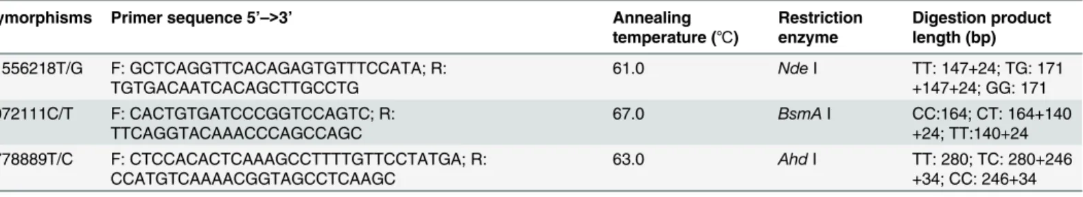
Thers11556218,rs4072111, andrs4778889polymorphism genotypes were determined by the polymerase chain reaction-restriction fragment length polymorphism (PCR-RFLP) method. The primer sequences, reaction conditions, restriction enzymes used, and length of digestion products are listed inTable 1. To confirm the genotyping results, a total of 30 (10%) PCR-am-plified DNA samples were randomly selected and genotyped by DNA sequencing with an ABI Prism 3100 (Applied Biosystems, Shanghai Sangon Biological Engineering Technology & Ser-vices Co., Ltd., China). The results were 100% concordant.
Table 1. Primer sequences and reaction conditions for genotypingIL-16polymorphisms.
Polymorphisms Primer sequence 5’–>3’ Annealing temperature (℃)
Restriction enzyme
Digestion product length (bp)
rs11556218T/G F: GCTCAGGTTCACAGAGTGTTTCCATA; R: TGTGACAATCACAGCTTGCCTG
61.0 NdeI TT: 147+24; TG: 171
+147+24; GG: 171 rs4072111C/T F: CACTGTGATCCCGGTCCAGTC; R:
TTCAGGTACAAACCCAGCCAGC
67.0 BsmAI CC:164; CT: 164+140
+24; TT:140+24 rs4778889T/C F: CTCCACACTCAAAGCCTTTTGTTCCTATGA; R:
CCATGTCAAAACGGTAGCCTCAAGC
63.0 AhdI TT: 280; TC: 280+246
+34; CC: 246+34
Serum IL-16 levels
Serum samples were available for all patients and healthy controls. Following blood sample col-lection, the serum was allowed to clot for 30 min at 4°C before centrifugation at 3,000 rpm for 10 min at 4°C. Total serum was isolated and stored at—20°C until further use. Serum IL-16 concentrations were detected using a sandwich ELISA with the same batch of reagents accord-ing to the manufacturer’s instructions. The minimum level of detection for IL-16 was 5 pg/mL. The intra-assay coefficients of variation were 10%.
Sample size consideration
We estimated the sample size using Quanto software (version 1. 2.4). We based on probability ofα= 0.05 andβ= 0.1 and assuming that the prevalence of the risk allele (rs11556218G) in the control group was 40% [30], and estimated odds ratio (OR) was 0.5 Approximately 1 to 1 case– control ratio was chosen. According to the above parameters, estimated 134 sample size had enough power to assess the effect ofIL-16genetic polymorphisms on the risk of knee OA.
Statistical analysis
Student’st-test (for continuous variables) orχ2test (for categorical variables) were used to evaluate differences in the distributions of selected demographic variables, and frequencies of genotypes ofIL-16polymorphisms between cases and controls. Agreement with the Hardy-Weinberg equilibrium for each SNP was tested using a goodness-of-fitχ2test. Genotype, allele, and haplotype distributions ofIL-16were compared among different groups using theχ2test and Fisher’s exact test when appropriate. The Haploview software [33] was used to calculate the degree of pairwise linkage disequilibrium (LD) for each pair of SNPs as well as for haplo-type analysis. We developed binary logistic regression models to estimate odds ratios (ORs) with corresponding 95% confidence intervals (CIs) to test the association of the various geno-types of interest and the risk of knee OA. All ORs were adjusted for age, gender, BMI, smoking and drinking state. Statistical significance was assumed at two-sidedPvalues at<0.05 level. All
of the statistical analyses were performed in the Statistical Package for Social Sciences (SPSS, version 13.0).
Results
Characteristics of the study population
Table 2summarizes the characteristics of the 150 knee OA patients and 147 control subjects included in this study. The mean ages (SD) of the control group and knee OA group were 58.3 ± 9.6 and 59.5 ± 8.9 years, respectively. There were no significant differences for sex, mean age, BMI, smoking and drinking status between cases and control groups, suggesting that sub-jects matching based on these variables was adequate.
Genotype and allele distribution of
IL-16
polymorphisms
The distribution of each allele and genotype is shown inTable 3. All three SNPs were within the Hardy-Weinberg equilibrium. For thers11556218polymorphism, there was a significant differ-ence in the genotype and allele frequencies among knee OA patients and control subjects. The frequencies of the TT, TG, and GG genotypes ofrs11556218were 36.7%, 51.1%, and 12.2% in healthy controls, and 55.3%, 37.3%, and 7.4% in patients with knee OA, respectively. Binary lo-gistic regression analyses adjusting for age, gender, BMI, smoking and drinking status showed that the TG and GG genotypes ofrs11556218were both associated with a statistically significant decreased risk of knee OA compared with the TT genotype (OR, 0.69; 95% CI, 0.53–0.89;
IL-16 Polymorphisms and Knee OA Risk
p= 0.006 for TG genotype; OR, 0.64; 95% CI, 0.45–0.90;p= 0.042 for GG genotype). Under the dominant model, the combined genotypes GG + TG appeared to have lower susceptibility to OA (OR = 0.68, 95% CI 0.29–0.87,p= 0.002). The data also revealed that subjects with the G allele appeared to have a lower susceptibility to knee OA compared with those bearing the T allele (OR, 0.58; 95% CI, 0.41–0.82;p= 0.002).
Regarding thers4072111polymorphism, the frequencies of the CC, CT, and TT genotypes were 60.5%, 33.3%, and 6.1% for control subjects and 80%, 18%, and 2% in knee OA patients, respectively. The CT and TT genotypes were associated with a significantly decreased risk of knee OA compared with patients with the CC genotype (OR, 0.66; 95% CI, 0.53–0.83;
p= 0.002 and OR, 0.57; 95% CI, 0.40–0.82;p= 0.027, respectively). The combined CC+TC ge-notypes were also associated with a significantly decreased risk of knee OA (OR, 79; 95% CI, 063–0.98;P= 0.045). Using the C allele as a reference, a significant correlation was detected be-tween the presence of the T allele and a lower risk of developing knee OA (OR, 0.69; 95% CI, 0.58–0.81;p<0.001).
For genotype and allele frequencies of theIL-16 rs4778889T/C polymorphisms, we found that subjects with the C allele and combined CC+TC genotypes (dominant model) appeared to have a slightly lower risk of knee OA compared with those bearing the T allele (OR, 0.68; 95% CI, 0.45–0.99;p= 0.044, and OR, 0.79; 95% CI, 0.63–0.98;P= 0.044, respectively).
Stratified analysis
When analyses of genotype and allele frequencies were stratified by gender, significant differ-ences in the distributions ofIL-16polymorphisms among patients with knee OA and control groups were observed (Table 3). Women who carried theIL-16(rs11556218T/G) G allele had a significantly decreased risk of knee OA compared with those carrying the T allele (OR, 0.74; 95% CI, 0.60–0.91;p= 0.007). Similarly, women who carried theIL-16(rs4072111C/T) T allele showed a lower susceptibility to knee OA compared with those carrying the C allele (OR, 0.68; 95% CI, 0.55–0.85;p= 0.004). Men who carried theIL-16(rs4072111C/T) T allele showed a de-creased risk of knee OA compared with those carrying the C allele (OR, 0.71; 95% CI, 0.55–0.92;
p= 0.038), but no significant differences were found for thers11556218T/G polymorphism. Re-garding thers4778889SNP, we found a significant difference of the genotype and allele frequen-cies between knee OA patients and controls in women, but not in men.
Table 2. Demographic characteristics of the study population.
Variables Healthy control (n = 147) n(%) Knee osteoarthritis patients (n = 150) n(%) Pvalue
Age(mean±SD) 58.3±9.6 59.5±8.9 0.13
Gender
Male 51 41 0.07
Femle 96 109
Body mass index (kg/m2) 23.7±2.5 24.2±3.3 0.19
Smoking
No 118 (80.3%) 129(86.0%) 0.187
Yes 29 (19.7%) 21(14.0%)
Drinking
No 121(82.3%) 119 (79.3%) 0.514
Yes 26 (17.7%) 31 (20.7%)
IL-16 concentration (x±S, pg/mL) 36.70±6.72 44.32±8.78 0.001
Table 3. Distributions ofIL-16SNPs genotypes in each group and logistic regression analyses of associations between these polymorphisms and knee OA risk.
Genotypes Overall Women Men
Controls n = 147(%) OA cases n = 150(%) OR (95% CI)a p Controls n = 96 OA cases n = 109 OR (95% CI)b p Controls n = 51 OA cases n = 41 OR (95% CI)b p
rs11556218
TT 54 (36.7) 83(55.3) 1.00ref 34 61 1.00ref 20 22 1.00ref
TG 75(51.1) 56(37.3) 0.69(0.53–0.89) 0.006 51 41 0.65(0.47–0.89) 0.011 24 15 0.77(0.52–1.16) 0.301 GG 18(12.2) 11(7.4) 0.64(0.45–0.90) 0.042 11 7 0.59(0.37–0.92) 0.047 7 4 0.75(0.43–1.29) 0.544 Dominant model
TT 54 83 1.00ref 34 61 1.00ref 20 22 1.00ref
GG+TG 93 67 0.68(0.29–0.87) 0.002 62 48 0.64(0.46–0.87) 0.005 31 19 0.79(0.52–1.13) 0.241 Recessive model
TG+TT 129 139 1.00ref 85 102 1.00ref 44 37 1.00ref
GG 18 11 0.78(0.57–1.06) 0.219 11 7 0.74(0.50–1.11) 0.306 7 4 0.85(0.52–1.39) 0.795
T allele 183 (62.2) 222 (74.0) 1.00ref 119 163 1.00ref 64 59 1.00ref
G allele 111 (37.8) 78(26.0) 0.77(0.66–0.90) 0.003 73 55 0.74(0.60–0.91) 0.007 38 23 0.84(0.65–1.08) 0.246 rs4072111
CC 89 (60.5) 120(80.0) 1.00ref 61 88 1.00ref 28 32 1.00ref
CT 49(33.3) 27(18.0) 0.66(0.53–0.83) 0.002 29 19 0.68(0.50–0.91) 0.029 20 8 0.65(0.46–0.93) 0.048 TT 9 (6.1) 3(2.0) 0.57(0.40–0.82) 0.027 6 2 0.82(0.30–2.22) 0.718 3 1 0.62(0.33–1.17) 0.561 Dominant model
CC 89 120 1.00ref 61 88 1.00ref 29 32 1.00ref
TT+CT 58 30 0.65(0.52–0.80) <0.001 35 21 0.66(0.50–0.87) 0.009 23 9 0.66(0.47–0.93) 0.043 Recessive model
CT+CC 138 147 1.00ref 90 107 1.00ref 48 40 1.00ref
TT 9 3 0.31(0.08–1.18) 0.071 35 21 0.73(0.57–0.94) 0.039 3 1 0.73(0.40–1.32) 0.771
C allele 227(75.7) 267(89.0) 1.00ref 151 195 1.00ref 76 72 1.00ref
T allele 67(23.3) 33(11.0) 0.69(0.58–0.81) <0.001 41 23 0.68(0.55–0.85) 0.004 26 10 0.71(0.55–0.92) 0.038 rs4778889
TT 82 (55.8) 101 (67.3) 1.00ref 50 71 1.00ref 32 30 1.00ref
TC 56 (38.1) 43 (28.7) 0.79(0.63–1.00) 0.079 37 35 0.80(0.59–1.10) 0.226 19 8 0.73(0.52–1.03) 0.158 CC 9 (6.1) 6 (4.0) 0.75(0.48–1.16) 0.387 9 3 0.55(0.37–0.82) 0.041 0 3 6.46(0.37–11.62) 0.248 Dominant model
TT 82 101 1.00ref 50 71 1.00ref 32 30 1.00ref
CC+TC 65 49 0.79(0.63–0.98) 0.045 46 38 0.76(0.57–1.10) 0.079 19 11 0.62(0.57–1.17) 0.403 Recessive model
TT+TC 138 144 1.00ref 87 106 1.00ref 51 38 1.00ref
CC 9 6 0.82(0.53–1.25) 0.569 9 3 0.60(0.42–0.86) 0.086 0 3 2.43(0.34–4.54) 0.170
T allele 220(74.8) 245(81.7) 1.00ref 137 177 1.00ref 83 68 1.00ref
C allele 74(25.2) 55(18.3) 0.83(0.69–0.98) 0.047 55 41 0.76(0.62–0.94) 0.026 19 14 0.96(0.69–1.32) 0.936
OA, osteoarthritis,ORodds ratio,CIconfidence interval,refreference
Boldindicated the difference was significant.
aAdjusted for age, sex, smoking and drinking status by logistic regression model.
bAdjusted for age, smoking and drinking status by logistic regression model.
doi:10.1371/journal.pone.0123442.t003
IL-16
Polymorphis
ms
and
Knee
OA
Risk
PLOS
ONE
|DOI:10.137
1/journal.p
one.0123442
May
8,
2015
6/1
Haplotype analyses of
IL-16
gene polymorphisms and knee OA risk
LD analyses were performed in knee OA patients and healthy controls using the Haploview ver.4.2 software. No statistically significant evidence of LD was observed among these three SNPs between knee OA patients and healthy controls (forrs11556218andrs4072111,D’= 0.22, r2= 0.006; forrs4778889andrs11556218,D’= 0.59, r2= 0.195; forrs4778889and
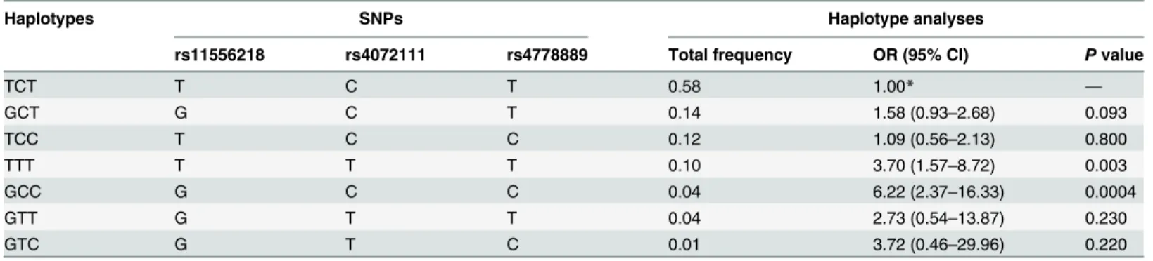
rs4072111,D’= 0.64, r2= 0.030). Further, haplotype analysis to evaluate the haplotype frequen-cies of polymorphisms located within the same chromosome regions was performed in order to derive haplotypes specifically correlated with knee OA. A total of seven haplotypes were de-rived from the observed genotypes. The haplotype distributions in knee OA patients and healthy controls are shown inTable 4. Two high-risk haplotypes were found: TTT (OR, 3.70; 95% CI, 1.57–8.72;p= 0.003) and GCC (OR, 6.22; 95% CI, 2.37–16.33;p= 0.0004). The re-maining haplotypes were not associated with risk of knee OA.
Serum IL-16 levels and polymorphisms
The median serum concentration of IL-16 detected was 36.70 ± 6.72 pg/mL in healthy controls and 44.32 ± 8.78 pg/mL in knee OA patients (Table 2). The serum levels of IL-16 detected in knee OA patients were significantly higher than those in healthy control subjects (p= 0.001). However, when studying the relationship between theIL-16polymorphisms present and IL-16 serum levels among patients with knee OA and healthy controls, no significant differences were observed.
Discussion
In the present study, we selected a commonIL-16SNP, namelyrs4778889, located 295 bp up-stream from the transcription start site and associated with altered levels of gene expression [23], as well as two other SNPs (rs11556218andrs4072111), to evaluate their association in pa-tients with knee OA and healthy controls. The latter two SNPs (rs11556218andrs4072111) are located in an exon region, and their single-nucleotide changes would result in an amino acid substitution. To the best of our knowledge, this is the first study to investigate whetherIL-16
gene polymorphisms are associated with the risk of knee OA and whether these correlate with serum levels of IL-16. The present results revealed that theIL-16 rs11556218polymorphism, representing anAsntoLyssubstitution in exon 6 of theIL-16gene, has a significant effect on the risk of knee OA; individuals carrying thers11556218G allele had a significantly decreased risk of developing knee OA compared with those carrying the T allele (OR, 0.77; 95% CI, 0.66– 0.90). In addition, the non-synonymous SNP,rs4072111C/T, representing aSertoPro substi-tution, was associated with a significantly decreased risk of developing knee OA. Regarding the
Table 4. Haplotype analysis between the case and control groups.
Haplotypes SNPs Haplotype analyses
rs11556218 rs4072111 rs4778889 Total frequency OR (95% CI) Pvalue
TCT T C T 0.58 1.00* —
GCT G C T 0.14 1.58 (0.93–2.68) 0.093
TCC T C C 0.12 1.09 (0.56–2.13) 0.800
TTT T T T 0.10 3.70 (1.57–8.72) 0.003
GCC G C C 0.04 6.22 (2.37–16.33) 0.0004
GTT G T T 0.04 2.73 (0.54–13.87) 0.230
GTC G T C 0.01 3.72 (0.46–29.96) 0.220
IL-16 rs4778889T/C polymorphism, we found that subjects with the C allele appeared to have a slightly lower risk of knee OA compared with those bearing the T allele (OR, 0.83; 95% CI, 0.69–0.98). We further evaluated the effect ofIL-16polymorphisms on knee OA risk stratified by sex and found that the association appeared stronger in female patient subgroups.
Despite the positive relationship betweenrs11556218andrs4072111polymorphisms and the risk of knee OA observed in this study, IL-16 serum levels did not show any significant dif-ferences other than being cumulatively higher in patients with knee OA relative to healthy con-trols. In addition, we found two haplotypes (TTT and GCC) to be significantly associated with susceptibility to knee OA. Thus, the above data indicates that there is no association between
IL-16polymorphisms and IL-16 serum levels. However, the results also suggest thatIL-16gene polymorphisms may be significantly associated with the risk of knee OA. Although the sample size is not large enough, this is the first case-control study evaluating the association between
IL-16polymorphisms and the risk of knee OA.
Previous attempts have been made to identify genetic factors involved in knee OA using ge-nome-wide association studies (GWAS). Recently, several OA susceptibility loci have been identified in GWAS with significance levels [34–40]. Among them, theDVWAgene (SNPs
rs11718863andrs7639618) and a region containing HLA class II/III genes (SNPsrs7775228
andrs10947262). However, this genome-wide significant association was shown in Asians but not in Europeans [34–36]. On the other hand, genome-wide significant loci were identified to have an association with knee OA in Europeans. These included thers3815148SNP inCOG5
on chromosome 7q22 [37,38],rs11842874inMCF2Lon chromosome 13 [40], andrs6976in
GNL3on chromosome 3 [39]. To date, theIL-16gene has not yet been identified through GWAS. In fact, recent GWAS in knee OA mainly focused on chromosome 3, chromosome 7, and chromosome 13 genes [34,37–39]. TheIL-16gene is located on chromosome 15q26.3. IL-16 is a pleiotropic cytokine whose functions include chemoattraction and modulation of T cell activation [13] and is an important mediator in inflammatory and autoimmune diseases as well as in tumor growth and progression [14,15]. In addition,IL-16can activate the secretion of tumor-associated inflammatory cytokines, such as TNF-α, IL-1β, IL-6, and IL-15, all of which are major factors involved in tumorigenesis [21,22]. In recent years,IL-16gene poly-morphisms have been associated with several human diseases, including gastric cancer [24], colorectal cancer [25], renal cell carcinoma [26], Graves’disease [27], coronary heart disease [28,41,42], and ischemic stroke [29]. Our previous study has shown that theIL-16 rs11556218
T/G polymorphism was significantly associated with susceptibility to both hepatocellular [30] and nasopharyngeal carcinoma [31].
Pathologically, various inflammatory components are involved in OA. In OA, the increased synthetic and anti-inflammatory activity of chondrocytes loses out to the increased degradative activity [43,44]. The increased synthetic activity is confined to the deeper cartilage layers, which allows the imbalance towards degradation to persist in the upper layer, near the synovial boundary. Ultimately, chondrocyte malfunction and apoptosis limit the response potential and hasten the progression of OA [43,44]. Therefore, we postulated thatIL-16polymorphisms may modulate the susceptibility to OA.
In the current study, we found that patients carrying the G (rs11556218T/G), T (rs4072111
C/T), and C (rs4778889T/C) alleles were associated with a significantly decreased risk for knee OA compared to individuals carrying the wild-type alleles. This finding is consistent with our hypothesis, suggesting thatIL-16 rs11556218,rs4072111, andrs4778889polymorphisms may play an important role in the pathogenesis of knee OA. Further, we found that the association betweenIL-16 rs11556218T/G andrs4778889T/C polymorphisms and knee OA risk appeared stronger in female patients. Nevertheless, this evidence is suggestive but not conclusive, and was unexpected and difficult to explain. It is possible that this result is due to the larger number
IL-16 Polymorphisms and Knee OA Risk
of female subjects (n = 205) compared to male subjects (n = 92), resulting in a limited statistical power and robustness. On the other hand, it might be attributed to the lower exposure to risk factors, such as tobacco smoking and heavy drinking, of female patients compared to males. Fi-nally, this association might also be the result of estrogen-related effects; estrogen can interact with IL-16 and reduce the possibility of developing knee OA [45–47]. Nevertheless, since the sample size of the current study was relatively small, these findings need to be confirmed by further larger sample size studies which also investigate the underlying mechanisms of this association.
A haplotype is a set of SNPs on a single chromatid which are likely to be inherited together in a block pattern more frequently than expected by chance owing to the presence of linkage disequilibrium [48]. In the current study, we found that two haplotypes (TTT and GCC) of the
IL-16gene were significantly associated with the susceptibility to knee OA (OR, 3.70; 95% CI, 1.57–8.72 for the TTT haplotype and OR, 6.22; 95% CI, 2.37–16.33 for the GCC haplotype). We also evaluated the influence ofIL-16polymorphisms on IL-16 serum levels in patients with knee OA versus healthy controls, and found that the median serum level of IL-16 in patients was significantly higher than in controls. Our data suggest that a higher serum IL-16 level might serve as a risk factor for knee OA. However, the serum levels of IL-16 detected in groups of patients with different genotypes did not show any significant differences, probably due to the relatively small sample size; further confirmation would be provided by additional patient data.
Several potential limitations of this study must be acknowledged. First, our patient sample size was relatively small and therefore the study’s statistical power may have been limited. Thus, additional studies with larger samples are desirable. Second, the study population was limited to the Guangxi population and therefore the findings may not be generalized to other populations. Continued study of the role ofIL-16polymorphisms in patient susceptibility to knee OA from other ethnic populations would also be of great value. Finally, the current re-search studied only three SNPs in theIL-16gene. It would be interesting to identify more SNPs and study their association with knee OA.
In conclusion, the present study showed that functional polymorphisms ofIL-16are associated with the risk of knee OA. We found that the variant allelesrs11556218T/G,rs4072111C/T, and
rs4778889T/C were associated with a decreased risk of knee OA compared with wild-type alleles. These findings suggest that theIL-16 rs11556218T/G,rs4072111C/T, andrs4778889T/C poly-morphisms might be markers for genetic susceptibility to knee OA. Furthermore, serum IL-16 levels were significantly associated with increased risk of knee OA. These findings, after validation by larger studies, might help identify at-risk populations for primary knee OA prevention.
Author Contributions
Conceived and designed the experiments: JMZ XQ. Performed the experiments: SXL SL XHZ JJZ. Analyzed the data: SXL YL XHZ GHL. Contributed reagents/materials/analysis tools: GFD WS YD YL. Wrote the paper: YL SXL.
References
1. Scott D, Kowalczyk A. Osteoarthritis of the knee. Am Fam Physician. 2008; 77: 1149–1150. PMID:
18481563
2. Sowers M. Epidemiology of risk factors for osteoarthritis: systemic factors. Curr Opin Rheumatol. 2001; 13: 447–451. PMID:11604603
4. Jiang L, Tian W, Wang Y, Rong J, Bao C, Liu Y, et al. Body mass index and susceptibility to knee osteo-arthritis: a systematic review and meta-analysis. Joint Bone Spine. 2012; 79: 291–297. doi:10.1016/j. jbspin.2011.05.015PMID:21803633
5. Goldring SR, Goldring MB. The role of cytokines in cartilage matrix degeneration in osteoarthritis. Clin Orthop Relat Res. 2004: S27–36.
6. Brooks P. Inflammation as an important feature of osteoarthritis. Bulletin of the World Health Organiza-tion. 2003; 81: 689–690. PMID:14710513
7. Honsawek S, Deepaisarnsakul B, Tanavalee A, Yuktanandana P, Bumrungpanichthaworn P, Malila S, et al. Association of the IL-6 -174G/C gene polymorphism with knee osteoarthritis in a Thai population. Genet Mol Res. 2011; 10: 1674–1680. PMID:21863560
8. Kaarvatn MH, Jotanovic Z, Mihelic R, Etokebe GE, Mulac-Jericevic B, Tijanic T, et al. Associations of the interleukin-1 gene locus polymorphisms with risk to hip and knee osteoarthritis: gender and subpop-ulation differences. Scand J Immunol. 2013; 77: 151–161. doi:10.1111/sji.12016PMID:23216199
9. Yigit S, Inanir A, Tekcan A, Tural E, Ozturk GT, Kismali G, et al. Significant association of interleukin-4 gene intron 3 VNTR polymorphism with susceptibility to knee osteoarthritis. Gene. 2014; 537: 6–9. doi:
10.1016/j.gene.2013.12.060PMID:24406619
10. Han L, Lee HS, Yoon JH, Choi WS, Park YG, Nam SW, et al. Association of IL-17A and IL-17F single nucleotide polymorphisms with susceptibility to osteoarthritis in a Korean population. Gene. 2014; 533: 119–122. doi:10.1016/j.gene.2013.09.113PMID:24096234
11. Hulin-Curtis SL, Bidwell JL, Perry MJ. Evaluation of IL18 and IL18R1 polymorphisms: genetic suscepti-bility to knee osteoarthritis. Int J Immunogenet. 2012; 39: 106–109. doi:10.1111/j.1744-313X.2011. 01060.xPMID:22136483
12. Ji B, Shi J, Cheng X, Zhou J, Zhou Q, Cao C, et al. Association analysis of two candidate polymor-phisms in the tumour necrosis factor-alpha gene with osteoarthritis in a Chinese population. Int Orthop. 2013; 37: 2061–2063. doi:10.1007/s00264-013-1931-4PMID:23748461
13. Smith AJP, Humphries SE. Cytokine and cytokine receptor gene polymorphisms and their functionality. Cytokine & Growth Factor Reviews. 2009; 20: 43–59.
14. Moss SF, Blaser MJ. Mechanisms of disease: Inflammation and the origins of cancer. Nat Clin Pract Oncol. 2005; 2: 90–97; quiz 91 p following 113. PMID:16264881
15. Lu H, Ouyang W, Huang C. Inflammation, a key event in cancer development. Mol Cancer Res. 2006; 4: 221–233. PMID:16603636
16. Kim HS. Assignment of human interleukin 16 (IL16) to chromosome 15q26.3 by radiation hybrid map-ping. Cytogenet Cell Genet. 1999; 84: 93. PMID:10343113
17. Baier M, Bannert N, Werner A, Lang K, Kurth R. Molecular cloning, sequence, expression, and pro-cessing of the interleukin 16 precursor. Proc Natl Acad Sci U S A. 1997; 94: 5273–5277. PMID:
9144227
18. Zhang Y, Center DM, Wu DM, Cruikshank WW, Yuan J, Andrews DW, et al. Processing and activation of pro-interleukin-16 by caspase-3. J Biol Chem. 1998; 273: 1144–1149. PMID:9422780
19. Cruikshank WW, Center DM, Nisar N, Wu M, Natke B, Theodore AC, et al. Molecular and functional analysis of a lymphocyte chemoattractant factor: association of biologic function with CD4 expression. Proc Natl Acad Sci U S A. 1994; 91: 5109–5113. PMID:7910967
20. Center DM, Kornfeld H, Cruikshank WW. Interleukin 16 and its function as a CD4 ligand. Immunol Today. 1996; 17: 476–481. PMID:8908813
21. Kai H, Kitadai Y, Kodama M, Cho S, Kuroda T, Ito M, et al. Involvement of proinflammatory cytokines IL-1beta and IL-6 in progression of human gastric carcinoma. Anticancer Res. 2005; 25: 709–713. PMID:15868900
22. Shanmugham LN, Petrarca C, Frydas S, Donelan J, Castellani ML, Boucher W, et al. IL-15 an immuno-regulatory and anti-cancer cytokine. Recent advances. J Exp Clin Cancer Res. 2006; 25: 529–536. PMID:17310844
23. Nakayama EE, Wasi C, Ajisawa A, Iwamoto A, Shioda T. A new polymorphism in the promoter region of the human interleukin-16 (IL-16) gene. Genes Immun. 2000; 1: 293–294. PMID:11196708
24. Zhang T, Wang H. Variants of interleukin-16 associated with gastric cancer risk. Asian Pac J Cancer Prev. 2013; 14: 5269–5273. PMID:24175812
25. Azimzadeh P, Romani S, Mohebbi SR, Kazemian S, Vahedi M, Almasi S, et al. Interleukin-16 (IL-16) gene polymorphisms in Iranian patients with colorectal cancer. J Gastrointestin Liver Dis. 2011; 20: 371–376. PMID:22187702
IL-16 Polymorphisms and Knee OA Risk
26. Zhu J, Qin C, Yan F, Wang M, Ding Q, Zhang Z, et al. IL-16 polymorphism and risk of renal cell carcino-ma: association in a Chinese population. Int J Urol. 2010; 17: 700–707. doi:10.1111/j.1442-2042.2010. 02559.xPMID:20529140
27. Tsai KH, Chang CY, Tsai FJ, Lin HJ, Yang YS, Lim YP, et al. Association of interleukin-16 polymor-phisms with graves' disease in a taiwanese population. Chin J Physiol. 2014; 57: 69–75. doi:10.4077/ CJP.2014.BAB150PMID:24694201
28. Tong Z, Li Q, Zhang J, Wei Y, Miao G, Yang X. Association between interleukin 6 and interleukin 16 gene polymorphisms and coronary heart disease risk in a Chinese population. J Int Med Res. 2013; 41: 1049–1056. doi:10.1177/0300060513483405PMID:23881440
29. Liu XL, Du JZ, Zhou YM, Shu QF, Li YG. Interleukin-16 polymorphism is associated with an increased risk of ischemic stroke. Mediators Inflamm. 2013; 2013: 564750. doi:10.1155/2013/564750PMID:
24288444
30. Li S, Deng Y, Chen ZP, Huang S, Liao XC, Lin LW, et al. Genetic polymorphism of interleukin-16 influ-ences susceptibility to HBV-related hepatocellular carcinoma in a Chinese population. Infect Genet Evol. 2011; 11: 2083–2088. doi:10.1016/j.meegid.2011.09.025PMID:22019522
31. Qin X, Peng Q, Lao X, Chen Z, Lu Y, Lao X, et al. The association of interleukin-16 gene polymorphisms with IL-16 serum levels and risk of nasopharyngeal carcinoma in a Chinese population. Tumour Biol. 2014; 35: 1917–1924. doi:10.1007/s13277-013-1257-2PMID:24101193
32. Altman R, Asch E, Bloch D, Bole G, Borenstein D, Brandt K, et al. Development of criteria for the classi-fication and reporting of osteoarthritis. Classiclassi-fication of osteoarthritis of the knee. Diagnostic and Thera-peutic Criteria Committee of the American Rheumatism Association. Arthritis Rheum. 1986; 29: 1039– 1049. PMID:3741515
33. Barrett JC, Fry B, Maller J, Daly MJ. Haploview: analysis and visualization of LD and haplotype maps. Bioinformatics. 2005; 21: 263–265. PMID:15297300
34. Miyamoto Y, Shi D, Nakajima M, Ozaki K, Sudo A, Kotani A, et al. Common variants in DVWA on chro-mosome 3p24.3 are associated with susceptibility to knee osteoarthritis. Nat Genet. 2008; 40: 994– 998. doi:10.1038/ng.176PMID:18622395
35. Nakajima M, Takahashi A, Kou I, Rodriguez-Fontenla C, Gomez-Reino JJ, Furuichi T, et al. New se-quence variants in HLA class II/III region associated with susceptibility to knee osteoarthritis identified by genome-wide association study. PLoS One. 2010; 5: e9723. doi:10.1371/journal.pone.0009723
PMID:20305777
36. Meulenbelt I, Chapman K, Dieguez-Gonzalez R, Shi DQ, Tsezou A, Dai J, et al. Large replication study and meta-analyses of DVWA as an osteoarthritis susceptibility locus in European and Asian popula-tions. Human Molecular Genetics. 2009; 18: 1518–1523. doi:10.1093/hmg/ddp053PMID:19181678
37. Kerkhof HJ, Lories RJ, Meulenbelt I, Jonsdottir I, Valdes AM, Arp P, et al. A genome-wide association study identifies an osteoarthritis susceptibility locus on chromosome 7q22. Arthritis Rheum. 2010; 62: 499–510. doi:10.1002/art.27184PMID:20112360
38. Evangelou E, Valdes AM, Kerkhof HJ, Styrkarsdottir U, Zhu Y, Meulenbelt I, et al. Meta-analysis of ge-nome-wide association studies confirms a susceptibility locus for knee osteoarthritis on chromosome 7q22. Ann Rheum Dis. 2011; 70: 349–355. doi:10.1136/ard.2010.132787PMID:21068099
39. Zeggini E, Panoutsopoulou K, Southam L, Rayner NW, Day-Williams AG, Lopes MC, et al. Identifica-tion of new susceptibility loci for osteoarthritis (arcOGEN): a genome-wide associaIdentifica-tion study. Lancet. 2012; 380: 815–823. doi:10.1016/S0140-6736(12)60681-3PMID:22763110
40. Day-Williams AG, Southam L, Panoutsopoulou K, Rayner NW, Esko T, Estrada K, et al. A variant in MCF2L is associated with osteoarthritis. Am J Hum Genet. 2011; 89: 446–450. doi:10.1016/j.ajhg. 2011.08.001PMID:21871595
41. Huang H, Zeng Z, Zhang L, Liu R, Li X, Qiang O, et al. The association of interleukin-16 gene polymor-phisms with susceptibility of coronary artery disease. Clin Biochem. 2013; 46: 241–244. doi:10.1016/j. clinbiochem.2012.11.009PMID:23195133
42. Hai-Feng T, Wei W, Yuan-Yuan Y, Jun Z, Su-Ping G, Hui-Ming L. Association between Polymorphisms in IL-16 Genes and Coronary Heart Disease risk. Pak J Med Sci. 2013; 29: 1033–1037. PMID:
24353682
43. Sandell LJ, Aigner T. Articular cartilage and changes in arthritis. An introduction: cell biology of osteoar-thritis. Arthritis Res. 2001; 3: 107–113. PMID:11178118
44. Pelletier JP, Martel-Pelletier J, Abramson SB. Osteoarthritis, an inflammatory disease: potential impli-cation for the selection of new therapeutic targets. Arthritis Rheum. 2001; 44: 1237–1247. PMID:
11407681
46. Jochems C, Islander U, Erlandsson M, Engdahl C, Lagerquist M, Gjertsson I, et al. Role of endogenous and exogenous female sex hormones in arthritis and osteoporosis development in B10.Q-ncf1*/*mice with collagen-induced chronic arthritis. BMC Musculoskelet Disord. 2010; 11: 284. doi: 10.1186/1471-2474-11-284PMID:21159208
47. Ma HL, Blanchet TJ, Peluso D, Hopkins B, Morris EA, Glasson SS. Osteoarthritis severity is sex depen-dent in a surgical mouse model. Osteoarthritis and Cartilage. 2007; 15: 695–700. PMID:17207643
48. Seng KC, Seng CK. The success of the genome-wide association approach: a brief story of a long struggle. Eur J Hum Genet. 2008; 16: 554–564. doi:10.1038/ejhg.2008.12PMID:18285837
IL-16 Polymorphisms and Knee OA Risk