Control of the rat angiotensin I
converting enzyme gene by CRE-like
sequences
1Laboratório de Genética e Cardiologia Molecular, Instituto do Coração,
Faculdade de Medicina, Universidade de São Paulo, São Paulo, SP, Brasil
2Laboratório de Bioquímica da Atividade Motora,
Escola de Educação Física e Esporte, Universidade de São Paulo, São Paulo, SP, Brasil
J. Xavier-Neto1,
A.C. Pereira1,
E.M. Oliveira2,
A.A. Miyakawa1,
M.L. Junqueira1
and J.E. Krieger1
Abstract
We characterized the role of potential cAMP-responsive elements (CRE) in basal and in induced angiotensin converting enzyme (ACE) gene promoter activity in order to shed light on the regulation of somatic ACE expression. We identified stimulators and repressors of basal expression between 122 and 288 bp and between 415 and 1303 bp upstream from the transcription start site, respectively, using a rabbit endothelial cell (REC) line. These regions also contained elements associated with the response to 8BrcAMP. When screening for CRE motifs we found pCRE, a proximal sequence between 209 and 222 bp. dCRE, a distal tandem of two CRE-like sequences conserved between rats, mice and humans, was detected between 834 and 846 bp. Gel retardation analysis of nuclear extracts of REC indicated that pCRE and dCRE bind to the same protein complexes as bound by a canonical CRE. Mutation of pCRE and dCRE in REC established the former as a positive element and the latter as a negative element. In 293 cells, a renal cell line, pCRE and dCRE are negative regulators. Co-transfection of ATF-2 or ATF-2 plus c-Jun repressed ACE promoter activity, suggesting that the ACE gene is controlled by cellular stress. Although mapping of cAMP responsiveness was con-sistent with roles for pCRE and dCRE, mutation analysis indicated that they were not required for cAMP responsiveness. We conclude that the basal activity of the somatic ACE promoter is controlled by proximal and distal CREs that can act as enhancers or repressors depending on the cell context.
Correspondence J.E. Krieger
Laboratório de Genética e Cardiologia Molecular InCor, FMRP, USP Av. Dr. Enéas C. Aguiar, 44 05403-000 São Paulo, SP Brasil
Fax: +55-11-3068-5048 E-mail: [email protected]
Research supported by FAPESP (No. 01/00009-0).
Received July 14, 2003 Accepted June 8, 2004
Key words
•Angiotensin converting enzyme
•Endothelium •cAMP
•Cyclic AMP responsive element
Introduction
Angiotensin converting enzyme (ACE) expression in cultured cells is modulated by hormones, ionophores, second messengers, and growth factors (1-5). The flexibility of modulation suggested by these properties supports the notion of regulated ACE
useful to understand several pathological processes such as myocardial ischemia-re-perfusion (6) and chronic inflammatory dis-eases such as sarcoidosis in which ACE is thought to play a role (7).
One of the most consistent stimulators of ACE activity and ACE mRNA expression is the cyclic AMP (cAMP)-dependent signal-ing pathway. cAMP analogues, adenylyl cy-clase activators (forskolin) and agents acting through the beta adrenergic signal transduc-tion pathway induce ACE activity and ACE mRNA expression in intact animals and in cell culture (8-10). Transduction of the intra-cellular cAMP signal is often performed by cAMP responsive element (CRE) genetic pathways. CRE pathways are activated upon phosphorylation of transcription factors bound to the prototypical CRE nucleotide sequence, TGACGTCA, or to its variants located in regulatory regions of target genes (11). Interestingly, non-canonical CREs me-diate either cAMP responsiveness or regula-tion by distinct signaling pathways. In fact, CRE-like sequences often conserve the abil-ity to bind transcription factors traditionally associated with cAMP signaling, while si-multaneously binding other classes of trans-cription factors. Therefore, CRE-like se-quences may confer multiple control possi-bilities to a regulatory region, integrating different signaling inputs (12,13).
We have shown that increases in intracel-lular cAMP levels induced by stimulation of ß-adrenergic receptors, activate transcription by the rat 1303-bp somatic ACE promoter in rabbit endothelial cells (REC) (10). Here we demonstrate that cAMP activates the CRE pathway but not the AP2 pathway, an alter-native route for cAMP regulation. This sug-gests that CRE participates in the regulation of the ACE gene in endothelial cells. We performed mutational and gel retardation analysis of the 5' flanking region of the ACE gene to assess the role of CRE regulation in basal as well as in cAMP-induced activity of the ACE promoter. We report evidence that
the basal activity of the ACE promoter in endothelial cells is controlled by both proxi-mal and distal CRE-like sequences that dis-play positive and negative regulatory activ-ity, respectively. Interestingly, these CRE-like sequences did not participate in the stim-ulation by 8BrcAMP, indicating that the ACE gene uses a different set of regulators to respond to cAMP. Thus, our data indicate that the basal regulation of ACE promoter activity is complex and suggest that the ACE gene is a target for signal transduction path-ways that use CRE-like sequences, but do not operate through the classic mechanisms utilized by cAMP.
Material and Methods
Material
Cell culture materials and media were from Gibco/Invitrogen (Carlsbad, CA, USA). 8BrcAMP was purchased from Sigma (St. Louis, MO, USA). Cell lysis buffers and the luciferase assay system were from Promega (Madison, WI, USA). The ß-galactosidase and renilla luciferase assay systems were from Tropix/Perkin Elmer (Boston, MA, USA) and Promega, respectively.
Plasmids and plasmid constructions
(15) and also by subcloning pTKCAT4CRE into pGL2. We synthesized a series of hybrid ACE-TK constructs to identify potential cAMP responsive sequences between the 1303- and 228-bp nucleotides of the ACE promoter. A Kpn-I/Sac-I fragment contain-ing ACE promoter sequences from 1281 to 228 bp was subcloned upstream from TK, generating the construct ACE1281-288-TK. Further deletion mutants, ACE1120-228-TK and ACE415-228-TK, were obtained by re-striction enzyme digestion. The ACE845-228-TK construct was synthesized by Pfu PCR. The PCR fragment was digested with Nhe-I and Bgl-II and subcloned into pGL2. A positive control for AP2 stimulation (AP2LUC) was constructed by subcloning a Sal-I/Bgl-II fragment from A2BCAT4 (16) containing three tandem repeats of a 19-bp hMtIIa AP2-binding site fused to the aden-ovirus E1b tata box, into pGL2. pact-CRE-BP1/ATF-2 (15) was a gift from Dr. Shunsuke Ishii, Laboratory of Molecular Genetics, RIKEN, Tsukuba, Ibaraki, Japan. The c-Jun and c-Fos expression vectors (17) were pro-vided by Dr. Moshe Yaniv, Gene Expression and Disease Unit, Department of Develop-mental Biology, Pasteur Institute, Paris, France.
Mutagenesis
Mutant ACE promoters mdCRE, mpCRE and mdCREpCRE, each respectively har-boring Bgl-II sites (AGATCT) in place of dCRE, pCRE or both, were synthesized us-ing extension of mutated oligonucleotides mdCRE, 5’AGTGTGGAGACCAGATCTA GATCTAAGCCGATCTGTCTCAGG3', mpCRE AGCGAGAGCTCGACCAGATC TCATCCTTCCACCC by a Pfu PCR and subsequent digestion of the 1303 ACE pro-moter plasmid template by Dpn-I with a mutagenesis kit from Stratagene (La Jolla, CA, USA). Mutated constructs were selected by restriction analysis and confirmed by se-quencing.
Transient transfections
REC cells (18) or human renal embry-onic cells (293; Invitrogen) were grown in 12- or 24-well plates on F12 medium (F12 Coon’s modification) or DMEM, respec-tively, and supplemented with 10% fetal bo-vine serum (FBS) and antibiotics (penicillin and streptomycin). Before reaching conflu-ence, REC and 293 were transfected (16-24 h) with 3.25 µg of luciferase reporter plas-mids and 0.48 µg of a control plasmid (pSV-ß-galactosidase; Promega) per well by the calcium phosphate method (19). Experiments employing expression vectors were per-formed with transfection of 1.78 µg of lu-ciferase reporter plasmids, 0.48 µg of LTR-ß-galactosidase and 0.48 µg of each expres-sion vector per well. Thus, we used the same amounts of DNA for each expression vector when they were co-transfected. Total DNA was maintained at 3.73 µg per well by ma-nipulating the amount of pBluescript SK+ (Stratagene).
cAMP treatment
Prior to stimulation, transiently trans-fected cells were serum-starved for 16-24 h on F12 supplemented with 0.5% FBS. 8BrcAMP was then administered to cells at a concentration of 5 mM for 4 h (10).
Enzyme assays
Luciferase and ß-galactosidase were measured in cell extracts from transient trans-fections with a model 2010 Monolight luminometer, Analytical Luminescence Lab-oratory (San Diego, CA, USA) according to the manufacturer’s instructions.
Gel shift binding assay
assayed by gel retardation analysis (20). The DNA used in these experiments was ob-tained by synthesis of complementary oligo-nucleotides representing the ACE promoter sequences of 796 to 776 bp (TGTGGA GACCTGAGGTGACTTGAAG), 199 to 174 bp (GAGCTCGACCTAACCTCATCC TTCCA) and oligonucleotides containing the canonical CRE (AGAGATTGCCTGACGT CAGAGAGCT). Protein-DNA complexes were visualized by labeling DNA with 32P by
a filling-in reaction using Klenow polymer-ase.
Sequence alignment and promoter
Rat, mouse (M34433), rabbit (M58580), and human (M34434) ACE promoters were aligned with CLUSTAL W at http:// transfac.gbf.de/. Detection of transcription factor binding sites was performed with MatInspector public domain software at http:/ /genomatix.gsf.de/free_services/.
Data presentation
Luciferase activity in extracts of tran-siently transfected cells was normalized for ß-galactosidase activity or renilla luciferase in order to control for differences in trans-fection efficiency. Data are reported as means ± SEM. Unless otherwise stated, data are reported as variation from control values. Where appropriate, results were analyzed by one-way analysis of variance. Statistical sig-nificance was set at P < 0.05.
Results
ACE promoter regions associated with basal and cAMP-induced regulation
To investigate the role of CRE-like se-quences in basal and cAMP-induced regula-tion of the ACE gene we first determined promoter regions containing potential regu-lators and then screened these sites for the
presence of CRE-like motifs.
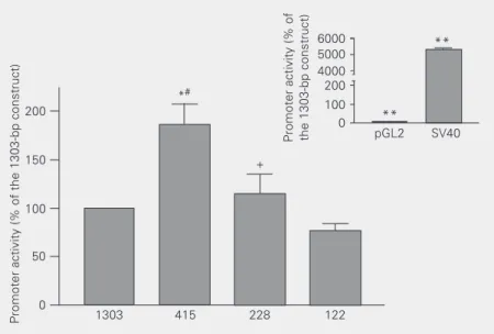
We transfected REC cells with luciferase constructs representing a nested set of frag-ments from the 5' regulatory region of the rat somatic ACE gene. The fragments contained 122, 228, 415, and 1303 bp (21). Luciferase expression from the 122-bp promoter was the smallest. Significant increases in lu-ciferase expression were detected with 228-and 415-bp promoter fragments, suggesting the presence of positive regulatory sequences between 122 and 228 bp, as well as between 228 and 415 bp upstream of the transcription start site. Luciferase expression by the 1303-bp promoter, however, was approximately half that by the 415-bp promoter, indicating the presence of negative sequences between 415 and 1303 bp upstream of the transcrip-tion start site (Figure 1). Luciferase expres-sion from all ACE promoter constructs was on average 50 times higher than the promot-erless pGL2 vector, but 50 times less than luciferase expression driven by the SV40 promoter (Figure 1, inset). Thus, deletion analyses of the 5' regulatory region of the ACE gene suggested the presence of posi-tive regulators between 122 and 415 bp and negative regulators between 415 and 1303 bp.
the same order of magnitude as those from CRETK, the positive control. Thus, deletion analysis of cAMP responsiveness suggested the presence of proximal CREs between 122 and 228 bp as well as additional distal CREs between 415 and 1303 (Figure 2A).
To establish whether putative distal CREs between 415 and 1303 bp operate autono-mously or only in association with the proxi-mal CREs located between 122 and 228 bp, we subcloned ACE promoter fragments 228 to 415, 228 to 845, 228 to 1120, and 228 to 1281 bp upstream from the heterologous TK promoter. Consistent with the above dele-tion analysis, addidele-tion of nucleotides 228 to 415 did not affect the cAMP responsiveness of the TK promoter. Remarkably, addition of nucleotides 228 to 845 doubled the induc-tion of the TK promoter by 8BrcAMP. How-ever, further addition of nucleotides did not change cAMP responsiveness of hybrid ACE-TK promoters, indicating that distal se-quences containing functional CREs must lie between 415 and 845 bp (Figure 2B). Thus, deletion analyses of distal ACE pro-moter regions in the context of the heterolo-gous TK promoter indicated that there are CREs acting in an autonomous fashion be-tween 415 and 845 bp.
pCRE and dCRE are CRE-like sequences of the 1303-bp ACE promoter
Inspection of the 1303-bp ACE promoter revealed the presence of two CRE-like se-quences representing 2 bp substitutions from the canonical CRE, TGACGTCA. One proxi-mal CRE-like sequence, TAACCTCA, (pCRE) was found between 209 and 222 bp upstream from the transcription initiation. Another distal CRE-like sequence, TGAGG TGACTTGA, (dCRE) represented 2 tandem overlapping repeats located between 801 and 819 bp upstream from the transcription ini-tiation (Figure 3). Alignment of rat, mouse, rabbit, and human ACE 5' regulatory regions indicated that only dCRE is evolutionarily
conserved in these three species.
As established by functional analysis, the position of pCRE is consistent with the loca-tion of a positive regulator of basal expres-sion and with a functional CRE mediating induction by 8BrcAMP. Likewise, the posi-tion of dCRE is consistent with the locaposi-tion of a negative regulator of basal transcription as well as a functional CRE mediating 8BrcAMP responsiveness.
Gel retardation analysis of pCRE and dCRE
To test the ability of pCRE and dCRE sequences to bind CRE-binding proteins we performed gel retardation assays. Figure 4 shows that REC nuclear extracts contain protein complexes that bind pCRE and dCRE (Figure 4A and B, respectively). Binding of these ACE CREs can be displaced by the
Promoter activity (% of the 1303-bp construct)
200
*#
+
**
150
100
50
0
1303 415 228 122 6000 5000 4000 200 100 0
Promoter activity (% of the 1303-bp construct) pGL2 SV40
**
Figure 1. Mapping of elements that regulate the basal expression of the angiotensin converting enzyme (ACE) promoter in an endothelial cell line (REC). The wild type 1303-bp ACE promoter, three deletion mutants (415, 228 and 122 bp) and controls for high (SV40) or low expression (pGL2) were transfected in REC to identify ACE promoter sequences with a role in basal expression. Cells were harvested 24 h after transfection. Luciferase activity in cell extracts was normalized for beta galactosidase expression from a control plasmid. Promoter activity is the ratio of luciferase/beta galactoside expression and is reported as percent of the activity of the 1303-bp ACE promoter. Data are reported as the mean ± SEM for 4 independent experiments (N = 4 in each). Data for the reporter plasmids pGL2 and SV40 (inset) were obtained in the same experiments, but displayed separately to accom-modate large variations in scale. *P < 0.05 compared to 1303 bp, **P < 0.0001 compared to 1303 bp, #P < 0.05 compared to 228 bp, +P < 0.05 compared to 122 bp (one-way analysis
addition of excess unlabeled canonical CRE, indicating that pCRE and dCRE bind to CRE-binding proteins in a reversible and competi-tive manner (Figure 4A,B). Interestingly, the protein complexes that bind to dCRE and pCRE (data for pCRE are not shown) appear to represent a subset of those binding the canonical CRE. ACE CRE-like motifs bind protein complexes with much lower affinity than the canonical CRE (Figure 4C), which
is consistent with both dCRE and pCRE being 2 bp substitutions from the canonical CRE.
Functional assessment of pCRE and dCRE in basal and cAMP-induced regulation of the ACE promoter
To test the function of pCRE and dCRE in basal and cAMP-stimulated transcription,
% Induction by cAMP
200 LUC
150
100
50
0
300
200
100
0
LUC
LUC
LUC
LUC
LUC
LUC
LUC
LUC
LUC
LUC
Response to cAMP
++ + + -++
+ +
-% Induction by cAMP
1303-bp ACE
415-bp ACE
228-bp ACE
122-bp ACE
TK
1303-bp ACE
ACE1281-228-TK
+ 1303 415 228 122 TK
* * *+ *+
* * *+
*+ *+ ACE1120-228-TK
ACE845-228-TK
ACE415-228-TK
TK
1303
ACE-TK
1120-228 ACE-TK 415-228
TK
1281-228 ACE-TK 845-228 ACE-TK
Figure 2. Mapping of the angiotensin converting enzyme (ACE) promoter response to cAMP in an endothelial cell line (REC). A, The wild type 1303-bp ACE promoter, three deletion mutants (415, 228 and 122 bp) and thymidine kinase (TK), a negative control, were transfected in REC to identify promoter sequences involved in the response to cAMP. After 24 h of transfection cells were serum-starved for 16-24 h in F12 supplemented with 0.5% FBS. 8BrcAMP was then administered to the cells at a concentration of 5 mM for 4 h. Luciferase (LUC) activity in cell extracts was normalized for beta galactosidase expression from a control plasmid. Promoter activity is the ratio of luciferase/beta galactoside expression. Data are reported as percent induction compared to control values for each construct. Data are reported as the mean ± SEM for 4 independent experiments (N = 4 in each). B, Mapping of cAMP responsiveness in distal ACE promoter sequences. The 1303-bp ACE promoter and hybrid ACE-TK constructs were transfected in REC to narrow the large distal interval containing cAMP-responsive elements between nucleotides 415 and 1303. Fragments 228 to 415, 228 to 845, 228 to 1120, and 228 to 1281 bp of the ACE promoter were placed upstream from the TK promoter. Transfections and stimulations were performed as above. Data are reported as percent induction compared to control values for each construct. Data are reported as the mean ± SEM for 5 independent experiments (N = 6 in each). *P < 0.05 compared to 1303 bp, +P < 0.05
compared to TK . A
A A A A A B B B B B
canCRE dCRE
NE NE + Cold NE NE
+
Cold NE NE NE NE NE NE
Figure 4. Gel retardation analysis of the cAMP-responsive elements pCRE and dCRE. Nuclear extracts of an endothelial cell line (REC) were combined with 32P-labelled duplex oligonucleotides containing sequences of ACE
CRE-like elements. Binding to the labeled oligonucleotide was reduced with addition of a 50-fold excess of an unlabeled duplex oligonucleotide containing the canonical CRE sequence. A, Nuclear extracts of REC produce two dCRE-retarded bands whose intensity is reduced by addition of a 50-fold excess of an unlabeled canonical CRE probe. B, Nuclear extracts of REC produce two pCRE-retarded bands that are also reduced by addition of a 50-fold molar excess of the unlabeled canonical CRE probe. C, Triplicate binding reactions for canonical CRE (canCRE) and dCRE suggest that dCRE binds with lower affinity to two out of three protein complexes binding the canonical CRE. NE = nuclear extract; Cold = 50 x unlabeled canonical CRE nucleotide. Black arrowheads indicate protein complexes (bands) binding to the labeled oligonucleotide. White arrowheads indicate bands whose intensities are reduced by excess of cold oligonucleotides (A and B).
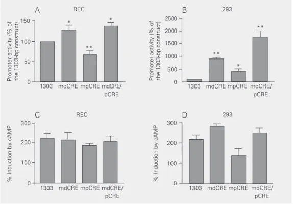
pCRE and dCRE were replaced with one Bgl-II site and two Bgl-II sites, respectively. As indicated in Figure 5A, mutation of pCRE reduced ACE promoter activity by 33% (P < 0.01), indicating that pCRE is a positive cis
element controlling basal transcription in REC. Mutation of the two tandem and over-lapping CREs within dCRE increased basal ACE promoter activity by 38% (P < 0.05), indicating that dCRE is a negative basal cis
element in REC. Simultaneous mutation of pCRE and dCRE increased ACE promoter activity by 43% (P < 0.01), indicating that the combined effect of both ACE CREs is to repress basal transcription in REC.
To test whether pCRE and dCRE are also important for ACE promoter regulation out-side the context of endothelial cells we
trans-fected the mutated constructs described above in 293 cells. In contrast to REC, mutation in either pCRE or dCRE greatly increased pro-moter activity, indicating that both sequences exert a negative role in the 293 context. As expected, simultaneous mutation of pCRE and dCRE further increased promoter activ-ity in 293 (Figure 5B).
Since mapping of functional cAMP-re-sponsive units in the ACE promoter was consistent with the position of both pCRE and dCRE, we tested whether individual or combined mutations in these motifs would affect cAMP responsiveness of the 1303-bp ACE promoter. Surprisingly, neither pCRE, dCRE, or double pCRE/dCRE mutants af-fected the cAMP responsiveness of the ACE promoter in REC or in 293 (Figure 5C,D).
A
A
A
A
Figure 5. Role of the cAMP-responsive elements dCRE and pCRE in basal and cAMP-induced transcription in endothelial and renal cells. A, Basal transcription in endothelial cells. The wild type 1303-bp promoter and mutants harboring Bgl-II restriction sites in place of dCRE (mdCRE), pCRE (mpCRE) and in both CRE-like sequences (mdCRE/pCRE) were transfected in rabbit endothelial cell (REC). Cells were harvested 24 h after transfection. Luciferase activity in cell extracts was normalized to beta galactosidase expression from a control plasmid. Promoter activity is the ratio of luciferase/beta galactoside expression and is reported as percent of the activity in the 1303-bp angiotensin converting enzyme (ACE) promoter. Data are reported as the mean ± SEM for 5 independent experiments (N = 6 in each). B, Basal transcription in renal cells: 293 cells were transfected with the same promoter constructs as in A and results were analyzed and reported as described for panel A. C, cAMP-induced transcription in endothelial cells. The wild type 1303-bp ACE promoter and the mutants harboring substitutions in CRE-like sequences were transfected in an endothelial cell line (REC) to identify promoter sequences involved in the response to cAMP. After 24 h of transfection, cells were serum-starved for 16-24 h on F12 supplemented with 0.5% FBS and then stimulated with 5 mM 8BrcAMP for 4 h. Data are reported as percent induction compared to control values for each construct. Data are reported as the mean ± SEM for 4 independent experiments (N = 6 in each). D, cAMP-induced transcription in renal cells. 293 cells were transfected with the same promoter constructs as in C and results were analyzed and reported as in panel A. *P < 0.05; **P < 0.01 compared to the 1303-bp ACE promoter (one-way analysis of variance and post hoc Tukey test).
This indicates that the 1303-bp ACE pro-moter can respond effectively to cAMP even in the absence of CRE-like sequences that regulate its basal expression.
Trans regulation of the somatic ACE
promoter by ATF-2 and its heterodimerization partner c-Jun
The presence of CRE-like sequences of the ACE promoter (Figure 4) suggested that the ACE gene is a target for regulation by
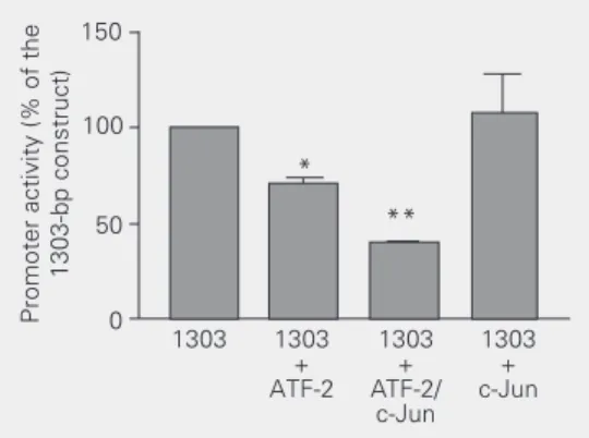
signaling pathways distinct from the classic cAMP pathway, such as those involving the stress-activated JNK or P38 kinases (22). To establish whether the ACE promoter is regu-lated by such pathways in endothelial cells we transfected the 1303-bp ACE promoter along with expression vectors for ATF-2 and for its heterodimerization partner, c-Jun. Co-transfection with ATF-2 reduced luciferase expression from the 1303 ACE promoter by 30%. Further decreases in luciferase expres-sion were obtained when ATF-2 was
co-Promoter activity (% of the 1303-bp construct)
150
Promoter activity (% of the 1303-bp construct)
2500
100
50
0
*
1303 mdCRE mpCRE mdCRE/ pCRE
1303 mdCRE mpCRE mdCRE/ pCRE
** *
2000
1500
1000
500
0
**
**
*
REC 293
% Induction by cAMP
300
200
100
0 % Induction by cAMP
300
200
100
0 1303 mdCRE mpCRE mdCRE/
pCRE
1303 mdCRE mpCRE mdCRE/ pCRE
REC 293
A B
expressed with c-Jun. In contrast, co-trans-fection with c-Jun alone did not change lu-ciferase expression by the 1303 ACE pro-moter. Thus, these results suggest that re-pression of the 1303 ACE promoter by ATF-2 is consistent with the ATF-ATF-2 protein acting as a homodimer or heterodimer together with c-Jun (Figure 6).
8BrcAMP activates CRE rather than AP2 pathways in REC
Mutational analysis of dCRE and pCRE indicated that both motifs are dispensable for cAMP induction of the 1303-bp ACE
promoter (Figure 5C,D). These results sug-gest that ACE promoter activation by cAMP is either due to extremely divergent CRE-like sequences, or alternatively to the poten-tial AP2 sites present in the 1303-bp pro-moter (Figure 3). In Figure 7 we compared the relative efficiency of the AP2 and CRE pathways in the transduction of the cAMP signal. In REC, 8BrcAMP efficiently induced CRETK, the positive control for the CRE pathway, but failed to affect luciferase ex-pression by AP2LUC, the positive control for the AP2 pathway. Co-transfection of an AP2 expression vector demonstrated that the AP2LUC construct is fully functional. Therefore, our data for REC suggest that induction of cAMP signaling does not lead to activation of the AP2 pathways.
Discussion
In the present study we used a transient transfection strategy in REC and 293 cells to assess the role of CRE-like motifs of the rat somatic ACE promoter in basal as well as in cAMP-stimulated transcription. Major find-ings include 1) description of a distal CRE (dCRE), conserved between rats, mice and humans, as a negative basal cis element in REC and 293; 2) description of a non-con-served proximal CRE (pCRE) as a positive basal cis element in REC and as a negative basal cis element in 293; 3) characterization of ATF-2 as a potential regulator of ACE gene expression; 4) mapping of cAMP responsive-ness to two independent domains within the 1303-bp ACE promoter; 5) assessment of the relative roles of CRE versus AP2 pathways in the response to cAMP in REC.
Functional analysis of the 1303-bp ACE promoter identified two regions containing regulators of basal and cAMP-induced ex-pression: a proximal one between 122 and 288 bp and a distal one between 415 and 1303 bp. Screening for CRE-like sequences in these regions revealed pCRE, between 209 and 222 bp and dCRE, between 801 and
Figure 6. ATF-2 inhibits the 1303-bp angiotensin converting zyme (ACE) promoter in an en-dothelial cell line (REC). The 1303-bp ACE promoter was co-transfected with expression vec-tors for transcription facvec-tors ATF-2 and c-Jun, separately or in combination. Cells were har-vested 24 h after transfection. Luciferase activity in cell ex-tracts was normalized for beta galactosidase expression from a control plasmid. Promoter
activ-ity is the ratio of luciferase/beta galactoside expression and is reported as percent of the activity of the 1303-bp ACE promoter. Data are reported as the mean ± SEM for 3 independent experiments (N = 6). *P < 0.05 compared to 1303 bp, **P < 0.01 compared to 1303 bp (one-way analysis of variance and post hoc Tukey test).
% Induction 500 1303 + cAMP 1303 + AP2 AP2 LUC + cAMP 400 300 0 200 100 -100 AP2 LUC + AP2 TATA + cAMP TATA + AP2 CRETK + cAMP
Figure 7. cAMP regulation of the 1303-bp angiotensin converting enzyme (ACE) promoter in an endothelial cell line (REC). The 1303-bp ACE promoter, positive controls for stimulation of the cAMP-responsive elements (CRETK) and AP2 (AP2LUC) pathways and a negative control (TATA) were co-transfected with expression vectors for AP2 when appropriate. After 24 h of transfection, cells were serum-starved for 16-24 h on F12
supplemented with 0.5% FBS. Transfected cells were then stimulated with 5 mM 8BrcAMP for 4 h when indicated. Luciferase (LUC) activity in cell extracts was normalized for beta galactosidase expression by a control plasmid. Promoter activity is the ratio of luciferase/ beta galactoside expression and is reported as percent induction compared to control values for each construct. Data are reported as the mean ± SEM for 4 independent experiments (N = 6). *P < 0.01 indicates statistically significant induction from unstimu-lated controls (one-way analysis of variance and post hoc Tukey test).
Promoter activity (% of the
819 bp. pCRE and dCRE represent the clos-est approximations to a canonical CRE within the 1303-bp ACE promoter. Although pCRE and dCRE contain substitutions at the CRE core binding site (CGTCA), inspection of labeled bands in the gelshifts of Figure 5C suggests that pCRE and dCRE bind two out of three protein complexes binding to the canonical CRE in REC nuclear extracts, al-beit at lower affinity. More importantly, bind-ing to pCRE and dCRE can be displaced by competition with cold oligonucleotides con-taining the canonical CRE. Thus, pCRE and dCRE share many properties with the ca-nonical CRE and therefore must be classi-fied as CRE-like sequences.
We established the roles of pCRE and dCRE in basal transcription by mutation anal-ysis. As shown in Figure 5A, mutation of pCRE reduces basal activity by about half, suggesting that it may participate in the in-crease in luciferase expression observed from the 122- to the 228-bp promoter construct. Conversely, mutation of dCRE produces a small but significant increase in basal activ-ity. As such, dCRE probably contributes to the inhibition of basal expression observed from the 415- to the 1303-bp promoter con-struct. Combined mutation of pCRE and dCRE produced a net increase in basal activ-ity. This indicates that pCRE and dCRE must interact to produce a net inhibitory regulation different from the simple addition of their individual effects in REC.
The 1303-bp rat ACE promoter responds to the activation of CRE and AP2 signaling pathways. However, in REC, 8BrcAMP in-duces the CRE, but not the AP2 reporter. This is in contrast to other systems where CRE and AP2 motifs transduce the cAMP signal either separately or in concert (13). As such, the resistance of the AP2 reporter to stimulation by cAMP is inconsistent with a major role of AP2 signaling in the REC response to cAMP. Moreover, all ACE AP2 sites, located between 228 and 415 bp as well as between 845 and 1120 bp (Figure 3),
fell outside the two domains of cAMP re-sponsiveness that we mapped (Figure 2). Thus, our data suggest that, in REC, the bulk of ACE promoter activation in response to cAMP is due to CRE mechanisms. It remains to be established whether the refractoriness of the AP2 pathway to cAMP stimulation in REC is shared by most endothelial cell types, or if it is a peculiar feature of the REC line. Because individual or combined mutation of pCRE and dCRE did not affect induction by 8BrcAMP, the CREs mediating activation of the ACE promoter by cAMP remains unac-counted for. Due to the pronounced diver-gence of the remaining candidates from the CRE consensus it is likely that identification of these putative elements will require the use of linker-scanning strategies (23).
The arrangement of CRE-like sequences in the ACE promoter is compatible with those of numerous promoters that display multiple CRE-like motifs scattered in their sequences. CRE-like motifs in these promoters are usu-ally multimers or single elements closely asso-ciated with other motifs and located in sepa-rated domains (24,25). Among those CRE-like sequences, some have effects on basal or on cAMP-stimulated transcription or on both. These CRE-like motifs play either posi-tive or negaposi-tive roles in basal transcription (26,27) and display redundant roles in the response to cAMP (24,28). In the present study we have shown that dCRE, a motif conserved between rats, mice and humans, is a negative cis regulator of ACE gene basal expression. This finding is consistent with work showing that conserved non-canonical CREs are regulators of basal transcription in the promoters of cystic fibrosis transmem-brane conductance genes from 8 mammalian species (29) as well as in the promoter for the testicular ACE isozyme (30,31).
mechanisms of gene regulation have been extensively modeled in heterologous con-texts and ACE regulation by cAMP, in par-ticular, is a conserved feature among mam-mals (for a review, see Ref. 10). REC is a cell line that maintains stable endothelial fea-tures such as uptake of 1,19-dioctadecyl-3',3',3',3'-tetramethylindocarbocyanine per-chlorate-labeled acetylated LDL and ACE gene expression (10). Thus, REC compares favorably with primary cultures of endothe-lial cells, which display limited growth char-acteristics, do not sustain expression of en-dothelial markers and are refractory to trans-fection in mammals (10).
The advantages and disadvantages of the heterologous approach are represented in our study. As shown in Figure 5, dCRE, which is conserved between rats, mice and men, is an inhibitory element in both rabbit and human cell lines, suggesting that this is its role in the native setting. In contrast, pCRE has a positive role in rabbits and a negative role in a human cell line, a finding which is consistent with the lack of evolu-tionary conservation in this motif between the aforementioned species. While the ac-tual role of pCRE in its native setting cannot be established by our experiments, we be-lieve nonetheless that the differences in the regulatory potential of pCRE can be easily accounted for by quantitative and qualitative cell-specific differences in transcription fac-tor content. This scenario is typical of the evolution of cis regulatory sequences, when one or more redundant units can diverge without risking basic promoter functions (32). What is then the relevance of pCRE and dCRE for ACE regulation in vivo? In
experi-ments that will not be documented here we generated transgenic mice for the wild type 1303-bp ACE promoter as well as for the double mutant mdCRE/pCRE. While the wild type 1303-bp promoter was not enough to drive reporter expression in kidneys, lungs and ovaries, the double mutant induced strong expression in all of these organs (Anéas I,
unpublished results). These results, taken together with our demonstration that pCRE and dCRE interact to repress basal transcrip-tion from the ACE promoter in endothelial and renal cell types, suggest that both CRE-like sequences are important targets for tis-sue-specific regulation of the ACE gene.
References
1. Friedland J, Setton C & Silverstein E (1977). Angiotensin converting enzyme: induction by steroids in rabbit alveolar macrophages in culture. Science, 197: 64-65.
2. Krulewitz AH, Baur WE & Fanburg BL (1984). Hormonal influence on endothelial cell angiotensin-converting enzyme activity. American Journal of Physiology,247: C163-C168.
3. Dasarathy Y, Lanzillo JJ & Fanburg BL (1982). Stimulation of bovine pulmonary artery endothelial cell ACE by dexamethasone: involve-ment of steroid receptors. American Journal of Physiology,263: H645-H649.
4. Dasarathy Y & Fanburg BL (1989). Calcium ionophore A23187 el-evates angiotensin-converting enzyme in cultured bovine endotheli-al cells. Biochimica et Biophysica Acta, 1010: 16-19.
5. Fishel RS, Thourani V, Eisenberg SJ, Shai SY, Corson MA, Nabel EG, Bernstein KE & Berk BC (1995). Fibroblast growth factor stimulates angiotensin converting enzyme expression in vascular smooth muscle cells. Possible mediator of the response to vascular injury. Journal of Clinical Investigation, 95: 377-387.
6. Kyriakis JM & Avruch J (2001). Mammalian mitogen-activated pro-tein kinase signal transduction pathways activated by stress and inflammation. Physiological Reviews,81: 807-869.
7. Weinstock JV (1986). The significance of angiotensin I converting enzyme in granulomatous inflammation. Functions of ACE in granu-lomas. Sarcoidosis,3: 19-26.
8. Lloyd CJ, Cary DA & Mendelsohn FAO (1987). Angiotensin convert-ing enzyme induction by cyclic AMP and analogues in cultured endothelial cells. Molecular and Cellular Endocrinology, 52: 219-225.
9. Dasarathy Y & Fanburg BL (1988). Elevation of angiotensin convert-ing enzyme by 3-isobutyl-1-methylxanthine in cultured endothelial cells: a possible role for calmodulin. Journal of Cellular Physiology, 137: 179-184.
10. Xavier-Neto J, Pereira AC, Junqueira ML, Carmona R & Krieger JE (1999). Rat angiotensin-converting enzyme promoter regulation by beta-adrenergics and cAMP in endothelium. Hypertension, 34: 31-38.
11. Montminy MR (2001). Transcriptional regulation by the phosphory-lation-dependent factor CREB. Nature Reviews. Molecular Cell Biol-ogy, 2: 599-609.
12. Roesler WJ (2000). What is a cAMP response unit? Molecular and Cellular Endocrinology, 162: 1-7.
13. Gao B, Chen J, Johnson C & Kunos G (1997). Both the cyclic AMP response element and the activator protein 2 binding site mediate basal and cyclic AMP-induced transcription from the dominant pro-moter of the rat alpha 1B-adrenergic receptor gene in DDT1MF-2 cells. Molecular Pharmacology, 52: 1019-1026.
14. Xavier-Neto J, Pereira AC, Motoyama AH & Krieger JE (1998). A luciferase-engineered cell line for study of cAMP regulation in endo-thelial cells. American Journal of Physiology,275: C75-C81. 15. Matsuda S, Maekawa T & Ishii S (1991). Identification of the
func-tional domains of the transcripfunc-tional regulator CRE-BP1. Journal of Biological Chemistry, 266: 18188-18193.
16. Williams T & Tjian R (1991). Analysis of the DNA-binding and activa-tion properties of the human transcripactiva-tion factor AP-2. Genes and Development, 5: 670-682.
17. Doucas V, Spyrou G & Yaniv M (1991). Unregulated expression of
c-Jun or c-Fos proteins but not c-Jun D inhibits oestrogen receptor activity in human breast cancer derived cells. EMBO Journal, 10: 2237-2245.
18. Buonassisi V & Venter JC (1976). Hormone and neurotransmitter receptors in an established vascular endothelial cell line. Proceed-ings of the National Academy of Sciences, USA, 73: 1612-1616. 19. Graham FL & van der Eb AJ (1973). A new technique for the assay
of infectivity of human adenovirus 5 DNA. Virology,52: 456-459. 20. Ribeiro RC, Apriletti JW, Yen PM, Chin WW & Baxter JD (1945).
Heterodimerization and deoxyribonucleic acid-binding properties of a retinoid X receptor-related factor. Endocrinology, 135: 2076-2085. 21. Krieger JE, Junqueira ML & Dzau VJ (1994). Characterization of the promoter from rat angiotensin-converting enzyme gene. Circula-tion, 90: 130 (Abstract).
22. Johnson GL & Lapadat R (2002). Mitogen-activated protein kinase pathways mediated by ERK, JNK, and p38 protein kinases. Science, 298: 1911-1912.
23. Gustin K & Burk RD (2000). PCR-directed linker scanning mutagen-esis. Methods in Molecular Biology, 130: 85-90.
24. Fisch TM, Prywes R, Simon MC & Roeder RG (1989). Multiple sequence elements in the c-fos promoter mediate induction by cAMP. Genes and Development,3: 198-211.
25. Soubt MK, Marksitzer R, Menoud PA & Nagamine Y (1998). Role of tissue-specific transcription factor LFB3 in a cyclic AMP-responsive enhancer of the urokinase-type plasminogen activator gene in LLC-PK1 cells. Molecular and Cellular Biology,18: 4698-4706. 26. Jungmann RA, Huang D & Tian D (1998). Regulation of LDH-A gene
expression by transcriptional and posttranscriptional signal trans-duction mechanisms. Journal of Experimental Zoology, 282: 188-195.
27. Murasawa S, Matsubara H, Kijima K, Maruyama K, Ohkubo N, Mori Y, Iwasaka T & Inada M (1996). Down-regulation by cAMP of angio-tensin II type 2 receptor gene expression in PC12 cells. Hyperten-sion Research, 19: 271-279.
28. Giono LE, Varone CL & Canepa ET (2001). 5-Aminolaevulinate syn-thase gene promoter contains two cAMP-response element (CRE)-like sites that confer positive and negative responsiveness to CRE-binding protein (CREB). Biochemical Journal,353: 307-316. 29. Vuillaumier S, Dixmeras I, Messai H, Lapoumeroulie C, Lallemand
D, Gekas J, Chehab FF, Perret C, Elion J & Denamur E (1997). Cross-species characterization of the promoter region of the cystic fibrosis transmembrane conductance regulator gene reveals mul-tiple levels of regulation. Biochemical Journal,327: 651-662. 30. Esther Jr CR, Semeniuk D, Marino EM, Zhou Y, Overbeek PA &
Bernstein KE (1997). Expression of testis angiotensin-converting enzyme is mediated by a cyclic AMP responsive element. Labora-tory Investigation,77: 483-488.
31. Kessler SP, Rowe TM, Blendy JA, Erickson RP & Sen GC (1998). A cyclic AMP response element in the angiotensin-converting en-zyme gene and the transcription factor CREM are required for transcription of the mRNA for the testicular isozyme. Journal of Biological Chemistry,273: 9971-9975.