JSCS–4004 543.57:615.277–188
Original scientific paper
Synthesis, thermal and antitumour studies of Th(IV) complexes
with furan-2-carboxaldehyde4-phenyl-3-thiosemicarbazone
GANGADHARAN RAJENDRAN1*, CHIRAKUZHI S. AMRITHA1,
RUBY JOHN ANTO2 and VINO T. CHERIYAN2
1Department of Chemistry, University College, Thiruvananthapuram-695034, Kerala
and 2Division of Cancer Research, Rajiv Gandhi Centre for Biotechnology,
Thiruvananthapuram-695014, Kerala, India
(Received 29 July 2009, revised 2 February 2010)
Abstract: Thorium(IV) complexes with the Schiff base
furan-2-carboxaldehyde4-phenyl-3-thiosemicarbazone (L) were synthesised and characterized. The composition and structure of the metal complexes were proposed based on elemental analysis, molar conductivity measurements, FTIR and 1H-NMR
spectroscopy. The Schiff base behaves as a neutral bidentate ligand coordinating through the azomethine N and the thioketo S atoms. From various studies, complexes were ascertained the general formula [ThL2X4] and
[ThL2Y2], where X represents NO3–, NCS–, CH3COO–, CH3CHOHCOO–,
ClO4– and Y SO
42–and C2O42–. The thermal behaviour of the nitrato and
oxalato complexes was studied and kinetic and thermodynamic parameters were calculated using the Coats-Redfern Equation. The ligand and a repre-sentative complex [ThL2(NO3)4] were screened in vitro for their antitumour
activity against the human cervical cancer cell line (HeLa).
Keywords: thorium(IV) complexes;
furan-2-carboxaldehyde4-phenyl-3-thiosemicarbazone; antitumour activity; thermal analysis.
INTRODUCTION
Complexes of thiosemicarbazones have been explored for a variety of
rea-sons, such as variable bonding properties, presence of several donor sites,
struc-tural diversity and pharmacological aspects.
1They present a variety of biological
activities, including anticancer and anti-inflammatory activities.
2–4Metal
thiose-micarbazonate complexes are emerging as a new class of experimental anticancer
and chemotherapeutic agents which exhibit inhibitory activities against most
can-cer through inhibition of a crucial enzyme obligatory for DNA biosynthesis and
cell division,
viz
. ribonucleotide diphosphate reductase (RDR).
5Some
carbazones even increase their antitumour activity by their ability to form
chela-tes with specific metal ions.
6It was reported that the anticancer activities of
thiosemicarbazones were closely related to the parent aldehyde or ketone group,
metal chelation ability and terminal amino substitution. Among them, the parent
aldehyde or ketone group was considered critical for the anticancer activity of
thiosemicarbazones. Heterocyclic thiosemicarbazone showed higher activity
com-pared with aromatic thiosemicarbazones.
7Heterocyclic thiosemicarbazones and
their metal complexes are among the most widely studied compounds for their
potential therapeutic uses, such as antitumoural, fungicidal, bactericidal or
anti-viral activity.
8The activity of these compounds is dependent on the nature of the
hetroaromatic ring and the position of attachment of the ring as well as on the
form of the thiosemicarbazone moiety.
9There were several studies involving
thiosemicarbazones with different metal ions.
10–13However, only a few reports
described studies on thorium thiosemicarbazone complexes. Hence as part of
on-going research regarding thiosemicarbazone complexes of thorium
14,15, the
syn-thesis, characterization and antitumour activity of Th(IV) complexes with furan-
-2-carboxaldehyde4-phenyl-3-thiosemicarbazone (Fig. 1) are reported herein.
Fig. 1. Schematic view of the ligand.
EXPERIMENTAL
Materials and analytical methods
All employed chemicals were of analytical grade purchased from Merck, Sisco (India),
etc. Commercial solvents were distilled and used for synthesis, but for the physicochemical
studies, they were purified by standard methods.
The IR spectral studies were performed using KBr discs on a Schimadzu 8201 PC FT in-frared spectrophotometer. The 1H-NMR spectra were recorded on a Bruker DRX-300 FT NMR
spectrophotometer employing TMS as the internal reference and DMSO-d6 as the solvent.
X-Ray diffraction studies were realized using a Philips X-ray PW 1710 diffractometer using K-α1 radiation of wavelength 1.54056 Å. Molar conductance measurements of 10-3 M
solu-tions in CH3CN and C6H5NO2 were performed at room temperature using a direct reading
The metal content was estimated gravimetrically by the oxalate–oxide method.16
Stan-dard gravimetric procedures were adopted for the estimation of the anions in the prepared com-plexes.17 The sulphate and thiocyanate present in the complexes were estimated
gravimetri-cally as BaSO4 and AgNCS, respectively, while the perchlorate content was determined by the Kurz method.18 The nitrate, oxalate, acetate and lactate contents were indirectly fixed by
per-forming elemental analysis for carbon, hydrogen and nitrogen by micro analytical methods.
Synthesis of ligand
The ligand, furan-2-carboxaldehyde4-phenyl-3-thiosemicarbazone, was prepared by the following method. Furan-2-carboxaldehyde (0.010 mol) in methanol (10 ml) was added dropwise to a hot solution of 4-phenyl-3-thiosemicarbazide (0.010 mol) in methanol (30 ml) under constant stirring. The resulting mixture was heated on a water bath for 3 h, concentrated and allowed to cool. The formed dark brown crystals of the ligand were washed, dried and recrystallized from ethanol. Yield: 84 %; m.p. 120 °C ; Anal. Calcd. for C12H11N3OS: C, 58.77; H, 4.49; N, 17.14; S, 13.06 %. Found: C, 58.56; H, 4.54; N, 17.63; S, 13.21 %. IR (KBr, cm-1): 3256, (m,–NH), 1623 (vs, C=N), 1526 (m, C–O furan ring), 1060 (m, (N–N),
868 (s, C=S).
Synthesis of metal complex
The metal complexes were prepared by refluxing a methanolic solution of the metal salt and the ligand in the stoichiometric ratio 1:2. For the preparation of the nitrato complex, the appropriate amount of the metal salt (0.0020 mol) dissolved in a minimum quantity of metha-nol (10 ml) was added to a solution of the ligand (0.0040 mol, 0.10 g) dissolved in methametha-nol (25 ml). The pH of the solution was raised to 7 and refluxed on a water bath for about 5 h. It was then concentrated and left standing over night. The separated complex was filtered, wash-ed with a methanol–water mixture (50 % v/v) and then with ether and driwash-ed over P4O10 in vacuo. The other anionic complexes were prepared from the nitrato complex by the
substi-tution method19 by refluxing stoichiometric amounts of the nitrato complex with the
respec-tive anionic salts of lithium.
Antitumour screening
The in vitro antitumour activities of the ligand and a representative complex were
exa-mined by the MTT assay method20,21 against human cervical cancer cell line (HeLa).
The human cervical cancer cell line (HeLa) was obtained from the National Centre for Cell Science Pune, India. The cells were grown in Dulbecco’s modified eagles medium (DMEM) containing 10 % foetal bovine serum (FBS), streptomycin (100 µg/ml), penicillin (100 units/ml)
and amphotericin B (2.5 µg/ml). The cells were incubated at 37 °C in a 5 % CO2 incubator in
a humid condition and harvested using trypsin–ethylene diamine tetraacetic acid.
The test samples were dissolved in DMSO and diluted to the required concentration for the biological experiments. Studies were undertaken with the test compounds in the concen-tration range from 10 to 100 µg/ml.
For the determination of the cytotoxic effects, cells harvested from the exponential phase were seeded equivalently (5000 cells/well) in a 96-well plate and incubated for 24 h. Test so-lutions of different concentrations were added in triplicate to the well plates. Six well plates were maintained in a drug free medium to determine the control, cell survival and the percentage of live cells after culture. Cells with various concentrations of the test samples were incubated at 37 °C for 72 h.
at 37 °C. MTT is metabolized in the presence of the pyridine cofactors NADH and NADPH, to give blue insoluble crystals. The cells were solubilised with 0.1 ml of extraction buffer (20 % sodium dodecyl sulphate in 50 % dimethylformamide) and then incubated for 4 h at 37 °C. Following the solubilisation of the cells, the colour intensity was read at 570 nm using an Elisa plate reader (Bio-Rad). The percentage viability of the cells or cell survival (CS) was
ex-pressed as mean optical density (drug exposed cell) divided by mean optical intensity (control). RESULTS AND DISCUSSION
All the prepared complexes were brown coloured, non-hygroscopic solids
stable at room temperature. They were soluble in DMSO and DMF but insoluble
in water and common organic solvents. The room temperature molar
conductivities of 10
–3M solutions of the complexes in CH
3CN and C
6H
5NO
2corresponded to those of non-electrolytes.
22The analytical data revealed that all
complexes possessed 1:2 metal to ligand stoichiometry. Based on elemental
analysis, the complexes were assigned the composition shown in Table I.
TABLE I. Molar conductance at room temperature and elemental analyses data of the com-plexes (L = C12H11N3OS)
Complex Found (Calcd.), %
Molar conductance S cm2 mol-1 Metal C H N S C6H5NO2 CH3CN
[ThL2(NO3)4] (1) 23.85
(23.92) 29.63 (29.69) 2.16 (2.26) 14.53 (14.43) 6.79 (6.59) 5.7 13.8
[ThL2(SO4)2] (2) 25.25
(25.38) (31.51)31.45 (2.41) 2.30 (9.19) 9.09 14.15 (14) 7.5 12.1 [ThL2(NCS)4] (3) 24.21
(24.32) (35.22)35.12 (2.31) 2.51 (14.67)14.47 (20.13)20.21 8.8 10.8 [ThL2(C2O4)2] (4) 25.64
(25.84) 37.22 (37.42) 2.15 (2.45) 9.55 (9.35) 7.03
(7.13) 6.9 9.6
[ThL2(CH3COO)4] (5) 24.30
(24.22) (40.08)40.02 (3.55) 3.25 (8.77) 8.17 (6.68) 6.28 7.1 11.1 [ThL2(C3H5O3)4] (6) 21.27
(21.52) (40.07)40.26 (3.89) 3.87 (7.79) 7.81 (5.93) 5.73 7.9 13.6 [ThL2(ClO4)4] (7) 20.67
(20.71) 25.76 (25.71) 1.50 (1.96) 7.63 (7.5) 5.49
(5.71) 12.9 17.1
Spectral studies
C=S.
7,25The bands observed at 3256 and 3423 cm
–1were assigned to N–H
vib-rations. This further indicates that the ligand remained in the thione form.
Fig. 2. Tautomeric forms of the ligand.
Fig. 3. IR spectrum of furan-2-aldehyde-N-phenylthiosemicarbazone.
The diagnostic IR spectral bands of the complexes are presented in Table II,
together with their tentative assignments. In the spectra of all the complexes, the
band due to the azomethine moiety (C=N) was shifted to a lower frequency by
≈
20–30 cm
–1, indicating its involvement in coordination with metal ion. The
ν
(C=S) stretching frequency was lowered by
≈
20–40 cm
–1in the spectra of the
complexes, indicating the involvement of the thioketo sulphur in the
coordina-tion. These findings are further supported by the appearance of new bands in the
far IR region at 495–505 and 359–370 cm
–1, which are assignable to
ν
(Th–N)
and
ν
(Th–S) vibrations, respectively.
1598 and 1596 cm
–1, respectively, and
ν
s(COO
–) at 1362 and 1352 cm
–1,
res-pectively, apart from the skeletal vibrations of the ligand. The separation between
the two frequencies adequately supports the monodentate coordination of the
ace-tate and lacace-tate group.
27The spectrum of complex
4
showed additional bands at
1665 and 1361 cm
–1, which were assigned to
ν
a(COO) and
ν
s(COO) modes of
the bidentately coordinated dicarboxylate ion.
28The IR spectrum of complex
3
had additional non-ligand bands at 2072, 777 and 493 cm
–1, assignable to
ν
(C–N),
ν
(C–S) and
δ
(NCS) of thiocyanate.
29The presence of these bands revealed the
N-coordinated nature of the thiocyanate ion.
29The spectrum of complex
2
exhi-bited additional non-ligand bands at 1248, 1177 and 1085 cm
–1, and the values
showed the bridging bidentate coordination of the sulphate group.
30For complex
7
, the spectral bands at 1110, 1071 and 627 cm
–1indicated the monodentate
coordination of the perchlorate group.
31The nature of the bonding of the various
anions is further supported by the non-electrolytic nature of all the complexes.
TABLE II. IR spectral data of the complexes (cm-1)Compound ν(N–H) ν(C=N) Furan ring ν(C–O) ν(N–N) ν(C=S) ν(Th–N) ν(Th–S)
[ThL2(NO3)4] (1) 3255 m 1598 vs 1520 m 1064 m 827 s 503 m 364 m
[ThL2(SO4) 2] (2) 3256 m 1601 vs 1522 m 1066 m 823 s 506 m 368 m
[ThL2(NCS)4] (3) 3252 m 1590 vs 1521 m 1062 m 834 s 507 m 363 m
[ThL2(C2O4)2] (4) 3524 m 1597 vs 1522 m 1066 m 846 s 504 s 369 m
[ThL2(CH3COO)4] (5) 3253 m 1593 vs 1524 m 1068 m 848 s 508 m 362 m
[ThL2(C3H5O3)4] (6) 3256 m 1596 vs 1527 m 1067 m 848 s 506 m 360 m
[ThL2(ClO4)4] (7) 3257 m 1591 vs 1523 m 1061 m 846 s 498 s 359 m
The
1H-NMR spectrum of the ligand recorded in DMSO-
d
6showed no peak
at 4 ppm attributable to SH protons
8but showed a peak at 9.87 ppm, which was
attributed to the N–H group, indicating that the ligand was in the thione form,
which is in conformity with the IR spectrum. A significant azomethine proton
signal due to CH=N was observed at 8.98 ppm, while that due to aromatic
tons were observed in the region 7.21–7.36 ppm. Signals for the furan ring
pro-tons were observed at 6.57, 7.38 and 7.41 ppm.
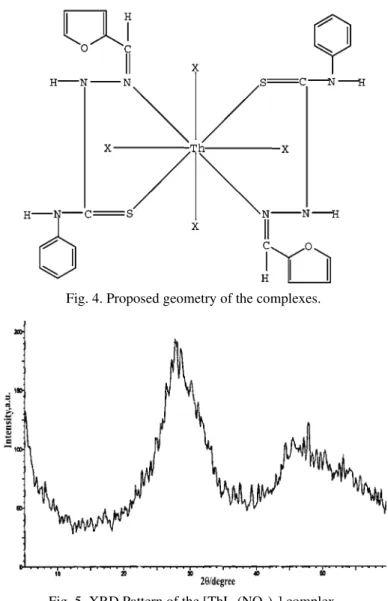
X-Ray diffraction study
The structure of [ThL
2(NO
3)
4] evaluated using powder X-ray diffraction
in-dicated the amorphous nature of the complex. The X-ray diffraction pattern is
given in Fig. 5.
Fig. 4. Proposed geometry of the complexes.
Fig. 5. XRD Pattern of the [ThL2(NO3)4] complex.
Thermal studies
The [ThL
2(NO
3)
4] complex showed a single stage decomposition, as shown
by the DTG curve. The TG curve showed the absence of water or any other
sol-vent molecules, as the complex was stable up to 190 °C (Fig. 6). Decomposition
started at 190 °C and ended at 270 °C with a peak temperature of 247 °C,
indi-cating the loss of the ligand and nitrate group. The residue 27.57 % (calcd. 27.82 %)
showed that the final product formed was ThO
2, which is in agreement with the
analytical result for the metal content.
Fig. 6. TG and DTG curves of [ThL2(NO3)4].
For the complex [ThL
2(C
2O
4)
2], the TG plateau up to 220 °C showed the
absence of coordinated water or any other solvent molecules and the stability of
complex (Fig. 7). Decomposition began at 220 °C and ended at 310 °C. The peak
temperature for the decomposition was 265 °C. The complex showed a single
stage decomposition, as evident from the DTG curve, and the decomposition
oc-curred with the loss of both ligand and oxalate molecules. The final product
formed was ThO
2and the residue obtained 29.55 % (calcd. 29.40 %) agreed well
with the analytical result obtained by an independent pyrolysis experiment.
Kinetic aspects
A kinetic evaluation of the thermal decomposition data of complexes was
carried out. The kinetic parameters,
viz
., the activation energy,
E
, and the
pre-ex-ponential factor,
A
, were calculated using the Coats-Redfern Equation.
32Compu-tational data for the evaluation of kinetic parameters are given in Tables III and
IV. Here the ln
g
(
α
)
/T
2vs
. 1000/
T
plots (Figs. 8 and 9) gave straight lines, from
∆
S
=
R
ln (
Ah
/
kT
s) (1)
where
R
is the gas constant,
A
is the pre-exponential factor,
k
is the Boltzmann
constant,
T
sis the DTG peak temperature and
h
is the Planck constant.
Fig. 7. TG and DTG curves of [ThL2(C2O4)2].
TABLE III. Computational data for the thermal decomposition of [ThL2(NO3)4] (n = 2;
r = 0.99295)
t / °C m / mg T/ K T / 10-3 K-1 Weight loss, % α g(α) ln (g(α)/T)2
200 3.33 473 – 0 0 0 –
210 3.30 483 2.07039 0.03 0.01240 0.01255 –16.73788 220 3.08 493 2.02840 0.25 0.10331 0.11521 –14.56204 230 2.24 503 1.98807 1.09 0.45041 0.81955 –12.64018 240 1.58 513 1.94932 1.75 0.72314 2.61194 –11.52046 250 1.03 523 1.91205 2.30 0.95041 19.1667 –9.56599
260 0.96 533 1.87617 2.37 0.97934 47.4 –8.69842
270 0.93 543 1.84162 2.40 0.99174 120 –7.80673
280 0.91 553 1.80832 2.42 1 – –
TABLE IV. Computational data for the thermal decomposition of [ThL2(C2O4)2] (n = 1.7;
r = 0.99566)
t / °C m / mg T/ K T / 10-3 K-1 Weight loss, % α g(α) ln (g(α)/T)2
220 3.45 493 2.0284 0 0 0 –-
230 3.42 503 1.98807 0.03 0.01240 0.01253 –16.82091 240 3.35 513 1.94932 0.10 0.04132 0.04283 –15.63107 250 3.25 523 1.91205 0.20 0.08264 0.08892 –14.93921 260 2.70 533 1.87617 0.75 0.30992 0.42356 –13.41611
270 1.76 543 1.84162 1.69 0.69835 1.8770 –11.96454
280 1.21 553 1.80832 2.24 0.92562 7.37952 –10.63201 290 1.08 563 1.77620 2.37 0.97934 20.16347 –9.66269 300 1.05 573 1.74520 2.40 0.99174 39.57784 –9.02350
The kinetic parameters determined for the thermal decomposition are listed
in Table V. The positive value of the entropy of activation in both cases indicates
that the activated state was less ordered than the reactants.
33Fig. 8. Coats-Redfern plot for [ThL2(NO3)4]. Fig. 9. Coats-Redfern plot for [ThL2(C2O4)2].
TABLE V. Kinetic parameters for the thermal decomposition of the complexes
Complex
Peak temp.
ts/ °C
Correlation coefficient Order n
Activation energy
E / kJ mol-1
Pre-exponential
term
A / s-1
Entropy of activation
ΔS / J K-1 mol-1
[ThL2(NO3)4] 247 0.99295 2 325 8.6×1031 362
[ThL2(C2O4)2] 265 0.99566 1.7 282 4.4×1025 241
Antitumour activity
The cell viability over the untreated control was determined using the MTT
assay, which is a very convenient method for assessing drug sensitivity even through
it does not discriminate between apoptosis and necrosis. The results showed that
both the ligand and complex possessed antitumour activity. The results are
sum-marized in Table VI.
of tumour cells and inhibiting tumour growth. The antitumour activity seems to
be due to an inhibition of DNA synthesis in cancer cells produced by
modifica-tion in reductive conversion of ribonucleotides to deoxyribonucleotides.
38 TABLE VI. Antitumour activity of the ligand and complexCompound Concentration, µg ml-1 Relative cell viability, %
L 10 88.2
25 85.5
50 89.4
100 83.3
[ThL2(NO3)4] 10 85.64
25 80.6
50 86.78 100 82.8
CONCLUSIONS
The coordination sites of the ligand and the coordination number of the
me-tal in the prepared complexes were confirmed by physicochemical studies.
Spec-tral analysis showed that the ligand in the thioketo tatutomer form acts as neuSpec-tral
bidentate with N and S atoms as the coordination sites. All the complexes were
neutral, amorphous solids stable at room temperature. From the research
fin-dings, the composition of the complexes can be ascertained as [ThL
2X
4] and
[ThL
2Y
2], where X represents NO
3–, NCS
–, CH
3COO
–, CH
3CHOHCOO
–and
ClO
4–, and Y SO
42–and C
2O
42–. A coordination number of 8 is proposed in all
these complexes. Antitumour studies indicated that complexation of the
thiosemi-carbazone with the metal ion lead to an enhancement of the activity of the
thio-semicarbazone.
, Њ Th(IV)
-2- -4-
-3-G. RAJENDRAN1, C. S. AMRITHA1, RUBY JOHN ANTO2 VINO T. CHERIYAN2
1
Department of Chemistry, University College, Thiruvananthapuram-695034, Kerala, India, 2Molecular medicine and Cancer research division, Rajiv Gandhi Centre for Biotechnology,
Thiruvananthapuram-695014, Kerala, India
(IV)
-2--4- -3- (L).
, , FT-IR 1
H-NMR .
N S .
[ThL2X4] [ThL2Y2], X NO3–, NCS–, CH3COO–,
CH3CHOHCOO– ClO
4–, a Y SO42– C2O42–.
,
Coats–Redfern- . [ThL2(NO3)4] in vitro
(HeLa).
REFERENCES
1. T. S. Lobana, Rekha, R. J. Butcher, A. Castineiras, C. Bermejo, P. V. Bharatam, Inorg. Chem.45 (2006) 1535
2. M. Ruan, Y. Ye, Y. Song, Q. Mauricio, F. Erben, C. O. D. Vedova, Spectrochim. Acta72 A (2009) 26
3. P. I. Das, M. F. Fernando, R. Pavan, C. Q. F. Leite, F. D. Sousa, A. A. Batista, O. R. As-cimento, J. E. Eduardo, E. Castellano, E. Niquet, V. M. Deflon, Polyhedron28 (2009) 205 4. Z. H. Chohan, TransitionMet. Chem. 34 (2009) 153
5. D. P. Saha, S. Padhye, E. Sinn, C. Newton, Indian J. Chem.41A (2002) 279 6. A. G. Quiroga, C. N. Ranninger, Coord. Chem. Rev.248 ( 2004) 119
7. H. Zhang, R. Thomas, D. Oupicky, F. Peng, J. Biol. Inorg. Chem.13 (2008) 47
8. E. M. Jouad, G. Larcher, M. Allain, A. Riou, G. M. Bouet, A. M. Khan, X. D. Thanh, J. Inorg. Biochem.86 (2001) 565
9. S. Chandra, M. Tyagi, M. S. Refat, J. Serb. Chem. Soc. 74 (2009) 907 10. M. J. M. Campbell, Coord. Chem. Rev.15 (1975) 279
11. M. A. Ali, S. E. Livingstone, Coord. Chem. Rev. 13 (1974) 101 12. S. Padhye, G. B. Kauffman, Coord. Chem. Rev.63 (1985) 127
13. J. S. Casas, M. S. Garcia-Tasenda, J. Sorda, Coord. Chem. Rev.209 (2000) 197 14. G. Rajendran, C. S. Amritha, Asian J. Chem.18 (2006) 2695
15. G. Rajendran, C. S. Amritha, Oriental, J. Chem.22 (2006) 365
16. I. M. Kolthoff, P. J. Elving, Treatise on Analytical Chemistry, Interscience, New York,
1963
17. A. I. Vogel, A Textbook of Quantitative Inorganic Analysis, 4th ed., ELBS, London, 1978
18. E. Kurz, G. Kober, M. Berl, Anal. Chem.30 (1958) 1983
19. P. Indrasean, G. Rajendran, Synth. React. Inorg. Met. Org. Chem.22 (1992) 715 20. T. Mosmann, J. Immunol. Methods65 (1983) 55
21. A. P. Wilson, Cytotoxicity and Viability Assays in Animal Cell Culture: A Practical Approach, 3rd ed., Vol. 1, Oxford University Press, Oxford, 2000
22. W. J. Geary, Coord. Chem. Rev.7 (1971) 81
23. A. K. Sen, G. Singh, K. Singh, R. K. Noren, R. M. Handa, S. N. Dubey, Indian J. Chem. 36A (1977) 891
24. K. S. A. Melha, J. Enzym. Inhib. Med. Chem. 23 (2008) 493 25. P. Bindu, M. R. P. Kurup, Transition Met. Chem.22 (1997) 578 26. S. G. Devi, P. Indrasenan, Inorg. Chim. Acta133 (1987) 157
27. B. W. Mistry, A Hand Book of Spectroscopic Data Chemistry, 1st ed., ABD Publishers,
Jaipur, India, 2000
28. D. N. Sathyanarayana, Vibrational Spectroscopy, Theory and Applications, New Age
In-ternational (P) Ltd., India, 2004
29. R. K. Agarwal, P. Kumar, H. K. Rawat, Thermochim. Acta 88 (1985) 397
30. K. Nakamoto, Infrared and Raman Spectra of Inorganic and Coordination Compounds,
Wiley, New York, 1987
31. M. Viswanathan, J. Indian Chem. Soc.82 (2005) 871
32. K. K. Aravindakshan, K. Muraleedharan, Thermochim. Acta 155 (1989) 247 33. A. A. Frost, R. G. Pearson, Kinetics and Mechanism, Wiley, New York, 1961
35. R. A. Finch, M. C. Liu, S. P. Grill, W. C. Rose, R. Loomis, K. M. Vasquez, Y. C. Cheng, A. C. Sartorelli, Biochem. Pharmacol. 59 (2000) 983
36. F. A. French, J. E. Blanz, J. Med. Chem.17 (1974) 172
37. M. B. Ferrari, F. Bisceglie, C. Casoli, S. Durot, I. M. Badarau, G. Pelosi, E. Pilotti, S. Pinelli, P. Tarasconi, J. Med. Chem.48 (2005) 1671


![TABLE II. IR spectral data of the complexes (cm -1 ) Compound ν(N–H) ν(C=N) ν(C–O) Furan ring ν(N–N) ν(C=S) ν(Th–N) ν(Th–S) [ThL 2 (NO 3 ) 4 ] (1) 3255 m 1598 vs 1520 m 1064 m 827 s 503 m 364 m [ThL 2 (SO 4 ) 2 ] (2) 3256 m 1601 vs 1522 m 10](https://thumb-eu.123doks.com/thumbv2/123dok_br/18296900.347244/6.892.176.716.556.707/table-spectral-complexes-compound-furan-ring-thl-thl.webp)
![Fig. 6. TG and DTG curves of [ThL 2 (NO 3 ) 4 ].](https://thumb-eu.123doks.com/thumbv2/123dok_br/18296900.347244/8.892.261.628.396.658/fig-tg-dtg-curves-thl.webp)
![Fig. 7. TG and DTG curves of [ThL 2 (C 2 O 4 ) 2 ].](https://thumb-eu.123doks.com/thumbv2/123dok_br/18296900.347244/9.892.275.619.323.590/fig-tg-dtg-curves-thl-c-o.webp)
![Fig. 8. Coats-Redfern plot for [ThL 2 (NO 3 ) 4 ]. Fig. 9. Coats-Redfern plot for [ThL 2 (C 2 O 4 ) 2 ]](https://thumb-eu.123doks.com/thumbv2/123dok_br/18296900.347244/10.892.178.716.325.550/fig-coats-redfern-plot-thl-fig-coats-redfern.webp)
