Metal Hyperaccumulation Armors Plants against Disease
Helen Fones, Calum A. R. Davis, Arantza Rico, Fang Fang¤, J. Andrew C. Smith*, Gail M. Preston*
Department of Plant Sciences, University of Oxford, Oxford, United Kingdom
Abstract
Metal hyperaccumulation, in which plants store exceptional concentrations of metals in their shoots, is an unusual trait whose evolutionary and ecological significance has prompted extensive debate. Hyperaccumulator plants are usually found on metalliferous soils, and it has been proposed that hyperaccumulation provides a defense against herbivores and pathogens, an idea termed the ‘elemental defense’ hypothesis. We have investigated this hypothesis using the crucifer Thlaspi caerulescens, a hyperaccumulator of zinc, nickel, and cadmium, and the bacterial pathogenPseudomonas syringae pv. maculicola (Psm). Using leaf inoculation assays, we have shown that hyperaccumulation of any of the three metals inhibits growth ofPsm in planta. Metal concentrations in the bulk leaf and in the apoplast, through which the pathogen invades the leaf, were shown to be sufficient to account for the defensive effect by comparison within vitrodose–response curves. Further, mutants ofPsmwith increased and decreased zinc tolerance created by transposon insertion had either enhanced or reduced ability, respectively, to grow in high-zinc plants, indicating that the metal affects the pathogen directly. Finally, we have shown that bacteria naturally colonizingT. caerulescensleaves at the site of a former lead–zinc mine have high zinc tolerance compared with bacteria isolated from non-accumulating plants, suggesting local adaptation to high metal. These results demonstrate that the disease resistance observed in metal-exposedT. caerulescenscan be attributed to a direct effect of metal hyperaccumulation, which may thus be functionally analogous to the resistance conferred by antimicrobial metabolites in non-accumulating plants.
Citation:Fones H, Davis CAR, Rico A, Fang F, Smith JAC, et al. (2010) Metal Hyperaccumulation Armors Plants against Disease. PLoS Pathog 6(9): e1001093. doi:10.1371/journal.ppat.1001093
Editor:Jeffery L. Dangl, The University of North Carolina at Chapel Hill, United States of America
ReceivedFebruary 23, 2010;AcceptedAugust 10, 2010;PublishedSeptember 9, 2010
Copyright:ß2010 Fones et al. This is an open-access article distributed under the terms of the Creative Commons Attribution License, which permits unrestricted use, distribution, and reproduction in any medium, provided the original author and source are credited.
Funding:This work was supported by a graduate studentship awarded to HF by the Natural Environment Research Council, and by grants from the Society for General Microbiology and the British Society for Plant Pathology. GMP was supported by a Royal Society University Research Fellowship. The funders had no role in study design, data collection and analysis, decision to publish, or preparation of the manuscript.
Competing Interests:The authors have declared that no competing interests exist.
* E-mail: [email protected] (GMP); [email protected] (JACS)
¤ Current address: Institute of Urban Environment, Chinese Academy of Sciences, Xiamen, China
Introduction
Metal hyperaccumulation is described as the accumulation of exceptionally high concentrations of metallic elements in the aerial parts of a plant [1,2]. The phenomenon has evolved in around 450 plant species, distributed across several families [2,3,4], and most hyperaccumulator species are endemic to metalliferous soils, either natural or anthropogenic in origin [5]. This unusual characteristic has attracted considerable interest, and a number of hypotheses have been proposed to explain the evolution of the hyperaccu-mulation phenotype [6,7]. The possibility that accumulated metal provides a defense against herbivores or pathogens, termed the ‘elemental defense hypothesis’, has received most attention and support [7,8,9].
A number of studies have reported findings consistent with a defensive effect of hyperaccumulated metals against herbivores [10,11,12], but the interpretation of these results remains controversial, as other studies have failed to support the defense hypothesis. For instance, Noret et al. [13,14] were unable to find a defensive effect attributable to metal hyperaccumulation in the field, despite that reported in laboratory trials. There is also some evidence that the importance of metal-based defense is dependent upon the mode of herbivory [15]. In the case of defense against pathogens, considerably fewer tests have been carried out [e.g. [16]] and only one study [17] has explicitly tested the defense hypothesis with regard to bacterial pathogens. So far, although
some evidence has been provided that plants exposed to high metal concentrations have reduced susceptibility to various pathogens, no study has demonstrated that the metal itself was directly responsible for this effect. It is therefore not possible, at present, to state that metal hyperaccumulation trait evolved as an antimicrobial defense.
Here, we test the possibility of a direct elemental defense against bacterial pathogens in the crucifer Thlaspi caerulescens, a hyper-accumulator plant characteristically associated with metalliferous soils [18]. In its natural habitats, this species can hyperaccumulate three different metals: zinc, nickel, and cadmium [19,20], and it has been widely used in studies of the hyperaccumulation trait [21,22,23,24,25].
growth ofPsmmutants with altered zinc sensitivity inT. caerulescens plants grown on different zinc regimes. This has allowed us to test whether zinc tolerance is important for bacterial growthin planta, as expected if metals are directly involved in plant defense. Finally, we have tested the zinc tolerance of endophytic bacteria found in the leaves ofT. caerulescensgrowing on a metal-rich soil at the site of a former lead–zinc mine to determine whether there is evidence that this form of defense may be effective under natural field conditions.
Results
Pseudomonas syringaepv. maculicola M4 is a pathogen ofThlaspi caerulescens
WhenThlaspi caerulescensplants were grown in the glasshouse, it was observed that occasional outbreaks of powdery mildew affected only those plants growing on low metal treatments (Figure 1A and B). This observation prompted us to consider the importance of metals in defendingT. caerulescensagainst pathogens. As powdery mildews are obligate biotrophs and therefore unculturable, we sought a more tractable model system for further study of this phenomenon. Bacterial plant pathogens are suitable for such investigations because they are easily culturedin vitroand are relatively straightforward subjects for genetic manipulations such as mutagenesis.
Of sixteen plant pathogenic bacteria tested, eight were found to cause necrotic symptoms inT. caerulescenswithin 2 to 4 days after inoculation [see Supporting Information Table S1]. Pseudomonas syringaepv. maculicola M4 (Psm), a rifampicin-resistant derivative ofPsm4326 [26], caused necrosis more rapidly and to a greater extent than any other tested strain. To confirm that Psm is pathogenic onT. caerulescens, we assessed the ability of Psm, Psm ES4326 (a streptomycin resistant derivative ofPsm4326) [30] and two type III secretion system (T3SS) mutants of ES4326 (gift of K. Schreiber and D. Desveaux) to grow inT. caerulescens. Over 5 days post-inoculation,PsmM4 andPsmES4326 both multiplied two to three logs in the leaves ofT. caerulescensplants grown on minimal (0.04mM) zinc. However, neither T3SS mutant was able to grow
inT. caerulescensat any of the zinc concentrations tested (Figure 2). Both T3SS mutants grew similarly to wild-type bacteriain vitro (Figure S1). This confirms that Psm is pathogenic towards T. caerulescens and shows that the ability of Psm to grow in T. caerulescensis T3SS-dependent.
Hyperaccumulation of zinc, nickel, or cadmium inhibits the growth ofPseudomonas syringaepv. maculicola M4 inThlaspi caerulescens
On inoculation of Psm into T. caerulescens plants treated with progressively higher concentrations of zinc, both symptom development and Psm growth were significantly reduced as the concentration of the zinc treatment increased (Figure 1C and D; Figure 2).
T. caerulescensis able to accumulate three metals, zinc, nickel and cadmium, in its shoots. To determine whether nickel and cadmium also conferred increased resistance to infection, bacterial growth was quantified in leaves ofT. caerulescensplants grown on nutrient solution supplemented with different concentrations of zinc, nickel, or cadmium. For all three metals, increasing metal concentration resulted in reduced bacterial growth. Zinc treat-ments at concentrations $30mM, and nickel and cadmium treatments at concentrations$10mM, caused significant inhibi-tion of bacterial growth at 2 and 5 days post-inoculainhibi-tion (Figure 3). Accumulation of any of these metals therefore inhibits bacterial growthin plantaand defendsT. caerulescensagainst disease.
Studies of plants that are not normally exposed to high concentrations of metals have shown that metal treatment can induce a range of stress responses, including up-regulation of genes associated with local plant defense responses and systemic acquired resistance (SAR) such asPR-1[31]. In order to determine whether Figure 1. High zinc concentrations suppress disease symptoms inThlaspi caerulescens. A. T. caerulescensplants growing on 10mM zinc during an outbreak of mildew (Erysiphesp.) in the glasshouse.B.T. caerulescensplants growing on 300mM zinc during the same outbreak of mildew in the glasshouse.C.T. caerulescensplants were grown for 10 weeks on nutrient solution containing 0.04, 10, 30, or 300mM ZnSO4.
Leaves were infiltrated withP. syringaepv. maculicola M4 suspended in 10 mM MgCl2 at 108cfu/ml and photographed 96 hours after
inoculation. Scale bars represent 10 mm. doi:10.1371/journal.ppat.1001093.g001
Author Summary
some of the protection conferred by high zinc was a consequence of the effect of high zinc concentrations on defense-associated gene expression, we compared the expression of PR-1in leaves of T. caerulescensplants grown on 0.04 and 300mM zinc by quantitative real-time PCR. Normalized PR-1 expression in the two metal treatments was similar to PR-1expression in healthy A. thaliana leaves (relative expression levels in 0.04mM, 1.42360.299; 300mM Zn, 1.94260.520; untreated A. thaliana, 2.10261.435), and there was no significant increase in expression in response to increased zinc (Figure S2). This indicates that the inhibitory effect of increasing metal treatments on Psm growth is not due to up-regulation of SAR.
Apoplast extracts fromThlaspi caerulescensplants grown on high metal concentrations show decreased ability to support bacterial growth
Psm infects the apoplastic phase between plant cells. To test whether metal accumulation reduces the suitability of this environment for bacterial growth, Psm was cultivated in vitro in apoplastic fluid extracted from plants treated with supplementary
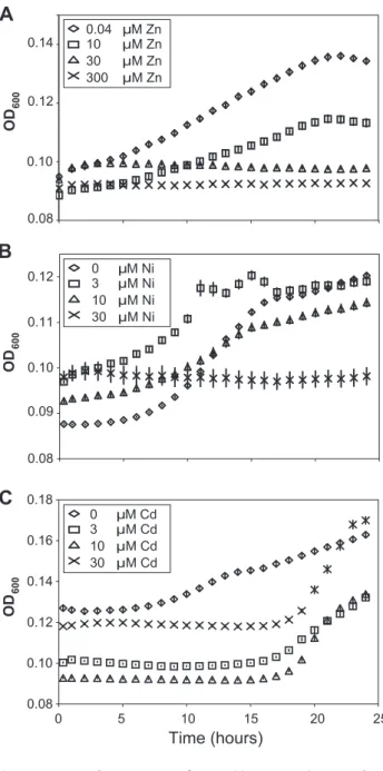
metals. Apoplast extracts from zinc- and nickel-treated plants supported significantly less bacterial growth than those from plants grown without supplementary metal (Figure 4A and B). Apoplast extracts from plants treated with higher cadmium concentrations also inhibited bacterial growth for up to 18 hours, although this inhibition was eventually released (Figure 4C). As such, it is clear that metal hyperaccumulation byT. caerulescensmakes the apoplast a more hostile environment for the growth ofPsm.
Metal concentrations inThlaspi caerulescensleaves and extracted apoplast are sufficient to explain the high disease resistance of metal-treated plants
To determine whether hyperaccumulated metal could be responsible for the observed reductions in bacterial growth in leaves, the ability ofPsm to tolerate each metal was tested in a range of synthetic media and in extracted apoplast (Figure S3) and compared with the metal concentrations of apoplast extracts and whole-leaf tissue samples (Figure 5; Table 1). For all metals tested, concentrations in whole-leaf tissue ofT. caerulescensplants grown at the two highest treatments were sufficient (i.e. higher than the IC50 Figure 2. T3SS mutants ofPseudomonas syringaepv. maculicola are unable to coloniseThlaspi caerulescens.P. syringaepv. maculicola M4 (PsmM4),P. syringaepv. maculicola ES4326 (Psm4326) and two T3SS mutants ofPsm(hrpS2andhrcN2) were inoculated into fully expanded leaves ofT. caerulescensat 106cfu/ml. Three samples were taken for each strain, zinc treatment and time point, each consisting of three leaf discs pooled
together. Values are means6SE (n= 3). The mean growth of the four strains at each zinc treatment over 5 days post-inoculation was compared in ANOVAs; growth of the four strains was found to differ significantly in all treatments except in the 300mM zinc treatment, where no strain was able to grow (P,0.0005 for 0.04 and 10mM Zn;P= 0.001 for 30mM Zn;P= 0.169 for 300mM Zn). Within each zinc treatment, Bonferroni simultaneous comparisons were used to determine which means differed significantly at 5 days post-inoculation, and these are marked with different letters. The inset shows symptoms observed in leaves fromT. caerulescensplants grown on 0.04mM zinc 72 hours after inoculation with wild-typePsmES4326 and thePsm hrcN2mutant at 106
cfu/ml compared with uninoculated control leaves. doi:10.1371/journal.ppat.1001093.g002
values for the respective metals) to explain the observed reduction in bacterial growth in planta. Apoplastic zinc and nickel concentrations from the highest treatments were also sufficient to explain bacterial growth reduction. Apoplastic cadmium concentrations were considerably lower, reaching around one-third of the IC50concentration forPsm in vitroin higher treatments, perhaps explaining the ability of Psm eventually to overcome inhibition by cadmium.
Pseudomonas syringaepv. maculicola zinc tolerance mutants show differential growth inThlaspi caerulescens
The importance of metal tolerance for bacteria growing in metal-hyperaccumulatingT. caerulescenswas assessed by screening a transposon mutant library of Psm to identify mutants with Figure 3. High metal concentrations inhibit bacterial growth in
Thlaspi caerulescens.T. caerulescensplants were treated with a range of zinc (A), nickel (B), or cadmium (C) concentrations. Nine leaves of each of six plants were infiltrated withP. syringaepv. maculicola M4 suspended in 10 mM MgCl2at 106cfu/ml and leaves sampled at 0, 2 and 5 days after
inoculation. Six samples were taken per time point and treatment, each sample consisting of three leaves pooled from one plant. Plant zinc, nickel and cadmium treatments were significant predictors of Psm growth at both day 2 and day 5 (ANOVAs; P,0.0005). Bonferroni simultaneous comparisons were carried out; means that were not significantly different are marked with the same letter. Values are means6SE (n= 6). The experiment was repeated twice with similar results. doi:10.1371/journal.ppat.1001093.g003
Figure 4. Apoplast extracts from Thlaspi caerulescens plants grown in high metal concentrations inhibit bacterial growth. Apoplast extracts obtained fromT. caerulescensplants treated with the same zinc (A), nickel (B), or cadmium (C) concentrations used in the bacterial colonization assays shown in Figure 3 were used as a growth medium for P. syringaepv. maculicola M4. Six samples of 100ml of apoplast were used for each treatment. Values are means6SE (n= 6). The experiment was performed three times with similar results, and the results shown are from one experiment representative of the three.
Concentrations of all three metals were significant predictors of growth at 18 and 24 hours (ANOVAs:P,0.0005 in all cases exceptP= 0.015 for cadmium at 24 h). Bonferroni simultaneous comparisons (a= 1%) show that all zinc and nickel treatments resulted in significantly less growth than the control at 18 h and 24 h, as did all cadmium treatments up to 18 h.
increased or decreased zinc tolerance relative to wild-type Psm. The performance of four representative mutantsin plantawas then compared to that of the wild-type strain under four zinc regimes. Two mutants with increased zinc tolerance (9A6 and 9A3) and two with reduced zinc tolerance (10C1 and 7C11) were used (Figure 6). These four mutants grew similarly to wild-typePsminA. thaliana (Figure S4) and were able to cause similar symptoms to wild-type PsminA. thalianaand pak choi,Brassica rapassp.chinensis(data not shown). Mutant 7C11 showed slightly reduced growth relative to wild-typePsmduring late log phase and stationary phase inin vitro growth assays in KB broth (Figure S5), but the in vitro growth kinetics of the other three mutants were not significantly different from wild-typePsm.
The disrupted genes were sequenced using an inverse PCR method and their predicted functions are described in Table 2. Interestingly, mutant 7C11 was found to contain an insertion in a TonB-dependent siderophore receptor (PSPTO_2152), which suggests that the slight growth defect observed in KB broth for this strain may be linked to impaired iron acquisition. The other mutation giving rise to reduced zinc tolerance, in mutant 10C1, was located in an NAD-dependent DNA ligase gene (PSPTO_0382); this does not have an obvious role in metal transport or metal tolerance, but is located close to two operons predicted to encode a heavy-metal-sensing two component regulatory system and com-ponents of a cobalt-zinc-cadmium (Czc) cation efflux system (PSPTO_0375 – PSPTO_0379) in the genome of P. syringaepv. tomato DC3000. The two mutations giving rise to increased zinc
tolerance were located inpslF(PSPTO_3533; 9A3), a gene within thepsloperon, involved in exopolysaccharide synthesis and biofilm formation in Pseudomonas aeruginosa [32], and in a proline iminopeptidase (pip) gene (PSPTO_5164; 9A6). The pip gene of Xanthomonas campestrispv. campestris has been shown to be induced during plant colonisation and to be essential for pathogenesis on cabbage [33]. However, our results indicate that thepipgene ofPsm is not required for pathogenesis inA. thaliana. Pip belongs to a family of metalloproteases and its enzymatic activity may be dependent on a metal cofactor such as zinc or cobalt. In the genome ofP. syringae pv. tomato DC3000,pipis located upstream of, and in a putative operon with, a predicted D-Tyr-tRNAtyr deacylase, which may have a role in protecting cells against D-tyrosine toxicity. InE. coli, zinc and D-tyrosine have been shown to have opposite effects on the phosphatase activity of the aromatic amino acid biosynthesis regulator TyrR, which is stimulated by zinc and suppressed by L-tyrosine and D-L-tyrosine [34].
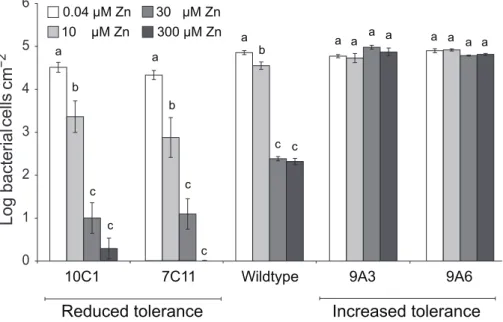
The ability of the four Psm mutants to grow inT. caerulescens plants cultivated on 0.04, 10, 30, or 300mM zinc was found to vary according to the zinc tolerance of the inoculated strain (Figure 7). Thus, while the wild-type showed significant growth reduction with plant zinc treatments of 30mM or higher, the two mutants with increased zinc tolerance, 9A6 and 9A3, were capable of growth in plants treated with 300mM zinc. The two mutants with decreased zinc tolerance, 7C11 and 10C1, were unable to grow even in plants treated with 10mM zinc, at which concentration the wild-type grew well.
Figure 5. Metal concentrations in zinc-, nickel- and cadmium-treated plants.Data represent average values from three independent sets of plants. Metal content was determined by atomic absorption spectrophotometry.A, C, E: Apoplastic concentrations of zinc, nickel and cadmium, respectively.B, D, F: Bulk-leaf concentrations of zinc, nickel and cadmium, respectively, expressed both as molar concentration calculated on the basis of leaf fresh biomass, and as mass concentration relative to leaf dry biomass. Values are means6SE (n= 6). Horizontal bars inA,CandF indicate metal concentrations representing the respective IC50values forP. syringaepv. maculicola M4 determined experimentally in extracted
apoplast (i.e. Zn = 0.12 mM; Ni = 0.025 mM; Cd = 0.01 mM); IC50values measured in LB were higher (Zn = 0.63 mM; Ni = 0.78 mM; Cd = 0.22 mM).
doi:10.1371/journal.ppat.1001093.g005
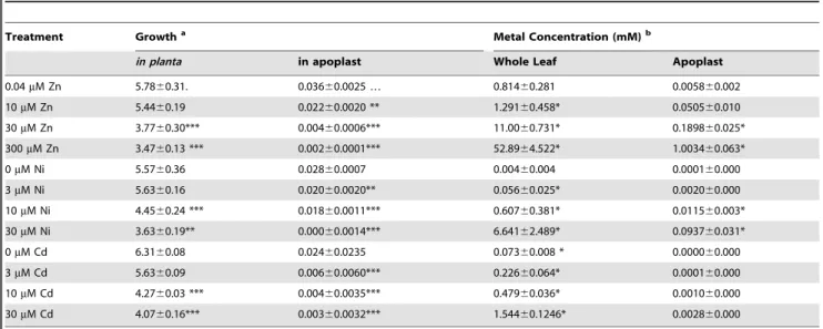
Table 1.Summary of bacterial growth across treatments in relation to metal concentration and metal-dependent growth inhibition ofP. syringaepv. maculicolain vitro.
Treatment Growtha Metal Concentration (mM)b
in planta in apoplast Whole Leaf Apoplast
0.04mM Zn 5.7860.31. 0.03660.0025 … 0.81460.281 0.005860.002
10mM Zn 5.4460.19 0.02260.0020 ** 1.29160.458* 0.050560.010
30mM Zn 3.7760.30*** 0.00460.0006*** 11.0060.731* 0.189860.025*
300mM Zn 3.4760.13 *** 0.00260.0001*** 52.8964.522* 1.003460.063*
0mM Ni 5.5760.36 0.02860.0007 0.00460.004 0.000160.000
3mM Ni 5.6360.16 0.02060.0020** 0.05660.025* 0.002060.000
10mM Ni 4.4560.24 *** 0.01860.0011*** 0.60760.381* 0.011560.003*
30mM Ni 3.6360.19** 0.00060.0014*** 6.64162.489* 0.093760.031*
0mM Cd 6.3160.08 0.02460.0235 0.07360.008 * 0.000060.000
3mM Cd 5.6360.09 0.00660.0060*** 0.22660.064* 0.000160.000
10mM Cd 4.2760.03 *** 0.00460.0035*** 0.47960.036* 0.001060.000
30mM Cd 4.0760.16*** 0.00360.0032*** 1.54460.1246* 0.002860.000
a‘Growth’ indicates mean log bacterial cells/cm2leaf 5 days after inoculation (
in planta) or increase in OD over 18 hoursin vitroin extracted apoplast. Means found to be significantly different from the control mean (Bonferroni simultaneous comparisons) are marked with *(a= 5%), **(a= 1%), or ***(a= 0.1%). In the case of Niin planta data, the 3mM treatment was used as a control for statistical analysis because of the relatively high variability of the 0mM treatment.
bMetal concentrations of either bulked whole leaf samples or of the apoplast are shown. * indicates that concentrations were equal to or greater than the IC
50values for
Because increased zinc tolerance could be correlated with reduced zinc uptake, we tested the ability of all four mutants to grow at low zinc concentrations. Wild-typePsmgrew equally well in M9 minimal medium and in M9 supplemented with 1 to 10mM Zn. Of the four mutants, only 9A6 had an increased zinc requirement relative to wild-type bacteria, showing optimal growth at concentrations ranging from 1 to 5mM zinc.
Naturally occurring endophytes ofThlaspi caerulescens
show high zinc tolerance
To test further the hypothesis that high metal tolerance is a prerequisite for bacterial growth in T. caerulescens, endophytic bacteria were collected from leaves of a natural population ofT. caerulescens plants at Hafna Mine, a former lead–zinc mine in North Wales, UK [35], and their zinc tolerance assessed. The
mean zinc content of the leaves of these plants was 16.261.39 g Zn per kg leaf dry mass (6s.e.m.,n= 23 leaves), slightly greater (p,0.05) than that of plants treated with the highest concentra-tion of zinc (300mM) used in the laboratory experiment (13.760.71 g Zn per kg dry mass (6s.e.m.,n= 18 leaves)). The average IC50for zinc of these naturally occurring endophytes was 9.4 mM (n= 86) in KB medium. In contrast, a set of plant pathogenic bacteria isolated from non-hyperaccumulating plants were found to have a significantly (p,0.001) lower mean IC50for zinc of 5.4 mM in KB (n= 7; Figure 8). When the zinc IC50 values for individual strains isolated from the Hafna mine plants were compared to the zinc IC50ofPsm, the most zinc tolerant of the plant pathogens used in this study, 65% were found to have a significantly higher zinc tolerance (p#0.05), while only a single strain out of 85 had a significantly lower zinc tolerance. Figure 6. Zinc tolerance ofP. syringaepv. maculicola M4 and four mutants.Fiveml of bacterial suspension at an OD600of 0.2 were inoculated
into 200ml of KB broth supplemented with zinc at 0 to 20 mM. The graph shows the percentage increase in OD60048 hours after inoculation, relative
to the increase in OD600over 48 hours observed for the same strain in the absence of zinc. At least four samples were analysed for each treatment.
Values are means6SE (n= 8). doi:10.1371/journal.ppat.1001093.g006
Table 2.P. syringaepv. maculicola M4 mutants with altered zinc tolerance.
Mutant ID
IC50(mM zinc) in KB Broth
Top BLAST Hit GenBank ID
Expectation Value
Best Match inP. syringae
pv. tomato DC3000 Predicted Function
9A3 12 NP_793313.1 3.00E242 PSPTO_3533 Glycosyl transferase
9A6 10 NP_794895 6.00E257 PSPTO_5164 Proline iminopeptidase
7C11 3.8 NP_791973 2.00E265 PSPTO_2152 TonB-dependent siderophore receptor
10C1 2.3 NP_790231.1 7.00E2124 PSPTO_0382 NAD-dependent DNA ligase
Wild-type 7.3 N/A N/A N/A N/A
doi:10.1371/journal.ppat.1001093.t002
Figure 7. P. syringaepv. maculicola M4 mutants with altered zinc tolerance show differential growth inThlaspi caerulescens.T. caerulescensplants were treated with 0.04, 10, 30, or 300mM zinc. Nine leaves of three plants were infiltrated with eitherP. syringaepv. maculicola M4 or one of four zinc-tolerance mutants (9A6, 9A3, 7C11, or 10C1) suspended in 10 mM MgCl2at 106cfu/ml. Leaves were sampled for bacterial counts
at 0 and 5 days after inoculation. Each replicate consisted of three leaves from one plant, giving a total of three replicates per time point and treatment. Values are means6SE (n= 3). The experiment was performed three times with similar results, and the results shown are from one experiment representative of the three. ANOVAs were used to test for a significant effect of plant zinc treatment on the growth of each bacterial strain. No effect was detected for the high tolerance mutant 9A3 (P= 0.13). For the other strains, growth was found to be dependent on zinc (9A6: P= 0.01; wild-type, 7C11 and 10C1:P,0.0005 in each case). Where a significant effect was found, Bonferroni simultaneous comparisons (a= 5%) were carried out. Within each strain, means marked with the same letter were not significantly different.
doi:10.1371/journal.ppat.1001093.g007
Figure 8. Bacterial endophytes isolated from a natural population ofThlaspi caerulescensexhibit high zinc tolerance.To determine IC50values for zinc, 5ml of bacterial suspension at an OD600of 0.2 were inoculated into 200ml of KB broth supplemented with zinc at 0 to 20 mM. OD600was measured after incubation, with continuous shaking, at 28uC for 48 hours, and IC50values were calculated from the resulting dose–
response curves. Values are means6SE (n= 3). Diamonds = Hafna mine endophytes; triangles = plant pathogenic bacteria isolated from non-metal-accumulating crop plants; squares = mutants of P. syringae pv. maculicola generated in the present work. Abbreviations: DC3000, Pseudomonas syringaepv. tomato DC3000;X.c.c.,Xanthomonas campestrispv. campestris 8004;P. cic3109,Pseudomonas cichoriiNCPPB3109;P. cic 907,P. cichoriiNCPPB907;P. cic943,P. cichoriiNCPPB943; Ea286,Erwinia amylovoraEa286; B728a,P. syringaepv. syringae B728a.
Therefore, in the field, as in the laboratory, only metal-tolerant bacteria can colonizeT. caerulescens.
Discussion
In this work, we have developed a model system to study the elemental defense hypothesis for plant metal hyperaccumulation using the bacterial pathogen Pseudomonas syringae pv. maculicola M4. We have shown that Psm displays T3SS-dependent pathogenesis in Thlaspi caerulescens plants grown in low metal concentrations and that metal hyperaccumulation byT. caerulescens at higher metal concentrations provides an effective defense againstPsm. Further, we have demonstrated that zinc tolerance is essential for bacterial colonization of zinc-hyperaccumulating plants. Thus, our work is consistent with the hypothesis that hyperaccumulation benefits plants by increasing their resistance to pathogens.
Although studies have found that zinc, nickel and cadmium are mainly stored in the leaf cell vacuoles of Thlaspi species, particularly in epidermal cells [27,28,36,37,38], metals are transported into the leaf through the extracellular spaces of the apoplast by means of the transpiration stream. By measuring metal concentrations in the apoplastic fluid, we were able to provide an approximation of the conditions experienced by invading pathogens in the leaves ofT. caerulescens. Comparison of the metal concentrationsin plantawith the IC50values measured forPsm in vitro in extracted apoplastic fluid allowed us to demonstrate that direct inhibition of bacterial growth by hyperaccumulated zinc or nickel is a realistic possibility.T. caerulescens, when grown on at least 10mM Zn or 30mM Ni, accumulated sufficient metal in apoplastic fluid to provide an elemental defense againstPsm. This result is particularly striking considering that, when grown at 10mM Zn,T. caerulescensaccumulated only an average of 0.3 g Zn per kg dry mass, while the threshold for designation as a zinc hyperaccumulating plant is 10 000 mg per kg [1]. This indicates that T. caerulescens could be protected against pathogens by zinc accumulation even when growing on relatively low-zinc soils. However, although T. caerulescens accumulates sufficiently high metal concentrations to account for its metal-dependent resistance toPsm, we cannot rule out the possibility that additional metal-dependent factors act in conjunction with metals to limit the growth ofPsminT. caerulescens. Accumulation of metals requires mechanisms by which the plant may tolerate elevated intracellular metal concentrations, which have not been fully elucidated, but which have been shown to involve redox-related compounds such as glutathione [39], enzymes such as superoxide dismutase [40], and metal-binding ligands such as organic acids and amino acids [27,41], as well as proteins (e.g. metallothioneins [42]). Such changes may affect the quality of the plant environment for growth of Psm or zinc tolerance in Psm, without having any expressly defensive function.
One case in which additional factors seem likely to contribute to metal-dependent defenses is in plants treated with cadmium. Cadmium was able to defendT. caerulescensagainstPsm in planta when plants were treated with $10mM cadmium. Bacterial growth was also inhibited for around 18 hours in apoplastic fluid extracted from these plants. After this time, inhibition was released, possibly as a result of changes in bacterial gene expression or in the composition of the apoplast during this time in vitro, which may include alterations in cadmium bioavailability. However, the cadmium concentrations detected in apoplast extracts from cadmium-treated plants were lower than those required for inhibition of bacterial growth in apoplast extracts from plants grown in the absence of cadmium, so it is possible that
cadmium concentrations are correlated with another defensive factor which may be unstable (such as ROS) or volatile.
To investigate further the possibility of a true elemental defense, we tested the importance of zinc tolerance for bacterial growthin planta in zinc-treated T. caerulescens. The results obtained using mutants ofPsmprovide the clearest evidence to date of a direct role for zinc as an elemental defense against pathogens in T. caerulescens. Mutants with reduced zinc tolerance were unable to multiply in plants grown at 10mM zinc, in leaves of which their wild-type counterpart was successful; conversely, mutants with increased zinc tolerance grew well in leaves of plants grown at 30mM or even 300mM zinc, in which the wild-type could not survive. This clear link between bacterial zinc tolerance and ability to colonize plants hyperaccumulating zinc provides strong support for the concept of an elemental defense by zinc inT. caerulescens.
Finally, we have compared the zinc tolerance of strains used in this work with that of bacteria isolated from the leaves of T. caerulescens plants growing under natural conditions in a zinc-polluted field site. We have shown that bacteria naturally colonizing these plants in the field had a range of zinc tolerances much higher than those of plant pathogenic strains isolated from non-accumulating crop plants, providing evidence for local adaptation of these endophytes to their environment [43,44,45]. This is in agreement with previous studies demonstrating that endophytic bacteria isolated from nickel hyperaccumulators exhibit high nickel tolerance [46,47]. Plant pathogens that have not been subject to this selection may find it difficult to grow and cause disease inT. caerulescens.
Metal-dependent resistance inT. caerulescensmay be particularly effective against airborne, foliar pathogens such asP. syringaeand powdery mildew, which may be deposited onto the surface ofT. caerulescens leaves by rain and wind having previously colonized non-accumulating or metal-excluding plants, with no prior selection for metal tolerance. Thus, hyperaccumulated metals may be functionally equivalent to the diverse array of anti-microbial secondary metabolites used by plants to provide protection against infection. Only pathogens that are able to tolerate or inhibit the chemical defenses present in a specific plant species or genotype can grow in plant tissues. Similarly, in the case of metal-hyperaccumulation, only a small number of organisms – those possessing high metal tolerance – are able to grow in these plants. When the plants are deprived of this form of defense by cultivation on a low-metal growth medium, they may become vulnerable to a wider range of pathogens, explaining the spontaneous outbreaks of mildew infection observed on such plants (Figure 1).
When considering the role of metals in protecting plants against infection in a natural setting it is important to note that metal concentrations in the leaves of hyperaccumulating plants are typically orders of magnitude higher than metal concentrations in the environment, as illustrated in Figure 5. Therefore, even though the environment surrounding these plants may favour the growth of moderately metal tolerant microorganisms, many of these organisms may have insufficient metal tolerance to be able to grow in the tissues of hyperaccumulating plants. In addition, the observation that T3SS mutants ofPsmwere unable to infect T. caerulescensplants grown on low metal concentrations indicates that the plants possess additional defense mechanisms that act in conjunction with metal hyperaccumulation to protect plants, but which can be suppressed by the action of T3SS effectors. Thus, successful pathogens ofT. caerulescensmust not only be adapted for growth in a metal-rich environment, but must also possess at least some of the pathogenicity mechanisms known to be required for infection of non-accumulating plants such asArabidopsis thaliana.
We have demonstrated that hyperaccumulation of any of three metals, zinc, nickel, or cadmium, by T. caerulescens provides the plant with an elemental defense against the hemibiotrophic pathogen Psm. The validity of the defense hypothesis has been challenged by some studies in which no evidence was found that metal hyperaccumulation defended T. caerulescensfrom herbivory in the field [13,14]. Our work, however, suggests that the defensive effect of metal hyperaccumulation against pathogens remains relevant in field conditions, with only metal-tolerant bacteria found growing naturally in the leaves ofT. caerulescens.Further, we have shown that metal concentrations in the leaves are sufficient to account for this defensive effect without invoking any other factors. For all of the metals hyperaccumulated byT. caerulescens, we have shown that growth is also inhibited in apoplastic fluid, and that both zinc and nickel are found in this specific compartment at concentrations sufficient to account for the defensive effect. Moreover, we have demonstrated that the zinc tolerance of Psm mutants is correlated with their ability to colonize zinc-hyperaccumulating T. caerulescens plants. This result is mirrored by our findings concerning the zinc tolerance of natural endophytes of T. caerulescens from a zinc-rich field site. We therefore believe that metal hyperaccumulation by T. caerulescens can provide an effective form of defense against a wide range of pathogens.
Materials and Methods
Plant material
Seeds ofThlaspi caerulescensJ. & C. Presl from Prayon, Belgium (provided by A.J.M. Baker and C. Lefe`bvre) were cultured hydroponically on modified 0.1-strength Hoagland solution [20] in a glasshouse. The Prayon population of T. caerulescens was chosen as it is a widely studied, well characterized population showing typical hyperaccumulation behavior and producing relatively large amounts of biomass under the growth conditions described [20,22,24,25]. Natural radiation was supplemented by sodium-vapor lamps for 14 hours per day. Night temperature was maintained at 14uC and day temperature at a minimum of 24uC. Two-week-old plants were transferred to modified 0.1-strength Hoagland solution containing 0.04, 10, 30, or 300mM ZnSO4, or 0, 3, 10, or 30mM NiSO4or CdSO4. The lowest zinc concentration used was 0.04mM, rather than 0mM for Ni and Cd. This is because zinc is an essential micronutrient without which the plants do not survive. The Hoagland solution used for all Ni and Cd assays contained 10mM Zn for this reason; at 0.04mM Zn, signs of zinc deficiency become apparent. T. caerulescens plants were grown on these metal treatments for a further 8 weeks, the nutrient solution being exchanged fortnightly for the first 6 weeks and weekly for the final 2 weeks. Arabidopsis thaliana(L.) Heynh. (Col-0) were sown on peat-based compost and grown in a glasshouse under the same conditions for 6 weeks.
Bacterial growth conditions, media and antibiotics Bacterial strains used in pathogenicity assays are listed in Table S1.PsmES4326,Psm hrpN2andPsm hrpS2were provided by K. Schreiber and D. Desveaux. Psm hrpN2 and Psm hrpS2 were isolated from a transposon library of Psm ES4326 constructed using a kanamycin-resistant derivative of mini-Tn5 ([48]; D. Desveaux, personal communication). All bacterial strains were streaked onto Luria–Bertani (LB) agar [49] from stocks kept in glycerol at280uC and incubated at 28uC (37uC forEscherichia coli) for 24 to 48 hours prior to use. Single colonies were transferred to LB broth and incubated at 28uC with shaking for most
applications. King’s B (KB) agar [50], supplemented with CFC (Cetrimide-Fucidin-Cephalosporin; Oxoid) at half the manufac-turer’s recommended concentration, was used to selectively culture bacteria isolated from plant tissues for in planta growth studies. LB and KB broths were used in metal-tolerance experiments. For zinc-requirement assays, the minimal medium M9 [49] was used.
Bacterial growthin vitro
To compare thein vitrogrowth of wild-type and mutant strains of Psm, bacteria were grown overnight on LB agar and resuspended in KB broth to give an OD600of 0.1. One hundred microliters of this suspension were added to a further 100ml of media in each of 16 wells of a 96-well microplate. Sixteen wells were inoculated with media alone as a media control. OD was measured every 20 min for the next 48 hours using an Infinite M200 plate reader (Tecan Group Ltd., Ma¨nnedorf, Switzerland).
Pathogenicity assays and bacterial growthin planta
For assays to examine the ability of bacteria to cause disease symptoms inT. caerulescens, bacteria were grown overnight on LB agar and re-suspended in sterile 10 mM MgCl2 at an optical density at 600 nm (OD600) of 0.35. Bacterial suspensions were infiltrated into at least three fully expanded leaves from 6-week-old T. caerulescensplants grown on nutrient solution (10mM zinc) or 6-week-oldA. thalianausing a blunt 1 ml syringe. Symptoms resulting from bacterial inoculation were monitored over 7 days. For growth assays, bacteria were resuspended in sterile 10 mM MgCl2 at an OD600of 0.2. This suspension was diluted 100-fold to give a suspension of approximately 106cfu/ml and infiltrated into fully expandedT. caerulescensorA. thaliana leaves through the abaxial surface using a blunt 1 ml syringe. Nine leaves on each of six plants were inoculated within each metal treatment. Leaf discs of 10-mm diameter were taken from three of the inoculated leaves immediately, and from three further leaves at 2 and 5 days after inoculation. Leaf discs were homogenized in 10 mM MgCl2and the resulting suspension spread onto agar plates with a minimum of three technical replicates used for each sample. After incubation at 28uC for 48 hours, the number of bacterial colonies was counted and used to estimate the number of bacterial cells per unit area of leaf.
Apoplast extracts
Apoplastic fluid was extracted fromT. caerulescensandA. thaliana leaves by a modification of the method described by Rico and Preston [51], using vacuum infiltration of the intercellular spaces with distilled water followed by centrifugation to extract apoplastic fluid. This fluid was centrifuged for a further 10 minutes at 4uC, filter-sterilized, and stored at 280uC. The degree of apoplast dilution was estimated as described by Rico and Preston [51]. For bacterial growth experiments, apoplast extract was freeze-dried and resuspended in an appropriate volume of distilled water to return it to its estimated concentrationin planta.
Bacterial growth in apoplast extracts
Metal content of whole-leaf and apoplast extracts of
Thlaspi caerulescens
For determination of whole-leaf metal concentrations, fresh leaf material was oven-dried at 80uC for 48 hours. Subsamples of 50 mg of dried leaf material were digested in 3 ml of concentrated (69%, v/v) nitric acid for 16 hours in glass vials. Samples were then diluted 10-fold with ultrapure water and filtered using Whatman grade 3 filter paper. Metal contents of samples were measured in an air–acetylene flame by atomic absorption spectrophotometry using a double-beam optical system with deuterium arc background correction (AAnalyst 100; Perkin-Elmer, UK). Samples were further diluted as necessary to fall within the linear range of calibration curves prepared using appropriate standard solutions and reagent blanks. The accuracy of the calibration curves was validated using Certified Reference Material LGC7162 (strawberry leaves; LGC Standards, Tedding-ton, UK). Three technical replicates were analysed for each measurement, and two samples from each of three plants were analysed for each metal treatment. Measurements of the fresh and dry biomass of 24 individual T. caerulescens plants were used to provide an average ratio between fresh and dry biomass, which allowed the metal content to be expressed as an approximate molar concentration in the fresh tissue. Apoplast samples were diluted appropriately with ultrapure water and measured without further treatment.
Metal-tolerance assays
The metal tolerance ofPsm in vitro was tested in LB. A 5ml aliquot of an overnight liquid culture was added to 200ml of media supplemented with zinc, nickel, or cadmium at a range of concentrations. OD600 was read using the plate reader. Metal tolerance was also tested in apoplast extracted fromT. caerulescens plants grown in 0.1-strength Hoagland solution (10mM zinc). For these experiments, 2ml of an overnight culture ofP. syringaepv. maculicola M4 was added to 75ml of apoplast extract or apoplast extract supplemented with metal.
Transposon mutagenesis of Pseudomonas syringaepv. maculicola M4
Transposon mutagenesis was carried out using the transposon miniTn5::gfp::luxcloned into pGP704 (generous gift of Phil Hill). The transposon was introduced into Psm by triparental mating using the helper plasmid pRK2013 [52]. The resultant bacterial mixture was spread onto LB agar containing 50mg/ml kanamycin and 50mg/ml rifampicin. Resulting colonies were inoculated into 150ml of KB broth in 96-well plates and incubated at 28uC overnight. Thirty microliters of 50% (v/v) glycerol was then added to each well and plates were stored at280uC as a mutant library.
Screening thePseudomonas syringaepv. maculicola mutant library for zinc tolerance mutants
Mutants were screened in a four-stage process. In round one, mutants were grown in KB in 96-well plates in which a wild-type was also included. A 5ml aliquot of overnight culture was transferred to 200ml of KB in a black, clear-bottomed 96-well plate and incubated at 28uC in the plate reader. Plates were maintained at 28uC with continuous shaking and the OD600and luminescence from each well was read at hourly intervals. After 3.5 hours, cultures were supplemented with 5ml of 0.5 mM ZnSO4. OD600and luminescence were read immediately and then at intervals of 3 to 9 hours for the next 48 hours. Changes in luminescence after the addition of zinc were recorded and growth was compared to wild-type.
A total of 866 strains were selected with markedly increased or decreased tolerance to zinc shock. Each of these mutants was transferred to 200ml KB in two separate wells of a 96-well plate and growth was monitored with and without 0.5 mM zinc over 48 hours. Of these, 134 mutants whose tolerance differed notably from the wild-type were selected and further screened for growth in 200ml KB in 96-well plates with zinc concentrations from 0 to 3.5 mM for 48 hours. IC50 values were then calculated and compared to wild-type. These results were validated in a second experiment for ten mutants that showed the largest consistent changes in zinc tolerance compared to wild-type. These ten mutants were then tested for their ability to cause symptoms inT. caerulescens,A. thalianaand pak choi (Brassica rapaspp.chinensis).
Inverse-PCR based sequencing of transposon mutants Genomic DNA was extracted using a DNeasy Blood and Tissue kit (Qiagen) according to the manufacturer’s protocol for Gram negative bacteria. Eight microliters of the subsequent DNA suspension were digested with 1ml of either the restriction endonucleaseSphI orNarI (New England BioLabs) and 1ml of the appropriate 106buffer according to the manufacturer’s specifica-tions.NarI cleaves the transposon DNA near the 39end, whileSphI cleaves it towards the 59 end; both also cleave the Psm genome frequently. Digested DNA was ethanol-precipitated, resuspended in 8ml of ultrapure water, and self-ligated overnight at 14uC using 1ml of T4 ligase and 1ml of 106buffer (New England BioLabs). Oneml of circularized DNA was then used as a template for inverse PCR. Primers designed to the ends of the short fragments of transposon resulting from NarI (AACAATCTAGCGAGGGCTTGGTAA-GGTGATCC and CTTGCAGTGGGCTTACATGACGATAG-CTAGAC) orSphI (GGAACGCCGCAGGAATG and CAGCA-GCTGTTACAAACTCAAGAAG) digestion, and facing outwards, were used to amplify the flanking DNA. This was then sequenced using a primer to the end of the transposon (CGGTTTACAAGC-TAAAGCTTGC for NarI-digested DNA and either CTTC-TTTAAAATCAATACC or TTCCAGTAGTGCAAATAA for SphI-digested DNA).
Assessment of zinc tolerance of naturally occurring endophytes ofThlaspi caerulescensfrom a zinc-rich site
Leaves ofT. caerulescenswere collected in the field at Hafna mine (Snowdonia, North Wales, UK: 53u079N, 3u499W). They were then returned to the laboratory and immediately surface-sterilized by immersion in 10% (w/v) sodium hypochlorite for 3 minutes, followed by immersion in 100% ethanol for 3 minutes. Sterile leaves were rinsed in ultrapure water and macerated in 1 ml of 10 mM MgCl2solution. The resulting suspension was plated onto KB-CFC agar. Plates were incubated at 28uC for 48 hours. Resultant colonies were transferred to 150ml of KB broth in 96-well plates. After 48 hours of growth at 28uC, 30ml of 50% (v/v) glycerol was added to each well and the plates stored at280uC. For zinc tolerance experiments, bacteria were grown in 200ml KB for 48 hours and replicated into 200ml KB supplemented with 0, 1, 2.5, 5, 7.5, 10, 12.5, 15, or 20mM ZnSO4. Bacterial growth at each of these zinc concentrations was determined by measuring OD600at 0 and 48 hours using the plate reader. Bacterial growth at 48 hours was then plotted against zinc concentration, from which the concentrations giving half-maximal inhibition (IC50) were estimated.
Quantitative RT-PCR ofPR-1gene expression
RNA for qRT-PCR analysis was isolated from leaves of 10-week-oldT. caerulescensgrown on either 0.04 or 300mM zinc and
from leaves of 6-week-old, non flowering A. thaliana. A. thaliana leaves inoculated with Psm at 106cfu/ml and incubated for 24 hours under standard plant growth conditions were used as a positive control forPR-1expression. Leaves were snap frozen in liquid nitrogen and RNA was extracted using the RNeasy kit (Qiagen) according to the manufacturer’s instructions. After elution, RNA was precipitated in 100ml of 8 M LiCl overnight. After two washes in 70% (v/v) ethanol, pellets were resuspended in 30ml ultrapure water. RNA concentration was measured using a Nanodrop-1000 spectrophotometer (Thermo Scientific) and integrity was checked by electrophoresis on an ethidium bromide gel. cDNA was prepared from 1mg RNA using the Bioline cDNA synthesis kit with oligo dT primer according to the supplier’s instructions. qRT-PCR was performed using SYBR green PCR master mix (Applied Biosystems) in a 7300 Realtime PCR machine (Applied Biosystems) and analysed by calibration to a standard curve of gene expression created from pooled cDNA from all samples under test, using the 7300 SDS system software v1.3.1 (Applied Biosystems). Four control genes were analysed in the same way, and PR-1 gene expression normalized to the geometric mean of the expression of these genes [53]. Gene-specific primers used are listed in Table 3.
Accession numbers
The GenBank (http://www.ncbi.nlm.nih.gov) accession num-bers for the genes and gene products discussed in this paper are: PSPTO_0382 (NP_790231.1); PSPTO_2153 (NP_791973); PSPTO_3533 (NP_793313.1); PSPTO_5164 (NP_794895).
Acknowledgments
We thank Prof. A.J.M. Baker for providing seeds of Thlaspi caerulescens; Prof. J.L. Dangl for providing Pseudomonas syringae pv. maculicolaM4, Dr. K. Schreiber and Dr. D. Desveaux for providinghrp mutants ofPseudomonas syringaepv. maculicola ES4326, Dr. P. Hill for proving the transposon used for mutagenesis, and Prof. J.A. Langdale, Prof. N.P. Harberd and Dr. A. Buckling for helpful discussions.
Supporting Information
Figure S1 In vitro growth of wild-type Pseudomonas syringae pv. maculicola ES4326 and hrcN- and hrpS- mutants. Strains were inoculated into KB broth to give an OD at 600 nm of 0.1, and 100ml of this suspension was added to a further 100ml of KB in a 96 well microplate. OD600was monitored over 24 h. Values are means6SE (n= 16).
Found at: doi:10.1371/journal.ppat.1001093.s001 (0.71 MB EPS)
Figure S2 Expression ofPR-1inThlaspi caerulescensin response to zinc. Quantitative real-time PCR was used to determine the
expression ofPR-1inT. caerulescensgrown on 0.04 or 300mM zinc, and in A. thaliana, either after no treatment or 24 hours after inoculation withPsmat 106cfu/ml. Expression was normalised to the geometric mean of the expression of four control genes: ubiquitin, EF-1a,b-tubulin and ornithine transcarbamylase. Within each species,t-tests were used to compare the two results obtained. These showed that there was no significant difference betweenPR-1 expression inT. caerulescensin the two zinc treatments (P= 0.2), while inoculation ofA. thalianawithPsmresulted in a significant increase in PR-1expression (P= 0.005). Values are means6SE (n= 12). Found at: doi:10.1371/journal.ppat.1001093.s002 (0.68 MB EPS)
Figure S3 Effect of increasing zinc, nickel and cadmium concentrations on growth of Pseudomonas syringae pv. maculicola M4in vitro. Fiveml of bacterial suspension at an OD600of 0.2 were inoculated into 200ml of LB supplemented with varying concentrations of metal. Graphs show the OD60024 hours after inoculation. A minimum of eight replicates were analysed for each treatment. Values are means6SE (n$8).
Found at: doi:10.1371/journal.ppat.1001093.s003 (0.77 MB EPS)
Figure S4 Growth of zinc tolerance mutants ofPseudomonas syringae pv. maculicola M4 inArabidopsis thaliana. Six leaves of three plants were infiltrated with eitherPsmor one of four zinc tolerance mutants ofPsm(9A6, 9A3, 7C11 and 10C1) suspended in 10 mM MgCl2at 106cfu/ml. Leaves were sampled at 0 and 5 days after inoculation. Each replicate consisted of three leaves from one plant, giving a total of three replicates per time point and treatment. Values are means6 SE (n= 5). The experiment was carried out twice with similar results. An ANOVA was used to test for an effect of strain upon growth. This was significant (P= 0.033), but Bonferroni simultaneous comparisons showed this significance to be due to a difference between the growth of mutants 9A6 and 9A3; no mutant grew to a level significantly different from that ofPsm(P= 1.000, 1.000, 0.612 and 1.000 for 9A6, 9A3, 7C11 and 10C1, respectively).
Found at: doi:10.1371/journal.ppat.1001093.s004 (0.47 MB EPS)
Figure S5 Growth of zinc tolerance mutants of Pseudomonas syringaepv. maculicola M4in vitro. Strains were added to KB broth to give an OD600of 0.1, and 100ml of this suspension was added to a further 100ml of KB in a 96 well microplate. OD600 was monitored over 24 h. Values are means6SE (n= 16).
Found at: doi:10.1371/journal.ppat.1001093.s005 (0.68 MB EPS)
Table S1 Bacteria that caused necrotic symptoms in Thlaspi caerulescens.
Found at: doi:10.1371/journal.ppat.1001093.s006 (0.05 MB DOC)
Table 3.Primers used in qRT-PCR.
Gene product Forward Primer Sequence Reverse Primer Sequence Accession numbera
PR-1 ACAACTACGCTGCGACGT TCACTTTGGCACATCCGAGTC NM_127557
EF1-a TGAGCACGCTCTTCTTGCTTTCA GGTGGTGGCATCCATCTTGTTACA X16430.1
b-tubulin CACCAGACATAGTAGCAGAAATCAAGT AAACTCACTACCCCCAGCTTTG M84702.1
Ornithine transcarbamoylase TGAAGGGACAAAGGTTGTGTATGTT CGCAGACAAAGTGGAATGGA AJ002524.1
UBQ10 (ubiquitin) AAAGCTCCGACACCATTGAC CTTATTCATCAGGGATTATACAAGG NM_178968.4
aGenBank accession numbers forArabidopsis thalianagene sequences used for primer design (www.ncbi.nlm.nih.gov).
Author Contributions
Conceived and designed the experiments: HF JACS GMP. Performed the experiments: HF CARD AR FF GMP. Analyzed the data: HF CARD
JACS GMP. Contributed reagents/materials/analysis tools: AR. Wrote the paper: HF JACS GMP.
References
1. Baker AJM, Brooks RR (1989) Terrestrial higher plants which hyperaccumulate metallic elements – a review of their distribution, ecology and phytochemistry. Biorecovery 1: 81–126.
2. Reeves RD, Baker AJM (2000) Metal-accumulating plants. In: Raskin I, Ensley BD, eds. Phytoremediation of Toxic Metals: Using Plants to Clean-up the Environment. New York: John Wiley & Sons. pp 193–230.
3. Brooks RR, ed (1998) Plants that Hyperaccumulate Heavy Metals. Wallingford: CAB International. 380 p.
4. Verbruggen N, Hermans C, Schat H (2009) Molecular mechanisms of metal hyperaccumulation in plants. New Phytol 181: 759–776.
5. Pollard AJ, Powell KD, Harper FA, Smith JAC (2002) The genetic basis of metal hyperaccumulation in plants. Crit Rev Plant Sci 21: 539–566.
6. Boyd RS, Martens SN (1992) Theraison d’eˆtrefor metal hyperaccumulation by plants. In: Baker AJM, Proctor J, Reeves RD, eds. The Vegetation of Ultramafic (Serpentine) Soils. Andover: Intercept Limited. pp 279–289.
7. Poschenrieder C, Tolra` R, Barcelo´ J (2006) Can metals defend plants against biotic stress? Trends Plant Sci 11: 288–295.
8. Boyd RS (2007) The defense hypothesis of elemental hyperaccumulation: status, challenges and new directions. Plant Soil 293: 153–176.
9. Vesk PA, Reichman S (2009) Hyperaccumulators and herbivores – a Bayesian meta-analysis of feeding choice trials. J Chem Ecol 35: 289–296.
10. Hanson B, Lindblem SD, Loeffler ML, Pilon-Smith EAH (2004) Selenium protects plants from phloem-feeding aphids due to both deterrence and toxicity. New Phytol 162: 655–662.
11. Behmer ST, Lloyd CM, Raubenheimer D, Stewart-Clark J, Knight J, et al. (2005) Metal hyperaccumulation in plants: mechanisms of defence against insect herbivores. Funct Ecol 19: 55–66.
12. Jiang RF, Ma DY, Zhao FJ, McGrath SP (2005) Cadmium hyperaccumulation protects Thlaspi caerulescens from leaf feeding damage by thrips (Frankliniella occidentalis).New Phytol 167: 805–814.
13. Noret N, Meerts P, Tolra` R, Poschenrieder C, Barcelo´ J, et al. (2005) Palatability ofThlaspi caerulescensfor snails: influence of zinc and glucosinolates. New Phytol 165: 763–772.
14. Noret N, Meerts P, Vanhaelen M, Dos Santos A, Escarre´ J (2007) Do metal-rich plants deter herbivores? A field test of the defence hypothesis. Oecologia 152: 92–100.
15. Jhee EM, Boyd RS, Eubanks MD (2005) Nickel hyperaccumulation as an elemental defense ofStreptanthus polygaloides(Brassicaceae): influence of herbivore feeding mode. New Phytol 168: 331–344.
16. Ghaderian YSM, Lyon AJE, Baker AJM (2000) Seedling mortality of metal hyperaccumulator plants resulting from damping off byPythiumspp. New Phytol 146: 219–224.
17. Boyd RS, Shaw JJ, Martens SN (1994) Nickel hyperaccumulation defends Streptanthus polygaloides(Brassicaceae) against pathogens. Am J Bot 81: 294–300. 18. Reeves RD, Schwartz C, Morel JL, Edmondson J (2001) Distribution and metal-accumulating behavior ofThlaspi caerulescens and associated metallophytes in France. Int J Phytorem 3: 145–172.
19. Baker AJM, Reeves RD, Hajar ASM (1994) Heavy metal accumulation and tolerance in British populations of the metallophyteThlaspi caerulescensJ & C Presl (Brassicaceae). New Phytol 127: 61–68.
20. Roosens N, Verbruggen N, Meerts P, Xime´nez-Embu´n P, Smith JAC (2003) Natural variation in cadmium tolerance and its relationship to metal hyperaccumulation for seven populations ofThlaspi caerulescens from western Europe. Plant Cell Environ 26: 1657–1672.
21. Hammond JP, Bowen HC, White PJ, Mills V, Pyke KA, et al. (2006) A comparison of theThlaspi caerulescensandThlaspi arvenseshoot transcriptomes. New Phytol 170: 239–260.
22. Assunc¸a˜o AGL, Schat H, Aarts MGM (2003)Thlaspi caerulescens, an attractive model species to study heavy metal hyperaccumulation in plants. New Phytol 159: 351–360.
23. Cobbett C (2003) Heavy metals and plants – model systems and hyperaccu-mulators. New Phytol 159: 289–293.
24. Peer WA, Mahmoudian M, Freeman JL, Lahner B, Richards EL, et al. (2006) Assessment of plants from the Brassicaceae family as genetic models for the study of nickel and zinc hyperaccumulation. New Phytol 172: 248–260.
25. Milner MJ, Kochian LV (2008) Investigating heavy-metal hyperaccumulation usingThlaspi caerulescensas a model system. Ann Bot 102: 3–13.
26. Debener T, Lehnackers H, Arnold M, Dangl JL (1991) Identification and molecular mapping of a singleArabidopsis thalianalocus determining resistance to a phytopathogenicPseudomonas syringaeisolate. Plant J. pp 289–302.
27. Ku¨pper H, Zhao FJ, McGrath SP (1999) Cellular compartmentation of zinc in leaves of the hyperaccumulatorThlaspi caerulescens. Plant Physiol 119: 305–311. 28. Ku¨pper H, Mijovilovich A, Meyer-Klaucke W, Kroneck PMH (2004) Tissue-and age-dependent differences in the complexation of cadmium Tissue-and zinc in the
cadmium/zinc hyperaccumulatorThlaspi caerulescens(Ganges ecotype) revealed by X-ray absorption spectroscopy. Plant Physiol 134: 748–757.
29. Cosio C, DeSantis L, Frey B, Diallo S, Keller C (2005) Distribution of cadmium in leaves of Thlaspicaerulescens. J Exp Bot 56: 765–775.
30. Dong X, Mindrinos M, Davis KR, Ausubel FM (1991) Induction ofArabidopsis defense genes by virulent and avirulentPseudomonas syringaestrains and by a cloned avirulence gene. Plant Cell 3: 61–72.
31. Chmielowska J, Veloso J, Gutie´rrez J, Silvar C, Dı´az J (2009) Cross-protection of pepper plants stressed by copper against a vascular pathogen is accompanied by the induction of a defence response. Plant Sci 178: 176–182.
32. Byrd MS, Sadovskaya I, Vinogradov E, Lu HP, Sprinkle AB, et al. (2009) Genetic and biochemical analyses of thePseudomonas aeruginosaPsl exopolysac-charide reveal overlapping roles for polysacexopolysac-charide synthesis enzymes in Psl and LPS production. Mol Microbiol 73: 622–638.
33. Zhang LL, Jia YT, Wang L, Fang RX (2007) A proline iminopeptidase gene upregulatedin planta by a LuxR homologue is essential for pathogenicity of Xanthomonas campestrispv. campestris. Mol Microbiol 65: 121–136.
34. Zhao S, Zhu Q, Somerville RL (2000) Thes70
transcription factor TyrR has zinc-stimulated phosphatase activity that is inhibited by ATP and tyrosine. J Bacteriol 182: 1053–1061.
35. Bennett J, Vernon RW (1990) Mines of the Gwydyr Forest: Part 2. The Hafna Mine, Llanrwst and some early ventures in Gwydyr Nant. Cuddington, Cheshire, UK: Gwydyr Mines Publications.
36. Kra¨mer U, Pickering IJ, Prince RC, Raskin I, Salt DE (2000) Subcellular localization and speciation of nickel in hyperaccumulator and non-accumulator Thlaspispecies. Plant Physiol 122: 1343–1354.
37. Vogel-Mikusˇ K, Regvar M, Mesjasz-Przybyłowicz J, Przybyłowicz WJ, Simcˇic J, et al. (2008) Spatial distribution of cadmium in leaves of metal hyperaccumulat-ingThlaspi praecoxusing micro-PIXE. New Phytol 179: 712–721.
38. Wo´jcik M, Vangronsveld J, D’Haen J, Tukiendorf A (2005) Cadmium tolerance inThlaspi caerulescens. II. Localization of cadmium inThlaspi caerulescens. Env Exp Bot 53: 163–171.
39. Freeman, JL, Persans MW, Nieman K, Albrecht C, Peer W, et al. (2004) Increased glutathione biosynthesis plays a role in nickel tolerance inThlaspi nickel hyperaccumulators. Plant Cell 16: 2176–2191.
40. Tuomainen MH, Nunan N, Lehesranta SJ, Tervahauta AI, Hassinen VH, et al. (2006) Multivariate analysis of protein profiles of metal hyperaccumulator accessions. Proteomics 6: 3696–3706.
41. Kra¨mer U, Cotter-Howells JD, Charnock JM, Baker AJM, Smith JAC (1996) Free histidine as a metal chelator in plants that accumulate nickel. Nature 379: 635–638.
42. Roosens NH, Leplae R, Bernard C, Verbruggen N (2005) Variations in plant metallothioneins: the heavy metal hyperaccumulatorThlaspi caerulescensas a study case. Planta 222: 716–729.
43. Frank SA (1992) Models of plant pathogen coevolution. Trends Genet 8: 213–219.
44. Kawecki TJ, Ebert D (2004) Conceptual issues in local adaptation. Ecol Lett 7: 1225–1241.
45. Nuismer SL, Gandon S (2008) Moving beyond common-garden and transplant designs: insights into the causes of local adaptation in species interactions. Am Nat 171: 658–668.
46. Idris R, Trifonova R, Puschenreiter M, Wenzel WW, Sessitsch A (2004) Bacterial communities associated with flowering plants of the Ni hyperaccumu-latorThlaspi goesingense. Appl Env Microbiol 70: 2667–2677.
47. Barzanti R, Ozino F, Bazzicalupo M, Gabbrielli R, Galardi F, et al. (2007) Isolation and characterization of endophytic bacteria from the nickel hyperaccumulator plantAlyssum bertolonii. Microbial Ecol 53: 306–316. 48. Alexeyev MF, Shokolenko IN, Croughan TP (1995) New mini-Tn5 derivatives
for insertion mutagenesis and genetic engineering in Gram-negative bacteria. Can. J Microbiol 41: 1053–1055.
49. Sambrook J, Russell DW (2001) Molecular Cloning. New York: Cold Spring Harbor Laboratory Press.
50. King EO, Ward MK, Raney DE (1954) Two simple media for the demonstration of pyocyanin and fluorescein. J Lab Clin Med 22: 301–307. 51. Rico A, Preston GM (2008) Pseudomonas syringae pv. tomato DC3000 uses
constitutive and apoplast-induced nutrient assimilation pathways to catabolize nutrients that are abundant in the tomato apoplast. Mol Plant–Microbe Interact 21: 269–282.
52. Figurski DH, Helinski DR (1979) Replication of an origin-containing derivative of plasmid RK2 dependent on a plasmid function provided intrans. Proc Natl Acad Sci USA 76: 1648–1652.
53. Larionov A, Krause A, Miller W (2005) A standard curve based method for relative real time PCR data processing. BMC Bioinformatics 6: 62.