Simon Brown
1,2Noorzaid Muhamad
3Lisa R. Walker
4Kevin C. Pedley
4David C. Simcock
1,4,5An
in silico
analysis of the glutamate
dehydrogenases of
Teladorsagia circumcincta
and
Haemonchus contortus
Authors’ addresses: 1
Deviot Institute, Deviot, Tasmania 7275, Australia. 2
School of Human Life Sciences,
University of Tasmania, Locked Bag 1320, Launceston, Tasmania 7250, Australia. 3
University Kuala Lumpur, Royal College of Medicine Perak, 3 Greentown Road, 30450 Ipoh, Perak, Malaysia.
4
Institute of Food, Nutrition and Human Health, Massey University, Private Bag 11222, Palmerston North, New Zealand. 5
Faculty of Medicine, Health and Molecular Sciences, James Cook University, Cairns, Queensland 4870, Australia.
Correspondence:
Simon Brown
School of Human Life Sciences,
University of Tasmania, Locked Bag 1320, Launceston, Tasmania 7250, Australia. Tel.: +61 3 63245400
e-mail: Simon.Brown@deviotinstitute.org
Article info:
Received: 19 October 2013
Accepted: 29 November 2013
ABSTRACT
Nematode glutamate dehydrogenase (GDH) amino acid sequences are very highly conserved (68-99% identity) and are also very similar to those of the bovine and human enzymes (54-60% identity). The residues involved in binding nucleotides or substrates are completely conserved and tend to be located in highly conserved regions of the sequence. Based on the strong homology between the bovine, Teladorsagia circumcincta and Haemonchus contortus GDH sequences, models of the structure of the T. circumcincta and H. contortus monomers were constructed. The structure of the T. circumcincta monomer obtained using SWISS-MODEL was very similar to that of the bovine enzyme monomer and the backbone of the polypetide deviated very little from that of the bovine enzyme monomer. Despite the sequence differences between the bovine and T. circumcincta enzymes, the relative positions and orientations of the residues involved in ligand binding were very similar. The reported Km for NADP+ of T. circumcincta is about 35 and times that of the bovine enzyme, whereas the Kms of the two enzymes for glutamate,
-ketoglutarate and NAD(P)H are much more similar. The residue corresponding to S267 of the bovine enzyme is involved in binding the 2′ -phosphate of NADP+ and is replaced in the T. circumcincta and H. contortus sequences by a tryptophan. The partial occlusion of the NAD(P)-binding site by the tryptophan sidechain and the loss of at least one potential H-bond provided by the serine may explain the lower affinity of the T. circumcincta for NADP+.
Key words: glutamate dehydrogenase, structure, Teladorsagia circumcincta, parasite, nematode
Introduction
Teladorsagia circumcincta and Haemonchus contortus are common nematode parasites of sheep. In some regions the burden of parasitism by these species and their growing resistance to current anthelmintics has compromised the viability of sheep farming (Waller et al., 1996; van Wyk et al., 1997), but the welfare of the sheep is at risk without reliable control of the parasite burden. These, and other considerations, have motivated a search for new targets for anthelmintics.
One target that has been suggested (Umair et al., 2011) is glutamate dehydrogenase (GDH, E.C. 1.4.1.3), which catalyses the reversible oxidative deamination of glutamate to
The suggestion that GDH might be a target for anthelmintics was based on undefined differences in amino acid sequence between the nematode and host enzymes and on their kinetic characteristics (Umair et al., 2011). Such suggestions are not unique. For example, it has been suggested that the GDH of Plasmodium spp. might be a target for antimalarial therapy (Werner et al., 2005). Plasmodium falciparum has three gdh genes, but it has been demonstrated that it can survive without GDH a (Storm et al., 2011), whereas Caenorhabditis elegans has only one on chromosome 4. As we show, the P. falciparum enzyme is very different from that of either T. circumcincta or H. contortus and the lifestyles of the parasites are also dissimilar.
The three most significant features of the kinetic properties of the T. circumcincta GDH (TcGDH) are (i) that it is active with either NAD(H) or NADP(H) (Muhamad et al., 2011), (ii) that the Kms for the dinucleotides tend to be greater for the TcGDH than for the bovine enzyme (BtGDH) (Frieden, 1959; Engel & Dalziel, 1969; Rife & Cleland, 1980; McCarthy & Tipton, 1985), and (iii) the Km(NADP+): Km(NAD+) ratio is much greater for the TcGDH than is the case for BtGDH (Frieden, 1959; Engel & Dalziel, 1969; Rife & Cleland, 1980; McCarthy & Tipton, 1985), although the kinetics of the latter are known to be very complex. That the enzyme is able to use both NAD(H) and NADP(H) prompts the suggestion that it might have more in common with mammalian enzymes, which behave similarly (Rife & Cleland, 1980; McCarthy & Tipton, 1985), rather than the plant, bacterial or Plasmodium spp. enzymes, which tend exhibit specificity for either NAD(H) or NADP(H) (Gore, 1981; Storm et al., 2011). The high ratio of Km(NADP+):Km(NAD+) reported for TcGDH is quite unlike BtGDH, for example, for which the ratio is less than 1 (Frieden, 1959; Engel & Dalziel, 1969; Rife & Cleland, 1980; McCarthy & Tipton, 1985) and the difference appears to result from the very high Km for NADP+ reported for the TcGDH (Muhamad et al., 2011; Umair et al., 2011). If the H. contortus GDH (HcGDH) has a similarly high Km for NADP+, it might explain the report by Rhodes and Ferguson (1973) that this enzyme could utilise only NAD(H). From these observations, we infer that there might be some significant structural difference between TcGDH and both HcGDH and BtGDH.
There has been no consideration of the structure of the nematode GDH, other than in a very preliminary form based on a partial T. circumcincta sequence (Green et al., 2004). In part this is due to the difficulty of obtaining sufficient
nematodes from which to purify the enzyme. However, cDNA sequences of both HcGDH and TcGDH have been reported (Skuce et al., 1999; Umair et al., 2011) and the latter has been expressed in Escherichia coli. This recombinant TcGDH (rTcGDH) appears to behave almost identically to the TcGDH in crude homogenates (Muhamad et al., 2011). Curiously, the specific activity of the rTcGDH is only about 4 times that of the enzyme in crude homogenates (Muhamad et al., 2011). We infer from this that either TcGDH is about 25% of the protein in T. circumcincta, which seems improbable, or that the rTcGDH was adversely affected by the six-histidine tag engineered into it, the expression in E. coli or the purification procedure. However, Kim et al. (2003) applied a very similar strategy to BtGDH and showed that the recombinant and the native enzyme had very similar kinetics, so the problem presumably lies elsewhere. Until this, and several others, issues are resolved, there is little point in attempting to determine the structure of the rTcGDH, but the sequence of TcGDH can provide some insight into the structural properties of the enzyme.
Here we report on the properties of the best of the models we have constructed using the TcGDH and HcGDH sequences and crystal structures of the homologous BtGDH. These models provide a possible explanation for the very high Km(NADP+) of TcGDH, but do not explain the lack of activity of HcGDH with NADP+.
Materials and Methods
Glutamate dehydrogenase amino acid sequences were obtained from GenBank for Caenorhabditis elegans (NP_502267.1), C. briggsae (XP_002633432.1), C. brenneri (EGT40056.1), C. remanei (XP_003100701.1), Ascaris suum (ADY42913.1), Brugia malayi (XP_001893113.1), H. contortus (AAC19750.1), Neospora caninum (CBZ49515.1), Plasmodium falciparum (XP_001348337.1), Toxoplasma gondii (XP_002370120.1) and T. circumcincta (AEO44571.1). The crystal structures of BtGDH (1HWY, 1HWZ and 3MW9), P. falciparum (2BMA) and human GDH (1L1F) were obtained from the Protein Databank (http://www.pdb.org/pdb/home/home.do) and the sequences used in the analysis are those of the crystals in order to maintain consistency between the sequence and structure analyses.
The mutability of the sequences was quantified at position i in the alignment using
i mm i
j x
j j
i
P
x
P
x
m
M
residue
ln
residue
05
.
0
ln
1
2
1
where Pj(·) is the probability of amino acid x appearing at position j and the inner summation is taken over all the amino acids aligned at position j and the values are averaged over a 2m + 1 residue window centred on residue i (Brown et al., 1993). If all the sequences are identical at position j, then Pj =
1 and contributes nothing to Mi, but if two or more amino
acids are aligned at position i, then Pj < 1 and Mi is increased.
Theoretical structures of the monomers of TcGDH and HcGDH were calculated from amino acid sequences and the structure of BtGDH (1HWY) using Phyre [http://www.sbg.bio.ic.ac.uk/phyre (Kelley & Sternberg, 2009)], ESyPred3D [http://www.fundp.ac.be/sciences/ biologie/urbm/bioinfo/esypred (Lambert et al., 2002)] and SWISS-MODEL [http://swissmodel.expasy.org (Kiefer et al., 2009)]. For comparison, a structure of TcGDH based on the P. falciparum structure (2BMA) was built using SWISS-MODEL. The quality of fit parameters were calculated using PDBeFold (Krissinel, 2007), which reports three specific scores:
1. The Q score measures the quality of the alignment of the C s and is calculated from the alignment length (Nalign) compared with the number of residues in the two amino acid sequences considered (N1 and N2) and the root mean square deviation (RMSD) between the Cs
1 22 0 2 align
RMSD
1
R
N
N
N
Q
where R0 is empirically set to 3 Å. The value of Q ranges from 1 for identical structures downwards as RMSD rises and Nalign falls.
2. The P score measures the probability (p) of achieving the same or better quality of match by randomly picking structures from the database
p
P
log
so the higher P, the more significant the match.
3. The Z score measures the probability (pz) that a
match of at least the same quality could be obtained by
randomly picking structures from the database assuming a normal distribution so
2
erfc
Z
p
z .If two structures are matched uniquely, then pz = p. The
higher Z-score, the higher is the statistical significance of the match.
The residues involved in ligating the reactants or effectors were identified from the relevant BtGDH crystal structures (1HWY, 1HWZ and 3MW9) using PDBsum [http://www.ebi.ac.uk/pdbsum (Laskowski, 2009)]. In each case all residues identified are given even if some are not identified in all the structures. Structural and functional domains were identified using CATH [http://www.cathdb.info (Cuff et al., 2011)] and SCOP [http://scop.mrc-lmb.cam.ac.uk/scop/index.html (Andreeva et al., 2008)].
Figures showing molecular structures were generated using PyMOL [http://sourceforge.net/projects/pymol/].
Results and Discussion
Sequence analysis
Figure 1. The bootstrap consensus phylogram derived by maximum likelihood from an alignment of 13 GDH amino acid sequences (432 gap-free positions). The maximum likelihood was based on the JTT matrix-based model (Jones et al., 1992) using 500 bootstrap replicates (Felsenstein, 1985). The percentage of replicate trees in which the associated taxa clustered together in the bootstrap test (500 replicates) are shown as integers next to the branch points. The branch lengths (number of substitutions per site) are indicated below the branches unless the value was less than 0.05 in which case it is not shown. This analysis was conducted using MEGA5 (Tamura et al., 2011).
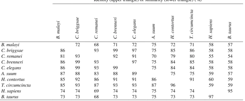
Table 1. Conservation of the amino acid sequence alignment shown in Figure 2. The values are the identity (upper triangle) or the similarity (lower triangle) expressed as a percentage and they were calculated using Clustal X (Thompson et al., 1997).
Identity (upper triangle) or similarity (lower triangle) (%)
B
.
ma
la
yi
C
.
b
rig
g
sa
e
C
.
rema
n
ei
C
.
b
ren
n
eri
C
.
eleg
a
n
s
A
.
su
u
m
H.
co
n
to
rtu
s
T.
circu
mcin
cta
H.
s
a
p
ien
s
B
.
ta
u
ru
s
B. malayi 72 68 71 72 75 72 71 58 57
C. briggsae 86 93 99 97 75 85 86 58 58
C. remanei 81 93 92 91 70 79 80 55 54
C. brenneri 86 99 93 97 75 84 85 58 58
C. elegans 86 99 93 99 75 84 84 58 58
A. suum 87 88 83 88 89 75 75 59 57
H. contortus 85 92 86 91 91 86 91 60 59
T. circumcincta 85 93 87 93 93 87 96 59 59
H. sapiens 74 74 69 74 74 75 74 74 95
Figure 2. Alignment of nematode, bovine and human GDH amino acid sequences. The sequences were aligned with Clustal X (Thompson et al., 1997) using the Gonnet matrix, a gap opening penalty of 10 and a gap extension of 0.2. The letters below the alignment indicate the positions of residues involved in ligand binding in the crystal structures (1HWY, 1HWZ and 3MW9) of the bovine enzyme (S –-ketoglutarate or glutamate; G – GTP; 1 – NADa or NADP; 2 – NADb; symbols in brackets indicate
As mentioned previously, the cDNA sequence of GDH from nematodes is very similar to that of mammals (Muhamad et al., 2011). From Figure 2 it is clear that the amino acid sequences are not only highly conserved among nematodes (68-99% identity), but they are also very similar (54-60% identity) to the bovine and human sequences (Table 1).
An interesting feature of the sequence alignment shown in Figure 2 is that HcGDH appears to have a glycine (G240, using the numbering for the H. contortus sequence) that is absent from all of the other sequences shown (position 276 of the alignment). This residue forms part of a tripeptide that is relatively poorly conserved between two relatively extensive regions of highly conserved sequence.
Structural models
The human GDH and BtGDH differ very little (Figures 1 and 2) and that other considerations would determine which of them should be used as a basis for the construction of a theoretical model of the TcGDH and HcGDH monomers. Consequently, the availability of kinetic data and several different crystal structures persuaded us to employ BtGDH.
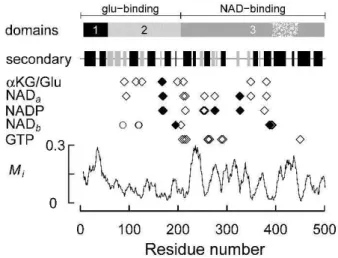
The essential properties of BtGDH are summarised in Figure 3. The glu-binding domain consists of a helical hairpin and a 3-layer(aba) sandwich (structural domains 1 and 2, respectively, in Figure 3) and the NAD-binding domain is a Rossmann fold with an internal region that forms the antenna domain (the stippled region in structural domain 3, Figure 3). Based on the PDBsum analysis of the BtGDH structures, residues involved in binding glutamate and -ketoglutarate are mostly located in domain 2, but residues in domain 3 are also involved. Similarly, NAD(P) (NADa and NADP)
binding in the cleft between the glu- and NAD-binding domains involves residues from both domains 2 and 3, although most of the ligands are located in domain 3. Of particular note is R211 in BtGDH that is involved in binding both glutamate/-ketoglutarate and NAD(P)(H). A second NAD-binding site (NADb) involves other residues in domains
2 and 3, as well as three residues in domain 2 of an adjacent subunit. Most of the residues involved in GTP binding are located in domain 3. All of these residues are located in relatively well conserved regions of the sequence and both the N-terminal portion of the NAD-binding domain and the antenna domain are relatively poorly conserved (Figure 3).
Of all the residues involved in binding NAD(P), -ketoglutarate, glutamate or GTP, only six in BtGDH (N168,
S170, H195, S276, S327, D388) differ from the corresponding residues in TcGDH (D, G, A, W, C, N) or HcGDH (D, G, S, W, C, N), as shown in Figures 2 and 3. Of
these, two (H195 ↔ A/S and D388 ↔ N) ligate NADb and will not be considered further, and another three are
relatively conservative substitutions (N168 ↔ D, S170 ↔ G, S327 ↔ C). The sixth difference (S276 ↔ W) is more
significant because a small polar residue (S) is replaced with a larger hydrophobic residue (W), but it is especially interesting as S276 in BtGDH (1HWZ) ligates the 2′ -phosphate of NADP(H).
Figure 3. Summary of the structure and ligand binding sites of BtGDH and sequence mutability (Mi) of selected GDHs.
The symbols (, , ○) indicate residues that are hydrogen bonded to the substrate, cofactor or effector. The residues at positions 87, 119 and 120, indicated by open circles (○), are located in another subunit. Those residues indicated by solid diamonds () differ between BtGDH and TcGDH or HcGDH. The three structural domains (1-3) were identified using CATH (http://www.cathdb.info (Cuff et al., 2011)) and the two functional domains were identified using SCOP [http://scop.mrc-lmb.cam.ac.uk/scop/index.html (Andreeva et al., 2008)]. The stippled region in domain 3 represents the
‘antenna’ domain. The secondary structure indicates the positions of -helices and -strands (black and grey rectangles, respectively).
Figure 4. Superimposition of BtGDH (1HWY in blue) and the model of TcGDH generated using SWISS-MODEL (in yellow).
Table 2. Quality of fit parameters for predicted structures obtained for the T. circumcincta and H. contortus sequences (AEO44571.1 and AAC19750.1, respectively) based on the BtGDH structure (1HWY). Structures were calculated using Phyre (Kelley & Sternberg, 2009), ESyPred3D (Lambert et al., 2002) and SWISS-MODEL (Kiefer et al., 2009) and the quality of fit parameters were calculated using PDBeFold (Krissinel, 2007).
Phyre ESyPred3D SWISS-MODEL
T. circumcincta
Q 0.5529 0.8863 0.9698
P 23.97 54.23 81.84
Z 15.34 22.26 27.34
RMSD (Å) 2.001 0.551 0.364
Number of residues aligned 446 480 497
Identical residues aligned (%) 58.74 65 63.78
H. contortus
Q 0.5542 0.8757 0.9753
P 22.92 54.58 77.77
Z 14.78 22.33 26.72
RMSD (Å) 2.016 0.404 0.251
Number of residues aligned 448 474 497
While all these structures were similar, those generated using SWISS-MODEL appeared to be the best (Table 2) because they had the smallest root mean square deviation (RMSD) from 1HWY, and the largest quality measures (Q, P and Z). For comparison, a theoretical structure of the TcGDH monomer based on the P. falciparum structure (2BMA) was built using SWISS-MODEL and this was clearly inferior (Q = 0.7618, P = 57.99, Z = 24.05, RMSD = 0.513 Å) to those based on the BtGDH structure (Table 2). Moreover, the models based on BtGDH yielded Ramachandran plots that were very similar to that of the bovine enzyme (Figure 5, A and B). This indicates that the backbone dihedral angles had not been greatly distorted in the model, which can be confirmed by inspection of Figure 4 in which the backbones of the aligned BtGDH and model T. circumcincta structures are supermposed. Naturally, there is rather more variation in the positions of the side chains, but those residues involved in ligand binding are largely in very similar positions even where the residues differ (Figure 6). The most significant exception to this generalisation is S276, which is located adjacent to the adenine ring of NAD(P)+ and provides three H-bonds to the 2′-phosphate of NADP+ in BtGDH (1HWZ). In the model TcGDH and HcGDH structures S276 is replaced with a tryptophan, the sidechain of which lies outside the electron density of the serine sidechain (Figure 6). This
difference involves the replacement of the polar sidechain with a hydrophobic sidechain that is significantly larger, resulting in the partial occlusion of the NAD(P)-binding site and removal of at least one potential H-bond to the 2′ -phosphate group.
The glycine residue that appears to be specific to HcGDH (G240) forms part of a loop that is distinct from the corresponding helix fragments found in TcGDH and BtGDH (indicated by G in Figure 7). This loop is exposed at the surface of the structure and is adjacent to the -ketoglutarate/glu-binding site (indicated by S in Figure 7) and is located in the vicinity of three ligands (Figure 2). In Figure 7 it is clear that the -ketoglutarate is parallel to a helix that is essentially identical in BtGDH and the model structures of TcGDH and HcGDH. The other side of the binding site is formed by loops that appear to protude further into the site in the model TcGDH and HcGDH structures than in the BtGDH structure (indicated by the three asterisks in Figure 7). The residues directly involved in binding -ketoglutarate or glutamate, based on the PDBsum analysis, are essentially identical in all three structures except for N168, which is replaced with an aspartate in both nematode sequences (Figure 2). It is clear from Figure 8 that the orientation of the sidechain differs between the BtGDH and the model structures and that this results in the loss of an H-bond.
Figure 5. Ramachandran plots for BtGDH (A, 1HWY) and SWISS-MODEL derived TcGDH (B) structures. The contours are
Figure 6. Structural alignment showing the NAD(P)-binding residues of BtGDH (1HWY, in blue) and the corresponding residues in the model of TcGDH generated using SWISS-MODEL (yellow). Also shown is the NAD+ bound in the BtGDH structure. Note that the tryptophan corresponding to S276 in BtGDH is very close to the 2′-OH of NAD+ and that the
tryptophan side-chain lies outside the electron density of S276.
Figure 8. Details of the ligands forming the -ketoglutarate/glu-binding site in BtGDH (1HWY, blue) and in the model TcGDH (yellow).
Structural implications
Both the structural and kinetic properties of TcGDH and HcGDH are more like those of the bovine enzyme than those of the P. falciparum GDH. The ligand binding residues are similar and the backbone of the monomer is not distorted relative to the bovine enzyme. However, six residues involved in ligating substrates or cofactors differ between BtGDH and TcGDH (Figures 2 and 3). The Km for NAD(P)H is about 0.02 mM for the bovine enzyme (Rife & Cleland, 1980; McCarthy & Tipton, 1985) and only very slightly larger (0.03-0.05 mM) for rGDH and HcGDH (Rhodes & Ferguson, 1973; Umair et al., 2011).
The most significant kinetic difference between BtGDH and the TcGDH is the Km for NADP+ (Table 3). This has been reported to be 0.028 mM for the bovine enzyme (Rife & Cleland, 1980; McCarthy & Tipton, 1985) and 1 mM for TcGDH (Umair et al., 2011). The 2′-phosphate group of NADP+ is ligated by S276 in the bovine enzyme, but this is replaced by a tryptophan in rTcGDH (Figure 1A). This has two effects: (a) the terminal hydroxyl group is absent from the side chain, removing one possible H-bond and (b) there is less space available for the NADP+. It might be speculated that the steric constraint is especially likely to provide some rationalisation for the lower affinity of TcGDH for NADP+, which might also explain the small decrease in the affinity of
TcGDH for NAD(H). Some limited support for this speculation is provided by a report (Yoon et al., 2002) with human GDH mutants of a residue equivalent to E275, in each of which the Km(NADH) was increased about 10-fold. Unfortunately, the similarity between TcGDH and HcGDH means that only one ligand binding residue is different (H195 is replaced with S rather than A). This does not explain the lack of activity of HcGDH with NADP(H) (Rhodes & Ferguson, 1973).
Table 3.
Values of the K
ms reported for BtGDH, r TcGDH, TcGDH and HcGDH.Km (mM)
Bos taurusa T. circumcincta H. contortusd
rTcGDHb TcGDHc
-ketoglutarate 0.36-2.4 0.07-0.1 0.06-0.09 0.74
glutamate 0.74-3 0.35-0.45 0.15-0.7 3.3
NAD+ 0.076-0.22 0.7 0.7 0.31
NADP+ 0.028 1 3 —
NADH 0.02 0.05 0.025 0.033
NADPH 0.02-0.022 0.03 0.10 —
NH3 6.5-50 37-40 18 42
a (Frieden, 1959; Engel & Dalziel, 1969; Rife & Cleland, 1980; McCarthy & Tipton, 1985) b (Umair et al., 2011) c (Muhamad et al., 2011) d (Rhodes & Ferguson, 1973)
Conclusion
It has been suggested that GDH is a potential target for anthelmintics (Umair et al., 2011) because there are
“significant differences” in amino acid sequence between the
host and the parasite enzyme. The structural models of TcGDH and HcGDH described here were based on the bovine enzyme, which differs in only 7 positions from the sequence of the sheep enzyme. We infer from this conservation of sequence (Figure 2) and, consequently, of
model structure (Figure 4) that the “significant differences”
to which Umair et al. (2011) refer, but do not define, are unlikely to render the parasite GDHs sufficiently different from that of the host to make them viable therapeutic targets. However, the replacement of S276 with a tryptophan in the nematode GDHs (Figure 6) provides a plausible explanation of their reduced affinity for NADP+ (Table 3) and this may have significant implications for amino acid metabolism.
References
Andreeva A, Howorth D, Chandonia J-M, Brenner SE, Hubbard TJP, Chothia C, Murzin AG. 2008. Data growth and its impact on the SCOP database: new developments. Nucleic Acids Res.,
36(Database issue):D419-D425.
Brown S, Moody AJ, Mitchell R, Rich PR. 1993. Binuclear centre structure of terminal protonmotive oxidases. FEBS Lett.,
316(3):216-223.
Cuff AL, Sillitoe I, Lewis T, Clegg AB, Rentzsch R, Furnham N, Pellegrini-Calance M, Jones D, Thornton J, Orengo CA. 2011. Extending CATH: increasing coverage of the protein structure universe and linking structure with function. Nucleic Acids Res.,
39(Database issue):D420-D426.
Engel PC, Dalziel K. 1969. Kinetic studies of glutamate dehydrogenase with glutamate and norvaline as substrates. Coenzyme activation and negative homotropic interactions in
allosteric enzymes. Biochem. J., 115(4):621-631.
Felsenstein J. 1985. Confidence limits on phylogenies: an approach
using the bootstrap. Evolution, 39(4):783-793.
Felsenstein J, Kishino H. 1993. Is there something wrong with the bootstrap on phylogenies? A reply to Hillis and Bull. Syst.
Biol., 42(2):193-200.
Frieden C. 1959. Glutamic dehydrogenase. III. The order of substrate addition in the enzymatic reaction. J. Biol. Chem.,
234(11):2891-2896.
Frigerio F, Casimir M, Carobbio S, Maechler P. 2008. Tissue specificity of mitochondrial glutamate pathways and the control of metabolic homeostasis. Biochim. Biophys. Acta,
1777(7-8):965-972.
Gore MG. 1981. L-glutamic acid dehydrogenase. Int. J. Biochem.,
13(8):879-886.
Green L, Simcock DC, Muhamad N, Page R, Patchett ML, Simpson HV, Brown S. 2004. Molecular properties of two
dehydrogenases active in L3 Ostertagia circumcincta. N. Z. J.
Zool., 31(4):376-377.
Hillis DM, Bull JJ. 1993. An empirical test of bootstrapping as a method for assessing confidence in phylogenetic analysis. Syst.
Biol., 42(2):182-192.
Hudson RC, Daniel RM. 1993. L-Glutamate dehydrogenases: distribution, properties and mechanism. Comp. Biochem.
Physiol., 106B(4):767-792.
Jones DT, Taylor WR, Thornton JM. 1992. The rapid generation of mutation data matrices from protein sequences. CABIOS,
8(3):275-282.
Kelley LA, Sternberg MJE. 2009. Protein stucture prediction on the web: a case study using the Phyre server. Nature Protocols,
4(3):363-371.
Kiefer F, Arnold K, Künzli M, Bordoli L, Schwede T. 2009. The SWISS-MODEL repository and associated resources. Nucleic
Acids Res., 37(Database issue):D387-D392.
Kim DW, Eum WS, Jang SH, Yoon CS, Kim YH, Choi SH, Choi HS, Kim SY, Kwon HY, Kang JH, Kwon O-S, Cho S-W, Park J, Choi SY. 2003. Molecular gene cloning, expression, and characterization of bovine brain glutamate dehydrogenase. J.
Krissinel E. 2007. On the relationship between sequence and
structure similarities in proteomics. Bioinform., 23(6):717-723.
Lambert C, Léonard N, De Bolle X, Depiereux E. 2002. ESyPred3D: prediction of proteins 3D structures. Bioinform.,
18(9):1250-1256.
Laskowski RA. 2009. PDBsum new things. Nucleic Acids Res.,
37(Database issue):D355-D359.
Lovell SC, Davis IW, Arendall WB, III, de Bakker PIW, Word JM, Prisant MG, Richardson JS, Richardson DC. 2003. Structure validation by Ca geometry: f, y and Cb deviation. Proteins,
50(3):437-450.
Marzluf GA. 1981. Regulation of nitrogen metabolism and gene
expression in fungi. Microbiol. Revs., 45(3):437-461.
Mayashita Y, Good AG. 2008. Glutamate deamination by glutamate dehydrogenase plays a central role in amino acid catabolism in
plants. Plant Signal. Behavior, 3(10):842-843.
McCarthy AD, Tipton KF. 1985. Ox glutamate dehydrogenase. Comparison of the kinetic properties of native and proteolysed
preparations. Biochem. J., 230(1):95-99.
Muhamad N, Simcock DC, Pedley KC, Simpson HV, Brown S.
2011. The kinetics of glutamate dehydrogenase of Teladorsagia
circumcincta and the lifestyle of the parasite. Comp. Biochem.
Physiol., 159B(2):71-77.
Newsholme P, Lima MMR, Procopio J, Pithon-Curi TC, Doi SQ, Bazotte RB, Curi R. 2003. Glutamine and glutamate as vital
metabolites. Braz. J. Med. Biol. Res., 36(2):153-163.
Owen OE, Kalhan SC, Hanson RW. 2002. The key role of anaplerosis and cataplerosis for citric acid cycle function. J.
Biol. Chem., 277(34):30409-30412.
Peterson PE, Smith TJ. 1999. The structure of bovine glutamate dehydrogenase provides insights into the mechanism of
allostery. Structure, 7(7):769-782.
Plaitakis A, Zaganas I. 2001. Regulation of human glutamate dehydrogenases: implications for glutamate, ammonia and
energy metabolism in brain. J. Neurosci. Res., 66(5):899-908.
Rhodes MB, Ferguson DL. 1973. Haemonchus contortus: enzymes.
III. Glutamate dehydrogenase. Exptl. Parasitol., 34(1):100-110.
Rife JE, Cleland WW. 1980. Kinetic mechanism of glutamate
dehydrogenase. Biochem., 19(11):2321-2328.
Skuce PJ, Stewart EM, Smith WD, Knox DP. 1999. Cloning and characterization of glutamate dehydrogenase (GDH) from the
gut of Haemonchus contortus. Parasitol., 118(3):297-304.
Soltis PS, Soltis DE. 2003. Applying the bootstrap in phylogeny
reconstruction. Statist. Sci., 18(2):256-267.
Storm J, Perner J, Aparicio I, Patzewitz E-M, Olszewski K, Llinas
M, Engel PC, Müller S. 2011. Plasmodium falciparum
glutamate dehydrogenase a is dispensable and not a drug target during erythrocytic development. Malaria J., 10:193.
Tamura K, Peterson D, Peterson N, Stecher G, Nei M, Kumar S. 2011. MEGA5: molecular evolutionary genetics analysis using maximum likelihood, evolutionary distance, and maximum
parsimony methods. Mol. Biol. Evol., 28(10):2731-2739.
Thompson JD, Gibson TJ, Plewniak F, Jeanmougin F, Higgins DG. 1997. The ClustalX windows interface: flexible strategies for multiple sequence alignment aided by quality analysis tools.
Nucleic Acids Res., 25(24):4876-4882.
Umair S, Knight JS, Patchett ML, Bland RJ, Simpson HV. 2011.
Molecular and biochemical characterisation of a Teladorsagia
circumcincta glutamate dehydrogenase. Exptl. Parasitol.,
129(3):240-246.
van Wyk JA, Malan FS, Randles JL. 1997. How long before resistance makes it impossible to control some field strains of Haemonchus contortus in South Africa with any of the modern
anthelmintics? Vet. Parasitol., 70(1-3):111-122.
Waller PJ, Echevarria F, Eddi C, Maciel S, Nari A, Hansen JW. 1996. The prevalence of anthelmintic resistance in nematode parasites of sheep in Southern Latin America: General overview.
Vet. Parasitol., 62(3-4):181-187.
Werner C, Stubbs MT, Krauth-Siegel RL, Klebe G. 2005. The
crystal structure of Plasmodium falciparum glutamate
dehydrogenase, a putative target for novel antimalarial drugs. J.
Mol. Biol., 349(3):597-607.
Yoon H-Y, Cho EH, Kwon HY, Choi SY, Cho S-W. 2002. Importance of glutamate 279 for the coenzyme binding of
human glutamate dehydrogenase. J. Biol. Chem.,


