Arch. Biol. Sci., Belgrade, 65 (3), 955-962, 2013 DOI:10.2298/ABS1303955G
955
MOLDS IN MUSEUM ENVIRONMENTS: BIODETERIORATION OF ART PHOTOGRAPHS AND WOODEN SCULPTURES
MILICA LJALJEVIĆ GRBIĆ*1, M. STUPAR1, JELENA VUKOJEVIĆ1, IVANA MARIČIĆ2 and NATAŠA BUNGUR1
1 University of Belgrade, Faculty of Biology, Institute of Botany and Botanical Garden “Jevremovac”, 11000 Belgrade, Serbia 2 Museum of Contemporary Art, Ušće 10, Blok 15, 11070 Novi Beograd, Belgrade, Serbia
Abstract - Pieces of art stored in museum depots and display rooms are subject to fungal colonization that leads to bio-deterioration processes. Deteriorated wooden sculptures and art photographs temporarily stored in the quarantine room of the Cultural Center of Belgrade were subject to mycological analyses. Twelve fungal species were identiied on the wooden substratum and ive species were detected on photograph surfaces. Trichoderma viride, Chaetomium globosum and Alternaria sp. were the fungi with proven cellulolytic activity detected on the examined cellulose substrata. Indoor air mycobiota were estimated to 210.09 ± 8.06 CFU m-3, and the conidia of fungus Aspergillus niger were the dominant fungal
propagules in the air of the examined room.
Key words: art objects, biodeterioration, cellulose substrata, fungi, indoor air, museum environment.
INTRODUCTION
he fungal colonization of pieces of art presented in display rooms of museums, galleries or stored in de-pots is nowadays a signiicant problem for cultural heritage conservators (Sterlinger, 2010). Pieces of art are made of all types of organic and inorganic materials. It is well known that fungi are capable of colonizing, altering and degrading all kinds of materials, and pieces of art are no exception. here are many reports concerning the fungal deteriora-tion of art objects such as paintings (Vukojević and Ljaljević Grbić, 2011), stone monuments and mason-ry (Ljaljević Grbić et al., 2010), wooden sculptures (Fazio et al., 2011), paper and parchment materials (Cappiteli and Sorlini, 2005), cinematographic ilms (Abrusci et al., 2005), textiles (Szostak-Kotowa, 2004) etc. Viable fungi isolated from pieces of art presented in an indoor environment in most cases come from
light (Saiz-Jimenez, 1993). A fungal infestation on a piece of art contains fungal structures and metabolic products, such as enzymes, citric acid cycle products, secondary metabolites, pigments, odors, etc. Mate-rials colonized by fungi usually undergo changes in their chemical and physical characteristics (Florian, 2002). Fungal infestation on pieces of art leads to bi-odeterioration and must not be neglected due to the increasing aesthetic value of art objects as well as the impact on health of conservators.
MATERIALS AND METHODS
Case report
he collection consisted of deteriorated art photo-graphs (Fig. 1B), 24 pieces of two artworks “Sky” and “Earth” made by the eminent photographer Aleksandar Rafajlović (part of the collection of the Museum of Contemporary Art in Belgrade), wood-en sculptures (Fig. 1A) made by the artist Marko Crnobrnja as well as diferent textile and terracotta artworks. he deteriorated wooden sculptures and
photographs were subjected to mycological analyses. he artworks were sent to Istanbul (Turkey) for an exhibition, “Belgrade’s experience: he October Sa-lon”, as reputable pieces of contemporary art in Ser-bia. During the process of preparing the exhibition Istanbul was devastated by a lood and seawater seri-ously impaired the condition of the photographs and sculptures and they lost their artistic value. he sam-pling took place in a quarantine room of the Cultural Center of Belgrade (CCB) where the deteriorated art objects were temporarily stored.
Sampling of aeromycobiota
Sampling of aeromycobiota was carried out in a tem-porary quarantine room of the CCB by the passive sedimentation method described by Omeliansky (1940). Petri dishes containing malt extract agar (MEA) were placed open at approximately 2 m above the loor and were exposed for 30 min. he antibiotic streptomycin was added during the preparation of the medium in order to suppress bacterial growth. Ater 7 days of cultivation at 25˚C the fungal
MOLDS IN MUSEUM ENVIRONMENTS 957
nies were counted. he CFU (colony-forming unit) number per cubic meter of air (CFU m-3) was esti-mated according to Omelyansky’s formula:
N=5a x 104(bt)-1
where N is fungal CFU m-3, a is the number of fun-gal colonies per Petri dish, b is the Petri dish surface (cm2), t is the exposure time (min). Relative fungal distribution was conducted according to Smith (1980) i.e. the number of colonies of genera or species/total number of colonies of all genera or species x 100.
Sampling of surface mycobiota
According to the surfaces, the examined samples were classiied as photographic paper and wooden samples. Sampling was performed from 2 cm2 of each photograph and each wooden sculpture with sterile cotton swabs.
Isolation and identiication of fungi
he swabs were immersed and homogenized in a sterile physiological solution and serial dilutions were made. Each dilution was inoculated (0.1 ml) on MEA with streptomycin added. Ater 7 days of cultivation at 25˚C, identiication of the fungi was performed. Cultural and micromorphological characteristics of fungal colonies were observed and identiication was performed using identiication keys (Ainsworth et al., 1973; Arx, 1974; Ellis and Ellis, 1997; Pitt, 1979; Raper, and Fennel 1965; Samson et al., 2004).
RESULTS
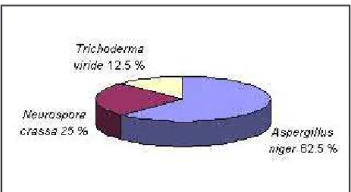
he fungal concentration of air in the quarantine room of the CCB was estimated at 210.09 ± 8.06 CFU m-3. he prevailing fungal species documented in the air was Aspergillus niger Tiegh (62.5%), followed by Neurospora crassa Shear & B.O. Dodge (25%) and Trichoderma viride Pers. (12.5%) (Fig 2).
he examined objects in quarantine room of the CCB showed clear signs of biodeterioration. Super-icial colonies of fungi were clearly visible and
abun-dant on the wooden sculptures and boxes (Fig 3A, B, C). he lightening of original dyes and discoloration phenomena occurred on some parts of the photo-graphs (Fig. 3D).
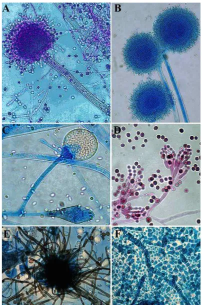
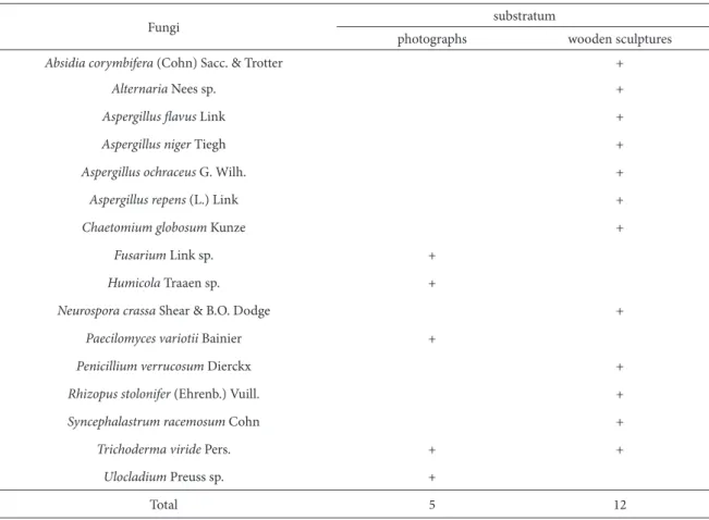
A total of 16 fungal species were identiied from all analyzed samples with 12 taxa identiied on the wooden substratum, followed by 5 taxa identiied on photographs (Table 1, Fig. 4). Fungi documented on the wooden substratum belonged to the genera Absidia, Alternaria, Aspergillus, Chaetomium, Neu-rospora, Penicillium, Rhizopus, Syncephalastrum and Trichoderma (Fig 4.), while Fusarium, Humicola, Paecilomyces, Trichoderma and Ulocladium species were isolated from the photographs (Fig. 4).
he most abundant group of fungi, regardless of the substratum, was Hyphomycetes with 11 taxa. Absidia corymbifera (Cohn) Sacc. & Trotter, Rhizo-pus stolonifer (Ehrenb.) Vuill. and Syncephalastrum racemosum Cohn were Zygomycetes identiied on wooden substratum. Chaetomium globosum Kunze and Neurospora crassa were the Ascomycetes identi-ied on the wooden substratum. During the cultiva-tion on MEA, the development of Chrysonilia crassa (Shear & B.O. Dodge) Arx, an anamorphic state of N. crassa, was recorded.
DISCUSSION
In this investigation, air sampling was done by passive sedimentation, and microbial prevalence was deter-mined using Omeliansky’s formula. Although there
are no data available to correlate Omeliansky’s meth-od with other active sampling methmeth-ods, passive sedi-mentation was used only to compare obtained results of indoor mycobiota concentration in the quarantine room of the CCB with international standards of in-door air quality. Omeliansky’s method is frequently reported for the estimation of fungal prevalence in
indoor air. Borrego et al. (2010) used Omeliansky’s method to determine fungal prevalence inside the building of the Photographic Library of the National Archive of the Republic of Cuba and of the Histori-cal Archive of the Museum of La Plata. Bogomolova and Kirtsideli (2009) estimated fungal prevalence in four stations of the St. Petersburg railway
MOLDS IN MUSEUM ENVIRONMENTS 959
ground system using this method. According to the recommendation of the World Health Organization (WHO) from 1990, the fungal density in an indoor environment should be lower than 500 CFU m-3. he obtained value (210.09 ± 8.06 CFU m-3), which is only approximate, is below WHO recommenda-tions. However, due to fungal ability to cause the bio-deterioration of art objects deposited in depots and have a negative impact on human health, this value
should not be neglected. Aspergillus niger was found to be the dominant air-borne fungus in the air of the CCB quarantine room. his species produces small, globose or subglobose conidia up to 5 μm in diam-eter which are easily dispersed through the air and settle on diferent surfaces (Florian, 2002). A. niger is a common contaminant on various substrates and due to production of the toxic secondary metabolites naphtho-γ-pyrones and malformins (Samson et al.,
Fig. 4. Fungi isolated from wooden sculptures and art photographs in temporary quarantine room of the Cultural Center of Belgrade:
A. Aspergillus lavus, conidiogenous apparatus B. Aspergillus ochraceus, conidiogenous apparatus C. Absidia corymbifera,
2004) as well as the allergens Asp n 14 and Asp n 18 (Knutsen et al., 2011), the presence of this fungus in indoor environments is very important.
he signiicantly larger number of fungal taxa documented on wooden and photographic layer sur-faces than in the air suggested that the initial colo-nization of fungi occurred while the exhibition was stored in Turkey. Flooding damaged the art objects and increased the water activity (aw) which made these surfaces more suitable for fungal colonization. Abundant supericial fungal colonies were found on the surfaces of wooden sculptures during in situ servation. Wooden material in museum heritage ob-jects rarely supports active fungal growth unless the surface has been wet for a period of time (Florian, 2002). Opportunistic species that utilize available sugars, hemicellulose, proteins and amino acids are
the primary colonizers of wooden objects in art col-lections. Most of the wood-degrading fungi that di-gest cellulose and lignin require a long period of wet conditions in order to colonize successfully wooden substrata (Florian, 2002). Some wood-degrading fungi were found on the wooden sculptures inside the quarantine room of the CCB. Chaetomium glo-bosum is a sot-rot fungi capable of degrading cellu-lose in the S2 layer of the secondary cell wall of plants (Popescu et al., 2011). Hyphomycetes Trichoderma viride and Alternaria sp. produce a variety of en-zymes capable of hydrolyzing cellulose to glucose (Shaique et al., 2009; Sohail et al., 2011).
Biodeterioration is a common problem in pho-tograph collections, but only a few studies have been carried out on this topic. Photographs have a stratigraphic structure composed of three generic
Table 1. Fungi isolated from deteriorated art photograph surface and wooden sculptures in quarantine room of CCB.
Fungi substratum
photographs wooden sculptures
Absidia corymbifera (Cohn) Sacc. & Trotter +
Alternaria Nees sp. +
Aspergillus lavus Link +
Aspergillus niger Tiegh +
Aspergillus ochraceus G. Wilh. +
Aspergillus repens (L.) Link +
Chaetomium globosum Kunze +
Fusarium Link sp. +
Humicola Traaen sp. +
Neurospora crassa Shear & B.O. Dodge +
Paecilomyces variotii Bainier +
Penicillium verrucosum Dierckx +
Rhizopus stolonifer (Ehrenb.) Vuill. +
Syncephalastrum racemosum Cohn +
Trichoderma viride Pers. + +
Ulocladium Preusssp. +
MOLDS IN MUSEUM ENVIRONMENTS 961
components: paper support, image forming materi-als, and gelatin as a binder. he most biosusceptible photographic materials are the gelatin and the paper, because they are organic and hygroscopic (Lourenço and Sampaio, 2009). Some external “materials”, such as dust, grease from ingerprints and glues, are im-portant factors in encouraging microbial develop-ment on photographs (Eaton, 1985). Many ilamen-tous fungi exhibit cellulolytic and proteolytic activ-ity and they are capable of degrading the paper sup-port and gelatin binder of photographs. In the case presented here, it can be concluded that infestation of photographs occurred ater the lood in Turkey. he microfungi identiied on the photographic sur-faces were the causative agents of biodeterioration. According to Borego et al. (2010), the main cause of the biodeterioration of the photograph collec-tions in the Photographic Library of the National Archive of the Republic of Cuba and in the Histori-cal Archive of the Museum La Plata were the yeasts and ilamentous fungi of Aspergillus and Penicillium genera.
It can be concluded that the irreversible changes in the analyzed artworks was caused by fungal infes-tation. Due to the total devastation of the examined artworks, the collections were destroyed because of their complete loss of artistic value. In addition, contaminated artwork collections could be potential source for the spread of infestation to surrounding artworks and environment.
Acknowledgments - his work is a part of research realized
within the project No.173032 inancially supported by the Ministry of Education and Science of the Republic of Serbia. he authors are very grateful to Biljana Šipetić, MSc, for her precious collaboration, constructive advice and suggestions.
REFERENCES
Abrusci, C., Martin-Gonzales, A., Del Amo, A., Catalina, F., Col-lado, J., and G. Platas (2005). Isolation and identiication of bacteria and fungi from cinematographic ilms. Int.
Bio-deter. Biodegr.56, 58-68.
Ainsworth, G.C., Sparrow, F.K., and S.A Sussman (1973). he
Fungi, Volume IVA, Taxonomic Review with Keys:
Asco-mycetes and Fungi Imperfecti. Academic Press, New York and London.
Arx, J.A. von (1974). he Genera of Fungi. Sporulating in Pure
Culture. Vaduz: J. Cramer.
Bogomolova, E., and I. Kirtsideli (2009). Airborne fungi in four
stations of the St. Petersburg Underground railway system.
Int. Biodeter. Biodegr.63, 156-160.
Borrego, S., Guiamet, P., Gómez de Saravia, S., Batistini, P., Gar-cia, M., Lavin, P., and I. Perdomo (2010). he quality of air at archives and the biodeterioration of photographs. Int.
Biodeter. Biodegr.64, 139-145.
Bush, R.K., and J.M. Portnoy (2001). he role and abatement of fungal allergens in allergic diseases. J. Allergy. Clin. Immu-nol.107, 430-440.
Cappitelli, F., and C. Sorlini (2005). From papyrus to compact disc: the microbial deterioration of documentary heritage.
Cr. Rev. Microbiol.31, 1-10.
Eaton, G. (1985). Conservation of Photographs. Kodak
Publica-tion No. F-40. Eastman Kodak Company, Rochester, New York.
Ellis, M.B., and P.J. Ellis (1997). Microfungi on Land Plants. An
Identiication Handbook. he Richmond Publishing Co. Ltd.
Fazio, A.T., Papinutti, L., Gómez, B.A., Parera, S.D., Rodríguez Romero, A., Siracusano, A.G., and M.S. Maier (2010). Fun-gal deterioration of a Jesuit South American polychrome wood sculpture. Int. Biodeter. Biodegr. 64, 694-701.
Florian, M-L.E. (2002). Fungal facts: Solving fungal problems
in heritage collections. Archetype Publications LtD. Lon-don.
Knutsen, A.P.,Bush, R.K, Demain, J.G.,Denning,D.W.,Dixit,A.,
Fairs Greenberger,P.A.,Kariuki, B.,Kita, K., Kurup, V.P.,
Moss, R.B., Niven, R.M., Pashley, C.H., Slavin, R.G., Vijay, H.M., and A.J. Wardlaw (2011). Fungi and allergic lower respiratory tract diseases. Clin. Rev. Allerg. Immu. 129, 280-291.
Lourenço, M.J.L., and J.P. Sampaio (2009). Microbial
deteriora-tion of gelatin emulsion photographs: Diferences of sus-ceptibility between black and white and colour materials.
Int. Biodeter. Biodegr.63, 496-502.
Ljaljević Grbić, M., Vukojević, J., and M. Stupar(2008). Fungal colonization of air conditioning systems. Arch. Biol. Sci. 60, 201-206.
Niesler, A., Gόrny, R.L., Wlazlo, A., Ludzeń-Izbińska, B.,
Ławniczek-Wałczyk, A., Gołoit-Szymczak, M., Meres, Z.,
Kasznia-Kocot, J., Harkawy, A., Lis, D.O., and E. Anczyk (2000). Microbial contamination of storerooms at the Auschwitz-Birkenau Museum. Aerobiologia. 26, 125-133.
Omelyansky, V.L. (1940). Manual in Microbiology. USSR
Acad-emy of Sciences, Moscow, Leningrad.
Pitt, J.I. (1979). he Genus Penicillium and Its Teleomorphic
State Eupenicillium and Talaromyces, Academic Press.
Popescu, C-M.,Tibirna, C.M., and A. Manoliu (2011).
Micro-scopic study of lime wood decayed by Chaetomium glo-bosum. Cellulose. Chem. Technol. 45, 565-569.
Raper, B.K., and D.I Fennel (1965). he Genus Aspergillus, he
Williams and Wilkins Company, Baltimore.
Saiz-Jimenez, C. (1993). Deposition of airborne organic pollut-ants on historic buildings. Atmos. Environ.27, 77-85.
Samson, R.A.,Hoekstra, E.S., and J.C. Frisvad (2004).
Introduc-tion to food- and airborne fungi. Ponse & Looyen, Wagen-ingen, he Netherlands.
Shaique, S., Bajwa, R., and S. Shaique (2009): Cellulase
biosyn-thesis by selected Trichoderma species. Pak. J. Bot.41, 907-916.
Smith, G. (1980). Ecology and Field Biology, second ed. Harper
& Row, New York. 835.
Sohail, M., Ahmad, A., and S.A. Khan (2011). Production of
cel-lulases from Alternaria sp. MS28 and their partial charac-terization. Pak. J. Bot.43, 3001-3006.
Sterlinger, K. (2010). Fungi: heir role in deterioration of
cul-tural heritage. Fungal Biol. Rev. 24, 47-55.
Szostak Kotowa, J. (2004). Biodeterioration of textiles. Int.
Biode-ter. Biodegr.53, 165-170.
Vukojević, J. and M. Ljaljević Grbić (2010). Moulds on paintings
in Serbian ine art museums. Afr. J. Microbiol. Res. 13, 1453-1456.


