ORIGIN
AL RESEAR
CH
Corresponding address: Elaine Paulin – Department of Physical Therapy, Universidade do Estado de Santa Catarina (Udesc), Centro de Ciências da Saúde e do Esporte (Ceid) – Rua Pascoal Simone, 358, Coqueiros – Florianópolis (SC), Brazil. – Zip Code: 88080-350. – E-mail: [email protected] – Phone number: (48) 3664-8602 – Finance source: self-funding – Conlict of interest: nothing to declare – Presentation: Dec. 1, 2016 – Accepted for publication: Aug. 12, 2017 – Approved by the Ethics Committee, Certiicate of Appreciation: 08857612.2.0000.0118 (Udesc).
1Physical therapist, Master’s in Physical Therapy by Universidade do Estado de Santa Catarina (Udesc), Florianópolis (SC), Brazil. 2Undergraduate student in Physical Therapy, Universidade do Estado de Santa Catarina (Udesc), Florianópolis (SC), Brazil.
3PhD in Sports Sciences by Universidade do Porto, Portugal. Professor of the Graduate Program in the Human Movement Sciences at
Universidade do Estado de Santa Catarina (Udesc), Florianópolis (SC), Brazil.
4PhD in Sciences by Faculdade de Medicina da Universidade de São Paulo – FMUSP – São Paulo (SP) Brazil; Professor of the Undergraduate
and Graduate Programs in Physical Therapy in Universidade do Estado de Santa Catarina (Udesc), Florianópolis (SC), Brazil.
This research was performed in the Laboratory of Respiratory Physical Therapy (Lair), from Universidade do Estado de Santa Catarina (Udesc). ABSTRACT | In the chronic obstructive pulmonary disease
(COPD), patients may have reduced diaphragmatic mobility and a series of compensations in the thoracic spine, the scapular and pelvic girdles. However, the relation between diaphragmatic mobility and postural changes in these individuals’ vertebral column and pelvis is not clear. The aim of this study was to verify if there is a relation between diaphragmatic mobility and spinal curvatures in patients with COPD and in apparently healthy individuals. Were evaluated 22 patients with COPD and 22 apparently healthy individuals. The evaluations consisted of: anthropometry, spirometry, diaphragmatic mobility and postural evaluation. Four postural alterations were analyzed: cervical lordosis, thoracic kyphosis, lumbar lordosis, pelvic position. There was no statistically signiicant diference between the groups, in relation to the variables age, body mass, stature and BMI, conirming that the groups were paired. There was no statistically signiicant diference in any of the variables related to spinal curvatures and pelvic position between the studied groups. In the COPD group, there was a correlation between diaphragmatic mobility and thoracic kyphosis (r=-0.543; p=0.009). Regarding the group of apparently healthy individuals, there was no correlation of mobility as the apparently healthy individuals presented the same 245
angles of curvature of the vertebral column and the same position of the pelvis. However, there was a relation between diaphragmatic mobility and the angle of the thoracic curvature in patients with COPD.
Keywords | Pulmonary Disease Chronic Obstructive; Diaphragm; Spine.
RESUMO | Na doença pulmonar obstrutiva crônica (DPOC), os pacientes podem apresentar redução da mobilidade diafragmática e uma série de compensações na coluna torácica, nas cinturas escapular e pélvica. No entanto, não está clara a relação da mobilidade diafragmática com alterações posturais na coluna vertebral e na pelve desses indivíduos. Objetivou-se veriicar se existe relação entre a mobilidade diafragmática com as curvaturas da coluna vertebral de pacientes com DPOC e em indivíduos aparentemente saudáveis. Foram avaliados 22 pacientes com DPOC e 22 indivíduos aparentemente saudáveis. As avaliações foram: antropometria, espirometria, mobilidade diafragmática e avaliação postural. Foram analisadas quatro alterações posturais: lordose cervical, cifose torácica, lordose lombar, posição pélvica. Não houve diferença estatisticamente signiicativa entre os grupos, em relação às variáveis idade, massa corporal, estatura e IMC, conirmando que os grupos foram pareados. Não houve
The relation between diaphragmatic mobility and
spinal curvatures in patients with chronic obstructive
pulmonary disease
Relação entre a mobilidade diafragmática e as curvaturas da coluna vertebral em pacientes
com doença pulmonar obstrutiva crônica
Relación entre la movilidad diafragmática y las curvaturas de la columna vertebral en los
pacientes con la enfermedad pulmonar obstructiva crónica
Márcia Aparecida Gonçalves1, Bruna Estima Leal1, Gabriella Da Cunha Viegas2, Mariana Nunes Lúcio2,
diferença estatisticamente signiicante em nenhuma das variáveis referentes às curvaturas da coluna vertebral e à posição da pelve entre os grupos estudados. No grupo DPOC houve correlação da mobilidade diafragmática com a cifose torácica (r=-0,543; p=0,009). Já em relação ao grupo de indivíduos aparentemente saudáveis, não houve correlação da mobilidade quanto os indivíduos aparentemente saudáveis apresentaram os mesmos ângulos de curvatura da coluna vertebral e a mesma posição da pelve. Contudo, os pacientes com DPOC apresentaram relação entre a mobilidade diafragmática e o ângulo da curvatura torácica.
Descritores | Doença Pulmonar Obstrutiva Crônica; Diafragma; Coluna Vertebral.
RESUMEN | En la enfermedad pulmonar obstructiva crónica (EPOC), los pacientes pueden presentar reducción de la movilidad diafragmática y muchas compensaciones en la columna torácica, en las cinturas escapular y pélvica. No obstante, no está clara la relación de la movilidad diafragmática con las alteraciones posturales en la columna vertebral y en la pelvis de esos individuos. Se tuvo el objetivo de ver si hay relación entre la movilidad diafragmática con las curvaturas de la columna vertebral de los pacientes con la EPOC y en
los individuos aparentemente saludables. Fueron evaluados 22 pacientes con la EPOC y 22 individuos aparentemente saludables. Las evaluaciones fueron: la antropometría, la espirometría, la movilidad diafragmática y la evaluación postural. Fueron analizadas cuatro alteraciones posturales: la lordosis cervical, la cifosis torácica, la lordosis lumbar, la posición pélvica. No hubo diferencia estadísticamente signiicativa entre los grupos, en relación a las variables edad, la masa corporal, la estatura y el IMC, conirmando que los grupos fueron pareados. No hubo diferencia estadísticamente signiicante en ninguna de las variables referentes a las curvaturas de la columna vertebral y a la posición de la pelvis entre los grupos estudiados. En el grupo EPOC hubo correlación de la movilidad diafragmática con la cifosis torácica (r = -0,543; p = 0,009). Ya en relación al grupo de los individuos aparentemente saludables, no hubo correlación de la movilidad cuanto a los individuos aparentemente saludables que presentaron los mismos ángulos de curvatura de la columna vertebral y la misma posición de la pelvis. No obstante, los pacientes con la EPOC presentaron relación entre la movilidad diafragmática y el ángulo de la curvatura torácica.
Descritores | Enfermedad Pulmonar Obstructiva Crónica; Diafragma; Coluna Vertebral
INTRODUCTION
Chronic Obstructive Pulmonary Disease (COPD) is the fourth leading cause of death in the world. It is a common, preventable and treatable disease characterized by persistent limitation of airlow, which is usually progressive, associated with an increase in the chronic inlammatory response in the airways and lungs to the exposure of noxious particles or gases1. here is evidence
that COPD causes losses in respiratory mechanics and posture2,3.
In COPD, respiratory mechanics is impaired by several factors, including changes in lung volumes and capacities that occur due to the pathophysiological processes of the disease such as
loss of elastic recoil, airlow obstruction, air trapping4
and pulmonary hyperinlation5. Studies have shown
that these mechanisms are related to the reduction of diaphragmatic mobility that occurs by reducing the tension-length relation in the apposition zone and in
the radius of curvature of the diaphragm muscle6.
Previous studies have already established that patients with COPD have reduced diaphragmatic
mobility due to their disease7-12. However, few
studies have investigated the impairment in postural
alignment2,3. Despite the scarcity of quantitative
descriptions of postural changes in patients with COPD in the literature, postural impairments seem to be more evident in clinical practice, especially in patients at more advanced stages of the disease3.
Some evidence suggests that the postural attitude of the hyperinlamed thorax can lead to a series of alterations in the cervical spine, thoracic spine, scapular and pelvic girdles2,3,13. In addition, another hypothesis
would be that the biomechanics of the thoracic cavity can inluence the overall body mechanics and any abnormalities of the thoracic cavity may result in alterations in the posture and balance of the whole
body14. It is still not clear how much a variable can
inluence the other, thus, it is necessary to conduct more studies.
and thoracic curvatures in this population. herefore, the objective of the study was to verify if there is a relationship between diaphragmatic mobility and spinal curvatures in patients with COPD and apparently healthy individuals.
METHODOLOGY
he present study is an analytical, cross-sectional and quantitative approach. he study was approved by the Human Research Ethics Committee of Universidade do Estado de Santa Catarina, Florianópolis (CAAE: 08857612.2.0000.0118). All individuals were informed about the research and signed a free consent form, as determined by Resolution 466/12 of the National Health Council.
A total of 44 subjects (21 men and 23 women) of both sexes participated in the study. hey were divided into two groups: group 1, consisted of 22 patients with COPD, aged 65.8 (±8.0) years and group 2, consisted of 22 apparently healthy individuals, with 63.7 (±5.2) years of age.
he COPD group was comprised of patients with a diagnosis of COPD, according to the classiication
of the Global Initiative for Chronic Obstructive Lung
Disease (GOLD)1, and who met the following
inclusion criteria: 1) clinical stability in the last month and at the beginning of the evaluation protocol; 2) patients who did not use oxygen supplementation; 3) non-existence of other associated respiratory or cardiovascular diseases; 4) patients without involvement in pulmonary rehabilitation programs in 6 months prior to the beginning of the present study; 5) patients who did not undergo recent spinal or lower limb surgeries and/or who did not have fractures in the previous 6 months.
As exclusion criteria, the authors adopted: 1) presence of exacerbations of the disease during the research; 2) clinical intercurrences of cardiorespiratory nature during the evaluations; 3) inability to perform any of the study evaluations (lack of understanding or collaboration) and; 4) patient withdrawal during the evaluation period.
he study included apparently healthy individuals
with normal spirometry (FEV1/FVC ≥0.7, FEV1 ≥80%
of predicted, FVC ≥80% of predicted), without any associated comorbidities and with age, weight, and BMI compatible with patients with COPD. Individuals who
were unable to perform any of the study evaluations (lack of understanding or collaboration) and/or who withdrew from the study during the evaluation process were excluded from this group.
Evaluated parameters
Anthropometry
For the anthropometric measurements, a previously calibrated scale was used to measure body mass and a stadiometer was used to measure height. Once the anthropometric values were obtained, the body mass index (BMI) was calculated by means of the equation:
body mass/stature2. hus, the individuals were classiied,
according to BMI, in low weight (≤18,5 kg/m2), normal
(18,5-24,9 kg/m2), overweight (25-29,9 kg/m2) and
obese (≥30 kg/m2)15. Patients were instructed to wear
light clothing, to remove their shoes and remain upright until the measured values were assessed.
Spirometry
he spirometry was performed to verify the pulmonary capacity of the study subjects using the
Easy One ndd Medical Technologies portable digital spirometer, previously calibrated according to the
methods and criteria recommended by the American
horacic Society and European Respiratory Society16.
he following parameters were measured: forced vital capacity (FVC), forced expiratory volume in the irst
second (FEV1) and FEV1/FVC ratio before and 15
minutes after the inhalation of bronchodilator (BD) salbutamol (400μg) in COPD patients. At least three acceptable maneuvers and two reproducible maneuvers were performed. he spirometric variables are expressed in absolute values and in percentage values of the predicted values of normality, as determined by
Pereira et al.17. he normal lung function test criteria
consist of FVC and FEV1 ≥80% of the predicted values
and FEV1/FVC ≥0.7.
Diaphragmatic mobility
and they were previously instructed to perform two sets of ten repetitions of diaphragmatic breathing with the objective of developing proprioception of the diaphragmatic movement and enable the evaluation of the maximum diaphragm amplitude during the X-ray examination.
After the diaphragmatic breathing training, the subjects performed two slow vital capacity (SVC)
maneuvers using a Wright Respirometer Brit.® spirometer
Pat. 765206 – UK. he irst maneuver was close to the total lung capacity (TLC) and almost to the residual volume (RV); and the second, starting from RV and almost to the TLC. he highest value was recorded for comparison with the value measured during the diaphragmatic mobility test to verify if the individuals performed the same respiratory efort (inspiratory and expiratory) before and during the assessment of diaphragmatic mobility.
he maximum inspiratory and expiration images were recorded in the same ilm. he measurement of diaphragmatic mobility was determined by the method of distance (MDdist)18.
Postural evaluation
he Postural Analysis Software (SAPO) was used in an electronic medium and validated by Ferreira et al.19. he male subjects were instructed to wear shorts for the photographs and the female, shorts and a top. Initially, anatomical points were identiied, through palpatory anatomy, in the following regions of the body:
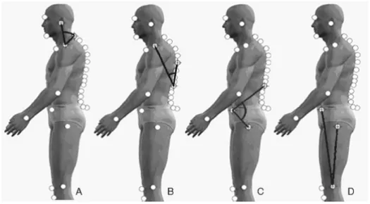
acromion, seventh cervical vertebra (C7), tragus of the ear, seventh thoracic vertebra (T7), irst lumbar vertebra (L1), anterior superior iliac spine (ASIS), greater trochanter (Figure 1). After identiication of the points, they were marked with styrofoam balls, with a diameter of 20 mm, ixed to the body parts with double-sided adhesive tape.
All subjects were placed in the static orthostatic position, remaining in the left side view, at a distance of 50 cm in front of a black wall and next to a plumb line marked with three styrofoam balls with a distance of 50 cm between each, enabling the calibration of the photograph. heir feet were placed freely on top of a black Ethylene-vinyl acetate (EVA) rug. he individual was informed to stay in a comfortable position, through a verbal command, with their gaze ixed on a point in their line of sight, keeping their posture relaxed.
A camera (Sanyo BD 200 14.1 mega pixels, DSC – W610) was used to take the photographs. It was positioned on a tripod (height of 97 cm) and at a distance of 2,30 m from the participant. he photographs were transferred to the computer and analyzed with the postural evaluation software (SAPO). he analysis of angles and measurements of the photographs were made with the Excel Program from the coordinates of the anatomical points obtained with SAPO. To mark the points and deine the postural changes that would be evaluated, the protocol of Yi et al.20 was used:
Source: Yi et al., 2008.
1) Cervical Lordosis: angle formed from three anatomical points: tragus of the ear, C7 and acromion, the acromion being the apex of the angle. he greater the angular measure, the more anterior the position of the head and the lower the cervical lordosis;
2) horacic kyphosis: angle formed from three anatomical points: acromion, T7 and L1, with L1 as the apex of the angle. he larger the angular measure, the greater the thoracic kyphosis;
3) Lumbar lordosis: angle formed from three anatomical points: L1, anterior superior iliac spine (ASIS) and greater trochanter, with ASIS as the vertex of the angle. he smaller the angular measure, the greater the lumbar lordosis;
4) Pelvic position: angle formed from three anatomical points: ASIS, greater trochanter and the midpoint of the knee joint interlining on the lateral face, with the midpoint of the articular interline as the apex of the angle. he larger the angular measure, the greater the anteversion of the pelvis.
Statistical analysis
Data were analyzed using SPSS for Windows, version 20.0 and treated with descriptive analysis as mean and standard deviation that were applied to all variables. To verify the normality of the data the Shapiro-Wilk test was applied. To compare the variables age, weight,
height, FEV1 (%prev), FVC (%prev), diaphragmatic
mobility, cervical lordosis, thoracic kyphosis, lumbar lordosis, pelvic position between COPD groups and apparently healthy individuals, Student’s t-test was
used. To compare the variables BMI and FEV1/FVC
(L) the Mann Whitney U test was used. To correlate diaphragmatic mobility with cervical lordosis, thoracic kyphosis, lumbar lordosis and pelvic position, Pearson’s linear correlation coeicient (r) was used. A signiicance level of 5% was adopted.
RESULTS
he anthropometric and pulmonary characteristics and diaphragmatic mobility of the studied groups are presented in Table 1. here was no statistically signiicant diference between age, body mass, stature and BMI, conirming that the groups were paired in relation to the anthropometric variables.
Regarding pulmonary function, there was a statistically signiicant diference in all variables of the spirometry, especially in the forced volume in the irst
second (FEV1), which characterizes the presence of
COPD. he COPD group showed a degree of severe obstruction, whereas apparently healthy individuals presented spirometric values within normal limits
(FEV1% predicted 49.6±15.7 and 95.0±10.3, p<0.001
respectively).
here was a signiicant diference in the values of diaphragmatic mobility between the studied groups, with COPD patients showing lower values when compared to healthy individuals (41.7±18.3 mm and 62.9±11.5 mm, respectively, p<0.001)
here was no statistically signiicant diference in any of the variables related to vertebral column curvatures and pelvic position, which indicates the similarity of the body posture of COPD patients and apparently healthy individuals.
Table 1. Comparison of pulmonary function, diaphragmatic mobility and postural changes between COPD groups and apparently healthy individuals (n=44)
Variables COPD
(n=22)
Healthy
(n=22) p value
Demographic and anthropometric data
Gender (M/F) 11/11 10/12
-Age (years) 65.8 ± 8.0 63.7 ± 5.2 0.312
Body mass (kg) 71.1 ± 16.4 75.5 ± 14.1 0.345
Stature (cm) 164.5 ± 8.0 164.9 ± 11.3 0.915
BMI (kg/m²) 26.2 ± 5.8 27.6 ± 3.6 0.110
Pulmonary Function
FEV1/FVC (L) 0.56 ± 0.10 0.79 ±
0.04 <0.001*
FEV1 (%prev) 49.6 ± 15.7 95.0 ± 10.3 <0.001* FVC (%prev) 68.2 ± 15.4 95.0 ± 10.7 <0.001*
DM (mm) 41.7 ± 18.3 62.9 ± 11.5 <0.001*
Cervical Lordosis (º) 85.3 ± 18.0 81.1 ± 13.1 0.380
Thoracic Kyphosis
(º) 29.5 ± 3.2 29.5 ± 4.2 0.975
Lumbar Lordosis (º) 96.3 ± 8.7 98.5 ± 8.6 0.413
Pelvic Position (º) 4.7 ± 2.4 4.6 ± 1.9 0.884
Values are expressed as mean and ± standard deviation; BMI (kg/m2): body mass index in kilograms per meter2; FEV1 (%prev): percentage of predicted forced expiratory volume in the irst second; FVC (%prev): percentage of predicted forced vital capacity; DM: diaphragmatic mobility. Mm: milimeters; º: degree. *p<0.05.
was no correlation of diaphragmatic mobility with the other studied variables (Table 2).
In relation to the group of apparently healthy individuals there was no correlation of diaphragmatic mobility with any of the variables of the vertebral column and pelvis position.
T
hor
acic k
yphosis angle (º)
Diaphragmatic mobility (mm) 20.00
34.00
32.00
30.00
28.00
26.00
24.00
22.00
20.00
40.00 60.00 80.00
Linear R2 = 0.295
Figure 2. Correlation of the thoracic kyphosis angle (º) with diaphragmatic mobility (mm) in the COPD group (n=22) (r=-0.543; p=0.009)
Table 2. Relation between diaphragmatic mobility and spinal curvatures in the COPD group (n=22) and the group of apparently healthy individuals (n=22)
Groups Correlation coeicient® p
Cervical Lordosis
COPD - 0.085 0.708
Healthy 0.122 0.589
Thoracic Kyphosis
COPD - 0.543 0.009*
Healthy 0.211 0.346
Lumbar Lordosis
COPD 0.031 0.892
Healthy 0.324 0.142
Pelvic Position
COPD 0.132 0.559
Healthy - 0.327 0.138
r: Pearson’s Correlation Coeicient. *p<0.05
DISCUSSION
he present study aimed to verify the relation between diaphragmatic mobility and spinal curvatures in patients with COPD and in healthy individuals. he results showed that there was a negative correlation between the diaphragmatic mobility and the angle of the thoracic curvature only in the COPD group, showing that the lower the diaphragmatic mobility, the
greater the degree of thoracic kyphosis. However, there was no correlation with the other vertebral curvatures evaluated. In the group of healthy individuals, there was no correlation with any of the studied variables.
he hypothesis for this inding would be the presence of reduced diaphragmatic mobility in patients with COPD. Some evidences suggest that the reduction of diaphragmatic mobility due to ineiciency of the diaphragm muscle can lead to an inevitable compensatory increase in the activity of the thoracic cavity muscles and accessory muscles of ventilation
that play an important role in patients with COPD21,22.
With the recruitment of accessory muscles and thoracic
cavity muscles23, the apical respiratory pattern occurs.
his respiratory pattern elevates the action potentials of muscles such as the sternocleidomastoid, resulting in postural changes24,25.
Regarding the thoracic kyphosis angle, there was no signiicant diference between the COPD groups and healthy individuals. Our results are consistent with the study by Dias et al.3 who evaluated the kinematics of the thoracic,
cervical, and scapular girdle of 19 patients with COPD and 19 healthy individuals and found only a greater elevation of the scapula. In contrast, Pachioni et al.2 compared 15
COPD patients with 15 healthy subjects and observed three important postural changes in patients with COPD: thoracic kyphosis, posterior pelvic unevenness and anterior pelvic tilting. he discrepancy between measurements may be related to diferences in methodologies for postural evaluation and lack of standardization in the thoracic curvature evaluation technique.
he traced relation between diaphragmatic mobility and thoracic kyphosis angle is of concern, as studies have shown that increased thoracic curvature may impair
lung function26,27,28, increase dyspnoea27, afect the
performance of daily life activities29,30, reduce quality of
life30 and predict mortality independent of underlying
vertebral osteoporosis31. As the patients already present
all these damages due to their disease, their condition can be aggravated in the presence of an increase in the angle of the thoracic curvature.
alterations. In the natural process of aging, several alterations can cause damage to the diferent systems
of the organism32. With the progression of age, postural
changes such as head anteriority, shoulder protrusion (antero-pulsion), increased thoracic kyphosis, reduced
lumbar lordosis and knee/hip lexion may arise33.
Patients with COPD already sufer from these natural processes due to aging, however, with the progression of their disease, these changes may be intensiied due to the pathophysiological factors of their disease. It is evident in the scientiic literature that the worsening of postural changes in the vertebral column can interfere with respiratory function, however, it should be taken into account that the patient with COPD already has respiratory impairments and their condition may be further aggravated by the postural alteration.
he methodological rigor to conduct the evaluations was a strong point of this research. However, there are limitations inherent to this study, it is possible to mention the fact that this cross-sectional feature makes it diicult to verify the cause-efect relationship between increased thoracic kyphosis angle and reduced diaphragmatic mobility. For this, it would be important to conduct a longitudinal prospective study to demonstrate the real inluence between these variables. Another limitation is the lack of standardization in the technique for evaluation of the curvatures of the vertebral column by the SAPO method. But despite this, the results obtained provide important subsidies regarding spinal curvatures and diaphragmatic mobility in patients with COPD and healthy individuals. It is also worth noting the clinical relevance of the present study in the early detection of postural changes alongside the evaluation of diaphragmatic mobility in patients with COPD, enabling professionals to plan and execute the most appropriate treatment.
CONCLUSION
In order to analyze the correlation between the diaphragmatic mobility and the angles of the vertebral curvatures and the position of the pelvis, the researchers conclude that only the COPD group presented a negative correlation between the variables. he researchers observed that lower the diaphragmatic mobility, the greater the angle of curvature of thoracic kyphosis.
Regarding the postural changes, it was veriied that COPD patients, as well as apparently healthy individuals, present the same angles of curvature of the vertebral column and the same position of the pelvis. However, only the COPD group presented reduction in diaphragmatic mobility.
REFERENCES
1. Global Initiative for Chronic Obstructive Lung Disease (GOLD). Global strategy for the diagnosis, management, and prevention of chronic obstructive pulmonary disease. 2015 [acesso em 10 ago. 2017]. Disponível em: https://goo.gl/SwBHGa
2. Pachioni CAS, Ferrante JA, Panissa TSD, Ferreira DMA, Ramos D, Moreira GL, et al. Avaliação postural em pacientes com doença pulmonar obstrutiva crônica. Fisioter Pesqui. 2011;18(4):341-5. doi: 10.1590/S1809-29502011000400008
3. Dias CS, Kirkwood RN, Parreira VF, Sampaio RF. Orientation and position of the scapula, head and kyphosis thoracic in male patients with COPD. Can J Respir Ther. 2009 Summer;45(2):30-4.
4. Russi EW, Stammberger U, Weder W. Lung volume reduction surgery for emphysema. Eur Respir J. 1997;10(1):208-18. 5. Laghi F, Tobin MJ. Disorders of the respiratory muscles.
Am J Respir Crit Care Med. 2003;168(1):10-48. doi: 10.1164/ rccm.2206020
6. Cassart M, Pettiaux N, Gevenois PA, Paiva M, Estenne M. Efect of chronic hyperinlation on diaphragm length and surface area. Am J Respir Crit Care Med. 1997;156(2 Pt.1):504-8. doi: 10.1164/ajrccm.156.2.9612089
7. Suga K, Tsukuda T, Awaya H, Takano K, Koike S, Matsunaga N, et al. Impaired respiratory mechanics in pulmonary emphysema: evaluation with dynamic breathing MRI. J Magn Reson Imaging. 1999;10(4):510-20. doi: 10.1002/ (sici)1522-2586(199910)10:4<510::aid-jmri3>3.0.co;2-g 8. Unal O, Arslan H, Uzun K, Ozbay B, Sakarya ME. Evaluation
of diaphragmatic movement with MR fluoroscopy in chronic obstructive pulmonary disease. Clin Imaging. 2000;24(6):347-50. doi: 10.1016/S0899-7071(00)00245-X 9. Iwasawa T, Kagei S, Gotoh T, Yoshiike Y, Matsushita K,
Kurihara H, et al. Magnetic resonance analysis of abnormal diaphragmatic motion in patients with emphysema. Eur Respir J. 2002;19(2):225-31. doi: 10.1183/09031936.02.00044602 10. Paulin E, Yamaguti WPS, Chammas MC, Shibao S, Stelmach R,
Cukier A, et al. Inluence of diaphragmatic mobility on exercise tolerance and dyspnea in patients with COPD. Respir Med. 2007;101(10):2113-18. doi: 10.1016/j.rmed.2007.05.024 11. Yamaguti WPS, Paulin E, Shibao S, Chammas MC, Salge
JM, Ribeiro M, et al. Air trapping: the major factor limiting diaphragm mobility in chronic obstructive pulmonary disease patients. Respirology. 2008;13(1):138-44. doi: 10.1111/j.1440-1843.2007.01194.x
with chronic obstructive pulmonary disease: a randomized controlled trial. Arch Phys Med Rehabil. 2012;93(4):571-7. doi: 10.1016/j.apmr.2011.11.026
13. Boulay C, Tardieu C, Hecquet J, Benaim C, Mouilleseaux B, Marty C, et al. Sagittal alignment of spine and pelvis regulated by pelvic incidence: standard values and prediction of lordosis. Eur Spine J. 2006;15(4):415-22. doi: 10.1007/s00586-005-0984-5
14. Butcher SJ, Meshke JM, Sheppard MS. Reductions in functional balance, coordination, and mobility measures among patients with stable chronic obstructive pulmonary disease. J Cardiopulm Rehabil. 2004;24(4):274-80. doi: 10.1097/00008483-200407000-00013
15. World Health Organization. Obesity: Preventing and Managing the Global Epidemic. Report of a WHO Consultation (WHO Technical Report Series 894). Geneva: World Health Organization. 2000 [acesso em 10 ago. 2017];284:256. Disponível em: https://goo.gl/GyeLBX
16. Miller MR, Hankinson J, Brusasco V, Burgos F, Casaburi R, Coates A, et al. Standardisations of spirometry. Series “ATS/ERS task force: standardisation of lung function testing”. Eur Respir J. 2005;26:319-38. doi:10.1183/09031936.05.00034805
17. Pereira CA, Sato T, Rodrigues SC. New reference values for forced spirometry in white adults in Brazil. J Bras Pneumol, 2007;33(4):397-406. doi: 10.1590/S1806-37132007000400008 18. Saltiel RV, Grams ST, Pedrini A, Paulin E. High reliability of
measure of diaphragmatic mobility by radiographic method in healthy individuals. Braz J Phys Ther, 2013;17(2):128-136. doi: 10.1590/S1413-35552012005000076
19. Ferreira EAG, Duarte M, Maldonado EP, Burke TN, Marques AP. Postural assessment software (PAS/SAPO): validation and reliability. Clinics. 2010;65(7):675-81. doi: 10.1590/ S1807-59322010000700005
20. Yi LC, Jardim JR, Inoue DP, Pignatari SSN. The relationship between excursion of the diaphragm and curvatures of the spinal column in mouth breathing children. J Pediatr. 2008;84(2):171-7. doi: 10.1590/S0021-75572008000200014 21 Martinez FJ, Couser JI, Celli BR. Factors inluencing ventilatory
muscle recruitment in patients with chronic airlow obstruction. Am Rev Respir Dis. 1990;142(2):276-82. doi: 10.1164/ ajrccm/142.2.276
22. Trevisan ME, Porto AS, Pinheiro TM. Inluência do treinamento da musculatura respiratória e de membros inferiores no desempenho funcional de indivíduos com DPOC. Fisioter Pesqui. 2010;17(3)209-13. doi: 10.1590/S1809-29502010000300004
23. Breslin EH, Garoutte BC, Kohlman-Carrieri V, Celli BR. Correlations between dyspnea, diaphragm, and sternomastoid recruitment during inspiratory resistance breathing in normal subjects. Chest. 1990;98(2):298-302. doi: 10.1378/chest.98.2.298 24. Pasinato F, Corrêa ECR, Peroni ABF. Avaliação da mecânica
ventilatória em indivíduos com disfunção têmporo-mandibular e assintomáticos. Rev Bras Fisioter. 2006;10(3)285-9. doi: 10.1590/S1413-35552006000300006
25. Corrêa ECR, Bérzin F. Efficacy of physical therapy on cervical muscle activity and on body posture in school-age mouth breathing children.Int J Pediatr Otorhinolaryngol. 2007;71(10):1527-35. doi: 10.1016/j.ijporl.2007.05.031
26. Teramoto S, Suzuki M, Matsuse T, Ohga E, Katayama H, Nagase T, et al. Inluence of kyphosis on the age-related decline in pulmonary function. Nihon Ronen Igakkai Zasshi. 1998 [acesso em 10 ago. 2017];35(1):23-7. Disponível em: https://goo.gl/ Vh5Ti5
27. Di Bari M, Chiarlone M, Matteuzzi D, Zacchei S, Pozzi C, Bellia V, et al. Thoracic kyphosis and ventilatory dysfunction in unselected older persons: an epidemiological study in Dicomano, Italy. J Am Geriatr Soc. 2004;52(6):909-15. doi: 10.1111/j.1532-5415.2004.52257.x
28. Loubresse CG, Vialle R, Wolf S. Cyphoses pathologiques: pathological kyphosis. EMC Rhumatol Orthop. 2005;2(3):294-334. doi: 10.1016/j.emcrho.2004.11.002
29. Ryan SD, Fried LP The impact of kyphosis on daily functioning. J Am Geriatr Soc. 1997;45(12):1479-86. doi: 10.1111/j.1532-5415.1997. tb03199.x
30. Takahashi T, Ishida K, Hirose D, Nagano Y, Okumiya K, Nishinaga M, et al. Trunk deformity is associated with a reduction in outdoor activities of daily living and life satisfaction in community-dwelling older people. Osteoporos Int. 2005;16(3):273-9. doi: 10.1007/s00198-004-1669-3
31. Kado DM, Huang MH, Karlamangla AS, Barrett-Connor E, Greendale GA. Hyperkyphotic posture predicts mortality in older community-dwelling men and women: a prospective study. J Am Geriatr Soc. 2004;52(10):1662-7. doi: 10.1111/j.1532-5415.2004.52458.x
32. Lacourt MX, Marini LL. Decréscimo da função muscular decorrente do envelhecimento e a inluência na qualidade de vida do idoso: uma revisão de literatura. RBCEH. Rev Bras Cienc Envelhec Hum.. 2006;3(1):114-21. 10.5335/rbceh.2012.51 33. Hinman, MR. Comparison of thoracic kyphosis and postural