DOI: http://dx.doi.org/10.18363/rbo.v76.2019.e1377 Literature Review/Oral Pathology
Analysis of cell proliferation index using the
Ki-67 Antigen in odontogenic keratocyst:
a systematic review
Juliana Portes,¹ Danielle Castex Conde,¹,² Eliane Pedra Dias¹,³
1Postgraduate Program in Pathology, Medical School, Fluminense Federal University (UFF), Niterói, RJ, Brazil 2Department of Dentistry, School of Dentistry, Veiga de Almeida University (UVA), Rio de Janeiro, RJ, Brazil 3Department Laboratory Medicine and Pathology, Institut Gustave Roussy, Paris, France
• Conflicts of interest: none declared.
A
bstrActObjective: this review aims to analyze scientific articles about cell proliferation index using Ki-67 in odontogenic keratocyst and compare these papers to estimate the average index of this lesion. Material and Methods: two researchers performed a literature search independently in the MEDLINE/PubMed database and 28 articles containing relevant data were selected. Results: the immunohistochemical analysis methodology showed great variability among all the papers, with unclear and unified methodologies, making the comparison among different studies difficult. Conclusion: considering odontogenic keratocyst as a lesion with an uncommon clinical behavior, an adequate classification for it is necessary, so an appropriate treatment with a good prognosis for the patient can be established according to its nature. A standardization is needed so immunohistochemical analyses will find reliable data to classify properly this lesion.
Keywords: Odontogenic keratocyst; Ki-67 antigen; Proliferation index.
Introduction
T
he odontogenic keratocyst (OKC) was first classified
as an odontogenic developmental cyst by Pindborg
& Kramer (1971), on the first official classification
of odontogenic cysts and tumors by the World Health
Or-ganization (WHO).
1However, this lesion has three
char-acteristics that raised discussions about its classification,
which are locally destructive and aggressive behavior, high
recurrence index and tendency toward multiplicity.
2In
2005,
the WHO changed the classification of OKC to an
odontogenic tumor, based on its high cell proliferation
ac-tivity when compared to other odontogenic cysts and the
presence of mutations in p53 and patched (PTCH) genes
that are also found in other neoplasms.
2Some authors did not accept this new classification,
claiming that the existing evidence are insufficient to
support the neoplastic origin of OKC and that further
researches are needed.
3The WHO published its new
clas-sification in 2017, and OKC was again categorized as an
odontogenic cyst.
3OKC represents about 10 to 20% of all odontogenic cysts,
usually occurring between the second and third decade of
life, with a slight predilection for men.
3The mandible bone
is often affected
4and the cyst can be solitary or multiple
– the last former is usually one of the components of
in-herited nevoid basal cell carcinoma syndrome (NBCCS).
4,5The evaluation of cell proliferation activity is one of the
most common methods to assess the biological behavior of
a lesion, being reported as an important prognostic marker
and indicator of biological aggressiveness.
5,6-8The Ki-67 antigen is a highly specific marker of
prolif-erating cells
4,6-8and has been widely used to evaluate the
proliferative activity of preneoplastic and neoplastic
le-sions.
5-7,9This protein is present in all phases of the cell
cycle, except in the G0 phase
4,6-12, with a 60–90 minutes
half-life.
11Over the years, many researches have evaluated the
proliferative activity of OKC cells, comparing it to other
cysts and tumors to better understand its pathogenesis and
behavior.
4,5,7,8,11-21However, each study presents a different
cell proliferation index (PI) and the methodology used is
not always clear, making the comparison between the OKC
index with other odontogenic lesions difficult, impairing
the improvement of our knowledge about its nature and
biological behavior, as well as the possibility of using Ki-67
as a prognostic marker for OKC.
This review aims to analyze and compare scientific
ar-ticles that used Ki-67 as reference to evaluate the cell
pro-liferation
activity in OKC, comparing their methodology
and results to define an average PI for OKC to facilitate
future studies and comparisons. To the best of our
knowl-edge, this is the first systematic review about this issue.
Material and Methods
Two researchers, independently, conducted a literature
search on the MEDLINE/PubMed database in June 2018.
The search had no year restriction, using the abstract
for-mat and sorted by most recent. The Medical Subject
Head-ings (MeSH) terms used were: Ki-67 antigen,
odontogen-ic cyst, keratocysts and mitotodontogen-ic index. Those words were
combined with other terms: odontogenic keratocyst,
ker-atocystic odontogenic tumor, Ki-67 and cell proliferation
index.
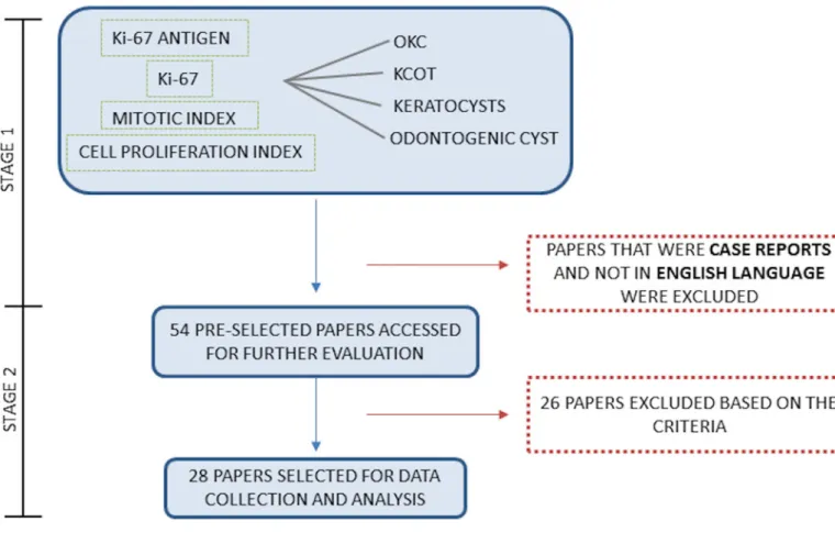
The search was performed in two stages: the first used
the keywords chosen in different combinations, and the
articles were selected based on their titles. Articles that
were case reports and that were not in English language
were excluded.
On the second stage, the researchers accessed the full
texts of articles that were considered eligible for inclusion
for further evaluation and selection. Some studies were
excluded according to the established exclusion criteria:
articles with an unclear methodology, articles without the
PI, and articles that used NBCCS-associated OKC as
sam-ples. However, for articles that included syndromic and
non-syndromic OKC in their samples with individualized
results, only sporadic OKC was considered.
10,19,22The same
approach was taken with the article that included
recur-rent and non-recurrecur-rent OKC, only the last one was
con-sidered.
9,22From the various keywords combinations, 54 papers
were pre-selected respecting the exclusion criteria for
stage one. All articles appeared repeatedly in the different
searches, these were only counted once, justifying the total
of papers selected in this stage (Figure 1).
After the analysis of all these papers on the second stage,
28 articles were selected according to the described criteria
and all relevant data were obtained from these papers: Ki-67
PI, number of analyzed cases and the
immunohistochem-istry analysis method in each study (Figure 1). Table 1
de-scribes the reasons for the exclusion of 26 articles.
Results
The selected papers were divided into two groups for
comparison: articles that show the index as mean (Table 2)
and articles that present it as median (Table 3), because it is
not possible to compare these different values.
Table 1. Number and reasons of excluded articles
REASONS FOR THE EXCLUSION N° OF ARTICLES
No cell proliferation index 6
Index expressed only as a graphic, making it impossible to precise the PI 3
Unclear methodology 2
Impossible to reach a single PI value with the information on the paper 11 PI calculated counting the positive cells per millimeters of basal membrane 2 Article with median and divided into two scores: more than 10% and less than 10% of
immunopositivity. 1
The authors did not provide the PI separately for syndromic OKC and sporadic OKC 1
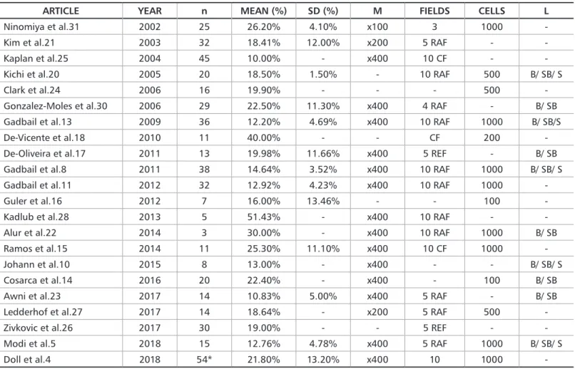
Table 2. Articles with PI as mean
ARTICLE YEAR n MEAN (%) SD (%) M FIELDS CELLS L
Ninomiya et al.31 2002 25 26.20% 4.10% x100 3 1000
-Kim et al.21 2003 32 18.41% 12.00% x200 5 RAF -
-Kaplan et al.25 2004 45 10.00% - x400 10 CF -
-Kichi et al.20 2005 20 18.50% 1.50% - 10 RAF 500 B/ SB/ S
Clark et al.24 2006 16 19.90% - - - 500
-Gonzalez-Moles et al.30 2006 29 22.50% 11.30% x400 4 RAF - B/ SB
Gadbail et al.13 2009 36 12.20% 4.69% x400 10 RAF 1000 B/ SB/S
De-Vicente et al.18 2010 11 40.00% - - CF 200
-De-Oliveira et al.17 2011 13 19.98% 11.66% x400 5 REF - B/ SB
Gadbail et al.8 2011 38 14.64% 3.52% x400 10 RAF 1000 B/ SB/ S
Gadbail et al.11 2012 32 12.92% 4.23% x400 10 RAF 1000
-Guler et al.16 2012 7 16.00% 13.46% - - 100
-Kadlub et al.28 2013 5 51.43% - x400 10 RAF -
-Alur et al.22 2014 3 30.00% - x400 10 RAF 1000 B/ SB
Ramos et al.15 2014 11 25.30% 11.10% x400 10 CF 1000
-Johann et al.10 2015 8 13.00% - x400 - - B/ SB/ S
Cosarca et al.14 2016 20 22.40% - x400 - 100 B/ SB
Awni et al.23 2017 14 10.83% 5.00% x400 5 RAF - B/ SB
Ledderhof et al.27 2017 14 18.64% - x200 5 RAF 500
-Zivkovic et al.26 2017 30 19.00% - - 5 REF -
-Modi et al.5 2018 15 12.76% 4.78% x400 5 RAF 1000 B/ SB/ S
Doll et al.4 2018 54* 21.80% 13.20% x400 10 1000
-M = magnification; RAF = randomly selected fields; REF = representative fields; CF = consecutive fields; L = evaluated layers; B = basal layer; SB = suprabasal layer; S = superficial layer; “-“= no data available.
*Authors did not say how many samples of sporadic OKC were evaluated, they consider that 54 lesions included sporadic and syndromic OKC. But the indexes were given separately.
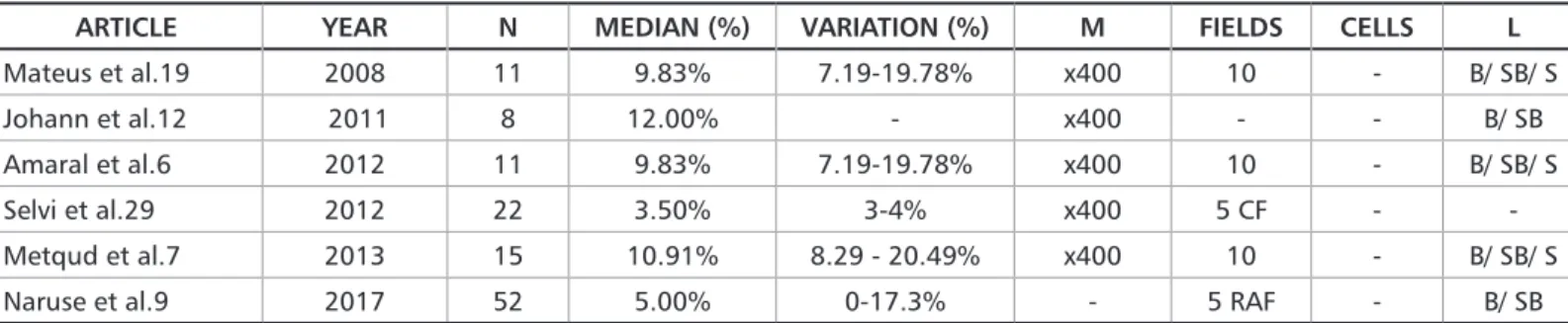
ARTICLE YEAR N MEDIAN (%) VARIATION (%) M FIELDS CELLS L Mateus et al.19 2008 11 9.83% 7.19-19.78% x400 10 - B/ SB/ S Johann et al.12 2011 8 12.00% - x400 - - B/ SB Amaral et al.6 2012 11 9.83% 7.19-19.78% x400 10 - B/ SB/ S Selvi et al.29 2012 22 3.50% 3-4% x400 5 CF - -Metqud et al.7 2013 15 10.91% 8.29 - 20.49% x400 10 - B/ SB/ S
Naruse et al.9 2017 52 5.00% 0-17.3% - 5 RAF - B/ SB
Table 3. Articles with PI as median
M = Magnification; RAF = randomly selected fields; CF = consecutive fields; L = evaluated layers; B = basal layer; SB = suprabasal layer; S = su-perficial layer; “- “= no data available.
Three papers divided the indexes into layers (basal and
parabasal).
14,17,23One provided only the number of positive
cells and the total number of cells counted but did not
pro-vide a PI.
24Two papers divided the OKC evaluation
accord-ing to inflammatory density
25and in follicular and
extra-follicular OKC.
21To include these papers in our study, an
overall PI was calculated for each study. All data collected
are shown in Tables 2 and 3.
Discussion
The classification of OKC has changed over the years and
many studies are being developed to determine a reliable
classification, if it is a cyst or a tumor, seeking to define the
best treatment and prognosis for this lesion. Some studies
try to determine the nature of OKC through the cell
prolif-eration index using Ki-67. But almost all papers present
dif-ferent methodologies, as well as difdif-ferent results. Therefore,
this study analyzed and compared these articles and their
methodologies to know how the PI was calculated.
Large variations of PI values can be observed in the
se-lected articles, from 10% to 51.43% as mean, and from 3.5%
to 12% as median. This can be explained by the different
methods used in each of the analyzed papers.
The magnification is a methodological variable. Most
au-thors used x400, others did the count at x200 or x100, which
can impact the total number of visualized cells. This
differ-ence can be observed in Tables 1 and 2, where the authors
indicate the number of cells that were evaluated by field.
The number of analyzed fields and how they were chosen
are also factors that may have impact on the calculation of
the percentage of positive cells: some authors select random,
continuous or representative fields. Any parameter that
changes the total number of evaluated cells might influence
the PI, i.e., the higher the number of analyzed fields, the
higher the number of cells. In addition, all tissues have fields
that are considered a “hotspot”, where greater
immunos-taining occurs; thus, depending on the selection of fields,
this may also influence the result. When papers mentioned
representative fields, the criteria used for such selection was
not explained, so we considered this feature as “hotspot”.
The location of immunopositive cells
is also an important
feature. Some authors analyzed all epithelial layers: basal,
suprabasal and superficial, while others analyzed only the
basal and suprabasal layer. This variable may also have
im-pact on the total number of evaluated cells.
Considering the papers that calculated the PI as mean
,the indexes varied from 10% to 51.43% and the mean of
these results is 20.70%. This shows a discrepancy among the
results, mainly in the PI obtained by Kadlub et al.
28(51.43%).
However, we can verify that most values are very close,
ranging from 10% to 25% (in 18 of 22 papers); therefore, the
influence of the variables mentioned above may not be
stat-ically significant.
Focusing our analysis on Ledderhof et al.
27and Kim et
al.,
21we find that they used the same magnification (x200)
and the same number of analyzed fields, and that cell
num-ber and the PI of both was similar: 18.64% and 18.41%,
re-spectively.
Despite such variance on OKC PI, it is interesting to note
that according to the WHO’s classification of odontogenic
cysts and tumors (2017),
3when using Ki-67, the PI of some
benign odontogenic tumors is low, like primordial
odonto-genic tumor (less than 2%) and dentinoodonto-genic ghost cell
tu-mor (less than 5%).
3Therefore, further exploration of OKC’s
cell proliferation index are needed to actually verify the
na-ture of this lesion.
Another example of how these variables may influence
immunohistochemical results can be observed on Table 3:
Amaral et al.
6and Mateus et al.
19used the same number of
analyzed samples, magnification, fields and the same
epi-thelial layers, so they found the same PI for OKC.
A high variation in the number of sample was also
ob-served, from 3 to 52 among all papers. However, some
ar-ticles showed similar PI using different number of sample,
but with similar quantities of evaluated cells, showing that
the number of sample may not have significant impact on
the results.
For example, Kim et al.
21and Ledderhof et al.
27used the
same magnification and number of analyzed fields, but a
dif-ferent number of sample, and achieved 18.41% and 18.64%
PI, respectively. Amaral et al.,
6Mateus et al.
19and Metqud
& Grupta
7used the same magnification, number of
ana-lyzed fields and epithelial layers, but as in the last
exam-ple, with different sample numbers. Their PI were 9.83%,
9.83% and 10.91%, respectively.
6,7,19Both of these examples
showed similar PI, which implies that the number of
sam-ples does not interfere on this calculation.
The main limitations of this systematic review are that
the collected data are not the same across all studies, as
well as the impossibility of performing a quantitative
anal-ysis of these results. Therefore, comparing the different
studies using Ki-67 to evaluate the biological behavior of
OKC is very difficult. Further and thorough research on
these methodological variables are needed to confirm if
they significantly influence PI results, from the statistical
point of view, but many authors do not provide all relevant
information of their methodology. For future researches,
we suggest a standard evaluation of the proliferative
activ-ity and calculation of the index of these lesions, using the
same magnification, number of cells and evaluating the
entire epithelium.
Conclusion
From the results of this review, it is clear that most
stud-ies involving the immunohistochemical analysis with
Ki-67 antigen for evaluating the proliferative activity in OKC
have unclear and non-standardized methodologies;
there-fore, the standardization of immunohistochemical
analy-ses is necessary to improve and optimize future research,
associating this antibody with the nature of these lesions.
16. Güler N, Çomunoglu N, Cabbar F. Ki-67 and MCM-2 in Dental Follicle and Odontogenic Cysts: The Effects of Inflammation on Proliferative Mark-ers. The Scientific World Journal. 2012;1-8.17. De-Oliveira MG, Lauxen IS, Chaves ACM, Rados PV, Filho MS. Odonto-genic epithelium: immunolabeling of Ki-67, EGFR and Survivin in Pericoro-nal Follicles, Dentigerous Cysts and Keratocystic Odontogenic Tumors. Head and Neck Pathol. 2011;5:1-7.
18. De-Vicente J, Torre-Iturraspe A, Gutiérrez A, Lequerica-Fernández P. Im-munohistochemical Comparative study of the odontogenic keratocysts and other odontogenic lesions. Med Oral Patol Oral Cir Bucal. 2010;15(5):709-15. 19. Mateus GCP, Lanza GHSP, Moura PHR, Marigo HA, Horta MCR. Cell proliferation and apoptosis in keratocystis odontogenic tumors. Med Oral Patol Oral Cir Bucal. 2008;13(11):697-702.
20. Kichi E, Enokiya Y, Muramatsu T, Hashimoto S, Inoue T, Abiko Y, et al. Cell proliferation, apoptosis and apoptosis-related factors in odontogenic keratocysts ans in dentigerous cysts. J Oral Pathol Med. 2005;34:280-6. 21. Kim DK, Ahn SG, Kim J, Yoon JH. Comparative Ki-67 expression and apoptosis in the odontogenic keratocyst associated with or without an im-pacted tooth in addition to unilocular and multilocular varieties. Yonsei Med J. 2003;44(5):841-6.
22. Alur J, Narayan TV, Mohanty L, Shenoy S, Jamadar S, Shetty S. Ki-67 and p53 expression in solitary sporadic, syndrome associated and recurrent kera-tocystic odontogenic tumor. J Oral Maxillofac Pathol. 2014;18(1):21-5. 23. Awni S, Conn B. Decompression of keratocystis odontogenic tumors lead-ing to increased fibrosis, but without any change in epitelial proliferation. Oral Surg Oral Med Oral Pathol Oral Radiol. 2017;123(6):634-44.
24. Clark P, Marker P, Bastian HL, Krogdahl A. Expression of p53, Ki-67, and EGFR in odontogenic keratocysts before and after decompression. J Oral Pathol Med. 2006;35:568-72.
25. Kaplan I, Hirshberg A. The correlation between epithelial cell pro-liferation and inflammation in odontogenic keratocyst. Oral Oncology. 2004;40:985-91.
26. Zivkovic ND, Mihailovic DS, Kostic MS, Cvetanovic AS, Mijovic ZZ, Mi-lentijevic MVJ, et al. Markers of proliferation and cytokeratins in the differ-ential diagnosis of jaw cysts. ENT-Ear, Nose & Throat Journal. 2017;96(9):376-83.
27. Ledderhof NJ, Caminiti MF, Bradley G, David K. Lam. Topical 5-Fluoro-uracil is a Novel Targeted Therapy for the Keratocystic Odontogenic Tumor. J Oral Maxillofac Surg. 2017;75:514-25.
28. Kadlub N, Coudert A, Gatibelza, Houmami NE, Soufir N, Ruhin-Poncet B, et al. PTCH1 mutation and local aggressiveness of odontogenic keratocys-tic tumors in children: is there a relationship? Human Pathol. 2013;44:1071-78.
29. Selvi F, Tekkesin MS, Cakarer S, Isler SC, Keskin C. Keratocystic odon-togenic tumors: predictive factors of recurrence by Ki-67 and AgNOR Label-ling. Int J Med Sci. 2012;9(4):262-8.
30. González-Moles MA, Mosqueda-Taylor A, Delgado-Rodríguez M,
References
1. Pindborg JJ, Kramer Jr. Histological Typing of Odontogenic Tumours. Jaw Cysts and Allied Lesions, 1th ed. Geneva: World Health Organization; 1971. 2. Barnes l, Eveson JW, Reichart P, Sidransky D. Pathology and Genetics of Head and Neck Tumors, 1th ed. Lyon: IARCPress; 2005.
3. El-Naggar AK, Chan JKC, Grandis Jr, Takata T, Slootweg PJ. WHO Classifi-cation of head and neck tumors, 4th edition. Lyon: IARCPress; 2017. 4. Doll C, Dauter K, Jöhrens, Hartwig S, Voss JO, Klein M, et al. Clinical char-acteristics and immunohistochemical analysis of p53, Ki-67 and cyclin D1 in 80 odontogenic keratocysts. J Stomatol Oral Maxillofac Surg. 2018;161:1-6. 5. Modi TG, Chalishazar M, Kumar M. Expression of Ki-67 in odontogenic cysts: A comparative study between odontogenic keratocysts, radicular cysts and dentigerous cysts. J Oral Maxillofac Pathol. 2018;22(1):142-6.
6. Amaral FR, Mateus GCP, Bonisson LA, Andrade BAB, Mesquita RA, Horta MCR, et al. Cell Proliferation and Apoptosis in Ameloblastomas and Kerato-cystic Odontogenic Tumors. Braz Dent J. 2012;23(2):91-6.
7. Metqud R, Gupta K. Expression of cell cycle and apoptosis-related proteins in ameloblastoma and keratocystic odontogenic tumor. Annals of Diagnostic Pathology. 2013;518-21.
8. Gadbail AR, Hande A, Chaudhary M, Nikam A, Gawande M, Patil S, et al. Tumor angiogenesis in keratocystic odontogenic tumor assessed by using CD-105 antigen. J Oral Pathol Med. 2011;40:263-9
9. Naruse T, Yamashita K, Yanamoto S, Rokutanda S, Matsushita Y, Sakamo-to Y, et al. HisSakamo-topathological and immunohisSakamo-tochemical study in keraSakamo-tocys- keratocys-tic odontogenic tumors: Predictive factors of recurrence. Oncology Letters. 2017;13:3487-93.
10. Johann ACBR, Caldeira PC, Caliari MV, Gomez RS, Aguiar MCF, Mesqui-ta RA. MeMesqui-tallothionein immunoexpression in non-syndromic and sundromic keratocystic odontogenic tumour. Med Oral Patol Cir Bucal. 2015;20(4):408-12.
11. Gadbail AR, Patil R, Chaudhary M. OKC-expression of ki-67 and p53 pro-tein in ameloblastoma and keratocystic odontogenic tumor. Act Odontologi-ca SOdontologi-candinaviOdontologi-ca. 2012;70:529-35.
12. Johann ACBR, Caldeira PC, Caliari MV, Abreu MHNG, Aguiar MCF, Mesquita RA. Metallothionein in the radicular, dentigerous, orthokerati-nized odontogenic cysts and in keratocystic odontogenic tumor. J Oral Pathol Med. 2011;40:270-6.
13. Gadbail AR, Chaudhary M, Gawande M. Actual Proliferating Index and p53 protein expression as prognostic marker in odontogenic cysts. Oral Dis-eases. 2009;15:490-8.
14. Cosarca AS, Mocan SL, Pacurar M, Fulop E, Ormenisan A. The evaluation of Ki-67, p53, MCM3 and PCNA immunoexpressions at level of the dental follicle of impacted teeth, dentigerous cysts and keratocystic odontogenic tu-mors. Rom J Morphol Embryol. 2016;57(2):407-12.
15. Ramos GO, Costa A, Meurer M, Vieira DSC, Rivero ERC. Immunohisto-chemical analysis of matrix metalloproteinases (1, 2 and 9), ki-67, and myofi-broblasts in keratocystic odontogenic tumors and pericoronal follicles. J Oral Pathol Med. 2014;43:282-8.
Submitted: 01/13/2019 / Accepted for publication: 03/15/2019 Corresponding Author
Danielle Castex Conde
E-mail: daniellecastex@yahoo.com.br
Mini Curriculum and Author’s Contribution
1. Juliana Portes – DDS; MSc. Contribution: effective scientific and intellectual participation for the study; data acquisition, data interpretation; preparation and draft of the manuscript; critical review and final approval. ORCID: 0000-0003-0038-8909
2. Danielle Castex Conde Portes – DDS; PhD. Contribution: effective scientific and intellectual participation for the study; data acquisition, data interpretation; pre-paration and draft of the manuscript; critical review and final approval. ORCID: 0000-0002-8492-9145
3. Eliane Pedra Dias – MD; PhD. Contribution: effective scientific and intellectual participation for the study; critical review and final approval. ORCID: 0000-0002-0917-6091
Martínes-Mata G, Gil-Montoya A, Días-Franco MA, et al. Analysis of p53 protein by PAb240, Ki-67 expression and Human Papillomavirus DNA de-tection in different types of odontogenic keratocyst. Anticancer research. 2006;26:175-82.
31. Ninomiya T, Kubota Y, Koji T, Shirasuna K. Marsupialization inhibits in-terleukin-1α expression and epithelial cell proliferation in odontogenic kera-tocysts. J Oral Pathol Med. 2002;31:526-33.
32. Dandena VK, Thimmaiah SY, Kiresur MA, Hunsigi P, Roy S, Rashmi M. A comparative study of odontogenic keratocyst and orthokeratinized odon-togenic cyst using Ki-67 and α smooth muscle actin. J Oral Maxillofac Pathol. 2017;21(3):458-9.
33. Dissanayake U, Johnson NW, Warnakulasuriya KAAS. Comparison of cell proliferation in the centre and advancing fronts of oral squamous cell carcinomas using Ki-67 index. Cell Prolif. 2003;36:225-64.