Ultrasound detects subclinical
joint inflammation in the hands and
wrists of patients with systemic lupus
erythematosus without musculoskeletal
symptoms
Carina A Ruano,1Rui Malheiro,2,3João F Oliveira,2,3Sofia Pinheiro,2,3 Luís S Vieira,1Maria Francisca Moraes-Fontes3,4
To cite: Ruano CA, Malheiro R, Oliveira JF, et al. Ultrasound detects subclinical joint inflammation in the hands and wrists of patients with systemic lupus erythematosus without musculoskeletal symptoms. Lupus Science & Medicine 2017;3:e000184. doi:10.1136/lupus-2016-000184
▸ Additional material is available. To view please visit the journal online (http://dx. doi.org/10.1136/lupus-2016-000184).
Received 17 August 2016 Revised 12 October 2016 Accepted 5 November 2016
For numbered affiliations see end of article.
Correspondence to
Dr Carina A Ruano; [email protected]
ABSTRACT
Objectives:To assess the prevalence and severity of ultrasonographic abnormalities of the hand and wrist of asymptomatic patients with systemic lupus erythematosus (SLE) and compare these findings with those from patients with SLE with musculoskeletal signs or symptoms and healthy controls.
Methods:We conducted a prospective cross-sectional study that evaluated bilaterally, with grey-scale and power Doppler (PD) ultrasound (US), the dorsal hand (2nd to 5th metacarpophalangeal and 2nd to 5th proximal interphalangeal joints) and wrist (radiocarpal, ulnocarpal and intercarpal joints) of 30 asymptomatic patients with SLE, 6 symptomatic patients with SLE and 10 controls. Synovial hypertrophy (SH) and intra-articular PD signal were scored using semiquantitative grading scales (0–3). Individual scores were graded as normal (SH≤1 and PD=0) or abnormal (SH≥2 or PD≥1). Global indexes for SH and PD were also calculated. US findings were correlated with clinical and laboratory data and disease activity indexes.
Results:US detected SH (score≥1) in 77% asymptomatic patients with SLE, mostly graded as minimal (score 1: 63%). 23% of the asymptomatic patients with SLE showed abnormal US PD findings (SH≥2 or PD≥1). SH was present in all symptomatic patients with SLE, mostly graded as moderate (grade 2: 67%), and with associated PD signal (83%). SH (score 1) was identified in 50% of controls, however, none presented abnormal US PD findings. SH index in the asymptomatic SLE group was higher than in the control group (2.0 (0–5) vs 0.5 (0–2), median (range), p=0.01) and lower than in the symptomatic SLE group (7.0 (4–23), median (range), p<0.001). No significant correlation was demonstrated between US PD findings and clinical or laboratory variables and disease activity indexes.
Conclusion:A small subgroup of asymptomatic patients with SLE may present subclinical joint inflammation. Global US scores and PD signal may be important in disease evaluation and therapeutic monitoring.
INTRODUCTION
Joint involvement is one of the most common features of systemic lupus erythema-tosus (SLE), with up to 95% of patients experiencing arthralgia or arthritis during the course of their disease.1 While multiple joints can be affected, nearly 50% of patients report difficulties in daily life performance due to hand symptoms.2 Traditionally, SLE arthritis is considered to be mild, reversible and non-erosive, with only 5–15% of cases progressing to deforming arthropathy, either erosive—as in rhupus syndrome (an overlap of SLE with rheumatoid arthritis (RA))—or non-erosive—as in Jaccoud’s arthropathy.3 4 In routine clinical practice, joint involvement is usually assessed with physical examination and radiographical studies. However, this approach has been shown to have a low sen-sitivity for joint abnormalities when com-pared with ultrasound (US) evaluation or MRI, suggesting that the burden of joint inflammation may be underestimated in the current clinical practice.5 The value of US with power Doppler (PD) in inflammatory arthritis has been extensively demonstrated in the literature, with similar sensitivity and specificity in the detection of synovitis in comparison with MRI, with the advantage of being more accessible and less expensive.6–8 While US shows the morphology of the syn-ovial membrane, PD identifies increased vas-cularisation within it, allowing for the detection of active joint inflammation.9 It is known that in RA synovial abnormalities are present before clinically evident arthritis develops, and that early treatment can limit erosive changes and avoid disease progres-sion.10 In this context, US PD has nowadays an established role in the detection of subclinical joint inflammation in RA, namely
to predict progression in patients with clinical remission.11 12
Although there is evidence that US PD is a valuable technique in the evaluation of musculoskeletal symp-toms in SLE, the prevalence of subclinical joint abnor-malities in SLE remains to be defined.13 14Furthermore, while it is known that minimal synovial proliferation may be seen in up to 50% of healthy subjects, the distinction between‘normal’ synovial hypertrophy (SH) and patho-logical subclinical synovitis in SLE still requires clari fica-tion.15–18
The aim of this study was to assess the prevalence and precise grading of US abnormalities of the hand and wrist in asymptomatic patients with SLE, while compar-ing thesefindings with a group of patients with SLE with musculoskeletal signs or symptoms and with healthy controls.
PATIENTS AND METHODS Patients and study design
We conducted a prospective cross-sectional study which included patients fulfilling the 1997 revised American College of Rheumatology (ACR) criteria for SLE,19 under standard of care, and on stable medication in the preceding 4 weeks. Patients were sequentially recruited from the population of the two outpatient autoimmune disease units from Centro Hospitalar de Lisboa Central (Lisbon, Portugal) between January 2014 and July 2015. A subset of these patients has been previously charac-terised.20 We excluded patients with a known diagnosis of rhupus syndrome (defined as patients with SLE also satisfying the classification criteria for RA)21or Jaccoud’s arthropathy and mild deforming arthropathy (according to the Jaccoud’s Arthropathy Index),4 22 as well as patients with known osteoarthritis, trauma or surgery of the hand or wrist. Healthy controls had no personal or familiar history of autoimmune diseases and denied pre-vious hand or wrist trauma and osteoarthritis. The study was approved by the Hospital Centre Ethics Committee and performed according to the principles of good clin-ical practice and to the Declaration of Helsinki. Informed consent was obtained from all enrolled patients.
Clinical and laboratory evaluation
Patients with SLE underwent a medical interview from which demographic, clinical data and previous history were recorded, together with a detailed physical examin-ation according to the European League Against Rheumatism (EULAR) recommendations.23 Patients were then classified as asymptomatic (if musculoskeletal symptoms or signs were absent) and symptomatic (if musculoskeletal signs or symptoms were present).
Disease activity was assessed using the SLE Disease Activity Index (SLEDAI-2K),24 and organ damage was assessed using the ACR/Systemic Lupus International Collaborating Clinics (SLICC) score.25Active disease was
defined as a SLEDAI score equal or superior to 3.26 Anti-nuclear antibodies (ANA) were detected by indirect immunofluorescence using HEp-2 epithelial cells as the substrate (American Type Culture Collection CCL 23). The serum dilution was 1/160, and a titre equal to or greater than 1:160 was considered positive. Serum samples were diluted 1/10 for detection of antibodies against double-stranded DNA (anti-dsDNA) on Crithidia luciliae. Positive results were quantified by ELISA for IgG. Laboratory evaluation also included erythrocyte sedimentation rate, C3 and C4 complement levels by nephelometry and ELISA for rheumatoid factor (RF) and anti-citrullinated protein IgG antibodies (anti-CCP). Grey-scale and PD ultrasonography
Patients and controls underwent bilateral musculoskel-etal US examination with PD evaluation of the hand and wrist, performed on the same week as the medical inter-view. All US scans were performed simultaneously by two radiologists with experience in musculoskeletal radi-ology, blinded to clinical and laboratory data, using a Logiq E9 machine (General Electric Medical Systems, Milwaukee, USA) with a 6–15 MHz linear array probe operating at 15 MHz. PD evaluation was performed with a PD frequency of 10 MHz, gain 50% and low wall filter.27 For each US evaluation, an adequate amount of
warm gel was used, and compression with the probe was avoided, to accurately evaluate synovial vascularisation. US examinations were performed with comparable tech-nical and environmental factors. Using a multiplanar scanning technique according to EULAR guidelines for musculoskeletal US in rheumatology,28 the following joints were scanned bilaterally, on the dorsal side: radio-carpal, ulnoradio-carpal, intercarpal; 2nd–5th metacarpopha-langeal (MCP) and 2nd–5th proximal interphalangeal (PIP) joints (total of 22 joints per person). The first MCP and interphalangeal joints were not scanned to avoid the bias of possible coexisting osteoarthritis.
SH was defined as hypoechoic intra-articular tissue that was non-displaceable and poorly compressible and which may or may not exhibit PD signal, according to definitions provided by the Outcome Measures in RA Clinical Trials (OMERACT) Special Interest Group for Musculoskeletal US in Rheumatology.29 Synovial vascu-larisation was assessed with PD evaluation. Each joint was evaluated individually and classified for SH and intra-articular PD using two semi-quantitative grading methods, the Szkudlarek grading method,30 and the OMERACT–EULAR composite PDUS synovitis score (figure 1).31 The Szkudlarek method grades each par-ameter individually as follows: SH: 0—no synovial thick-ening, 1—minimal synovial thickening (filling the angle between the periarticular bones, without bulging over the line linking tops of the bones), 2—synovial thicken-ing bulgthicken-ing over the line linkthicken-ing tops of the periarticular bones but without extension along the bone diaphysis, 3—synovial thickening bulging over the line linking tops of the periarticular bones and with extension to at least
one of the bone diaphysis; PD: 0—no flow in the syno-vium, 1—single vessel signals, 2—confluent vessel signals in less than half of the area of the synovium, 3—vessel signals in more than half of the area of the synovium.30 The OMERACT–EULAR composite PDUS synovitis score is a composite score of synovial hyperplasia and PD signal: 0 (normal joint)—no grey-scale-detected syn-ovial hyperplasia and no PD signal; 1 (minimal synovitis) —grade 1 synovial hyperplasia and ≤ grade 1 PD signal; 2 (moderate synovitis)—grade 2 synovial hyperplasia and ≤ grade 2 PD signal; or grade 1 synovial hyperplasia and grade 2 PD signal; 3 (severe synovitis)—grade 3 synovial hyperplasia and≤ grade 3 PD signal, or grade 1 or 2 syn-ovial hyperplasia and grade 3 PD signal.31 A reference atlas for grey-scale and PD semi-quantitative scoring of RA joints was also consulted for the purpose of grading abnormalities in the radiocarpal, ulnocarpal or intercar-pal joints.32 To compare the groups of patients with SLE and controls, individual scores of SH and PD were dichotomised: SH scores of 0 and 1 and PD score 0 were considered normal; SH scores 2 and 3 and PD scores
1–3 were considered pathological.30 33Decisions of indi-vidual joint scores were made by consensus between the two radiologists.
For the purpose of group comparisons, in addition to individual scores, we also calculated two global indexes for SH and PD, corresponding to the sum of individual scores for all joints assessed. SH index and PD index were calculated using the Szkudlarek grading method,14 34 and the Global OMERACT–EULAR Synovitis Score (GLOESS) was calculated using the com-posite PDUS synovitis score.31
Statistical analysis and sample size
Continuous variables were recorded as medians (range or IQR) and comparisons were made using Wilcoxon rank sum test. Dichotomous variables were examined by frequency distribution, recorded as proportions, and comparisons were made using the χ2 test or Fisher’s exact test. Correlations between variables were analysed with Spearman’s correlation coefficient or with simple logistic regression, as appropriate. Nominal two-sided
Figure 1 Representative ultrasound images of patients with SLE. Images (A–D) show dorsal longitudinal scans of the radiocarpal joint with different degrees of synovial hypertrophy and PD, according to the Szkudlarek semi-quantitative grading method: (A) no synovial hypertrophy (grade 0); (B) minimal synovial hypertrophy (grade 1) with minimal PD signal (grade 1); (C) moderate synovial hypertrophy (grade 2) with minimal PD signal (grade 1); (D) severe synovial hypertrophy (grade 3) with moderate PD signal (grade 2). Images (E and F) show dorsal longitudinal scans of the metacarpophalangeal joints: (E) absence of synovial hypertrophy (grade 0); (F) severe synovial hypertrophy (grade 3) with minimal PD signal (grade 1). In these
examples, the OMERACT–EULAR composite PDUS synovitis scores were equivalent to the synovial hypertrophy scores from the Szkudlarek method. EULAR, European League against Rheumatism; PD, power Doppler; OMERACT, Outcome Measures in RA Clinical Trials; SLE, systemic lupus erythematosus.
p values of<0.05 were considered statistically significant. Analyses were performed using STATA Statistical Software Release 13 (College Station, Texas, USA).
Based on data from Yoon et al (subclinical synovitis detected in 58.3% of asymptomatic patients with SLE and 5.6% of controls),14 for a 0.05 α-value and 90% power, and considering a 3:1 ratio, a sample size of 30 asymptom-atic patients with SLE and 10 controls was calculated.
RESULTS
Population characterisation
Thirty-six consecutive patients with SLE (34 females, 2 males) were enrolled in this study, with a median age of 41.5 years (IQR=35–53). Age and gender in the controls (n=10) were similar to those of the patients with SLE (median age: 33.5 years (IQR=29–49), p=0.2; female gender: 9 (90%), p=0.61).
The majority of patients with SLE (n=30, 83.3%) were asymptomatic, while the remaining six presented muscu-loskeletal signs or symptoms at enrolment. Demographic, clinical, laboratory and therapeutic features were homo-genous between the two groups (table 1).
All patients with SLE were positive for ANA, anti-dsDNA was present in 17 (47.2%), 3 (8.3%) were RF positive and 1 (2.8%) was anti-CCP positive. All patients with SLE have been treated with hydroxychloro-quine during the course of their disease. Of these, 19 asymptomatic (63.3%) and 4 symptomatic (66.7%) patients were receiving oral prednisolone at the time of enrolment (median dose 5 mg, range 2.5–25 mg in the asymptomatic group; median dose 5 mg, range 3.75– 10 mg in the symptomatic group; p=0.93).
Ultrasonographical findings US findings at joint level
When considering the total number of joints examined, US detected a significantly higher prevalence of joints with SH (grade≥1) in the SLE population, when com-pared with the control group (11.1% (88/792) vs 2.7% (6/220), p<0.001) (table 2).
In the asymptomatic SLE group, the prevalence of joints with SH (grade≥1) was also significantly higher than in the control group (8.2% (54/660) vs 2.7% (6/220), p=0.005) and significantly lower than in the symptomatic patients with SLE (8.2% (54/660) vs 25.7% (34/132), p<0.001).
The prevalence of abnormal SH (score ≥2) and posi-tive PD signal in the asymptomatic SLE population was 0.7% (5/660) and 1.4% (9/660), respectively. None of these findings were identified in the joints of the healthy controls.
US findings at population level
US detected SH (grade ≥1) in 29 of the 36 (80.6%) patients with SLE, and particularly in 23 of the 30 (76.7%) asymptomatic patients with SLE (table 3).
In the asymptomatic SLE group, most SH was graded as minimal (grade 1: 19 out of 30 patients,
63.3%). Abnormal US PD findings (SH ≥2 or PD signal ≥1) were seen in 7 (23.3%) asymptomatic patients with SLE.
In the symptomatic SLE group, SH was present in all the patients, and mostly graded as moderate (grade 2: 4 out of 6, 66.7%). PD signal was present in 5 out of 6 patients (83.3%).
SH (grade ≥1) was identified in 5 out of 10 healthy controls (50%), however none presented abnormal SH (score≥2) or a positive PD signal.
The SH index in the asymptomatic SLE group was higher than in the control group (2.0 (0–5) vs 0.5 (0–2), median (range), p=0.01) and lower than in the symp-tomatic SLE group (2.0 (0–5) vs 7.0 (4–23), median (range), p<0.001). GLOESS was equivalent to the SH index in asymptomatic patients with SLE and in the control group, being slightly higher than the SH index in the symptomatic patients with SLE (7.5 (4–23), median (range)).
PD index was higher in the symptomatic patients with SLE when compared with the asymptomatic patients with SLE (4.0 (0–8) vs 0 (0–3), median (range), p=<0.001) and to the controls (4.0 (0–8) vs 0 (0–0), median (range), p=0.001).
Clinical and laboratory associations with US findings
No correlation was demonstrated on univariate analysis between US PD variables (GLOESS, SH index, PD index, prevalence of abnormal US PD findings) and demographic, clinical and laboratory data (see online supplementary table SI). Disease activity and organ damage scores did not show significant correlation with US PD variables (see online supplementary tables SI and SII). Patients receiving oral prednisolone at the time of the evaluation had higher PD indexes compared with patients who were not taking this medication (see online supplementary table SIII).
DISCUSSION
The present study provides data on the US PD findings of a population of patients with SLE without musculosk-leletal signs or symptoms, in comparison with symptom-atic patients with SLE and healthy controls. In our study, US revealed SH of the hand or wrist in 8% of the exam-ined joints and in 77% of patients with SLE who denied arthralgia and did not present abnormalities on physical examination. The majority of these patients presented minimal SH (score 1) without associated PD, which could be considered normal, according to previous lit-erature.13 30 33 However, abnormal US findings (SH≥2 or PD signal) were seen in 23% of patients. We believe that these findings indicate subclinical joint inflamma-tion. Accordingly, while minimal SH was detected in 50% of the controls, none presented abnormal SH (score≥2) or positive PD signal.
While a number of studies have recently described US findings in the SLE population, only some of these
included patients who did not complain of musculoskel-etal symptoms at the time of the US evaluation,5 35–39 and, to our knowledge, only one focused on asymptom-atic patients.14 In these studies, the prevalence of US abnormalities in asymptomatic patients with SLE varied from 3% to 58%.5 13 36–38 This inconsistency is thought to be related to US reporting methodologies, mostly due to the lack of consensus about the definition of a posi-tive US examination. Most studies defined synovitis as SH grade 1 or higher; however, grade 1 synovitis without PD can be found in healthy subjects and in patients with
osteoarthritis.13 33 In addition, while the majority of studies described a semi-quantitative grading score in the methodology, results were most often presented as binary (synovitis/no synovitis), and the prevalence of each SH score was usually not addressed.13Furthermore, to our knowledge, only a limited number of studies described global US scores in patients with SLE.14 39
Our study contributes to a better understanding of subclinical joint disease in patients with SLE, as it pro-vides a detailed description of the US PD findings both at joint level and at population level and characterises
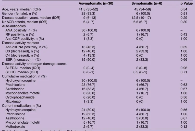
Table 1 Demographical, clinical, laboratory and therapeutic characteristics of asymptomatic and symptomatic patients with SLE
Asymptomatic (n=30) Symptomatic (n=6) p Value
Age, years, median (IQR) 41.5 (35–52) 45 (34–58) 0.54
Gender (female), n (%) 28 (93.3) 6 (100.0) 0.51
Disease duration, years, median (IQR) 9 (6–13) 12.5 (10–17) 0.29
Nr ACR criteria, median (IQR) 6 (4–7) 6.5 (6–7) 0.30
Auto-antibodies
ANA positivity, n (%) 30 (100.0) 6 (100.0) –
RF positivity, n (%) 2 (6.7) 1 (16.7) 0.43
Anti-CCP positivity, n (%) 1 (3.3) 0 (0) 1.00
Disease activity markers
Anti-dsDNA positivity, n (%) 13 (43.3) 4 (66.7) 0.39
C3 (decreased), n (%) 12 (40.0) 2 (33.3) 1.00
C4 (decreased), n (%) 4 (13.3) 0 (0) 1.00
ESR (increased), n (%) 15 (50.0) 2 (33.3) 0.66
Disease activity and organ damage scores
SLEDAI, median (IQR) 2 (0–4) 2 (0–8) 0.96
SLICC, median (IQR) 0 (0–1) 0.5 (0–1) 0.71
Cumulative medication, n (%) Hydroxychloroquine 30 (100.0) 6 (100.0) – Prednisolone 23 (76.7) 4 (66.7) 0.63 Azathioprine 16 (53.3) 4 (66.7) 0.67 Mycophenolate mofetil 6 (20.0) 1 (16.7) 1.00 Cyclophosphamide 6 (20.0) 0 (0) 0.56 Rituximab 1 (3.3) 0 (0) 1.00 Current medication, n (%) Hydroxychloroquine 24 (80.0) 6 (100.0) 0.56 Prednisolone 19 (63.3) 4 (66.7) 1.00 Azathioprine 12 (40.0) 3 (50.0) 0.67 Mycophenolate mofetil 5 (16.7) 1 (16.7) 1.00 Methotrexate 2 (6.7) 2 (33.3) 0.12
Statistical tests: Wilcoxon rank sum test,χ2test, Fisher’s exact test.
ANA, anti-nuclear antibodies; Anti-CCP, anti-citrullinated protein antibody; C3, C3 complement fraction; C4, C4 complement fraction; ds-DNA, anti-double-stranded DNA antibody; ESR, erythrocyte sedimentation rate; RF, rheumatoid factor antibody; SLEDAI, SLE Disease Activity Index; SLICC, Systemic Lupus International Collaborating Clinics.
Table 2 Number of joints with SH and PD signal in each group
Controls SLE SLE asymptomatic SLE symptomatic
A vs B p Value A vs C p Value A vs D p Value C vs D p Value Number of joints (n=220) (n=792) (n=660) (n=132) A B C D SH≥1, n (%) 6 (2.7) 88 (11.1) 54 (8.2) 34 (25.7) <0.001 0.005 <0.001 <0.001 SH≥2, n (%) 0 25 (3.1) 5 (0.7) 20 (15.1) 0.002 0.23 <0.001 <0.001 PD≥1, n (%) 0 26 (3.3) 9 (1.4) 17 (12.9) 0.002 0.07 <0.001 <0.001
Statistical tests:χ2test, Fisher’s exact test.
the groups of patients and controls by using two sets of global US scores. SH index and GLOESS scores were similar in the asymptomatic SLE group; however, in the symptomatic group GLOESS values were slightly higher. We believe GLOESS may be advantageous, as it includes information about SH and PD signal in the same score. Although the prevalence of asymptomatic patients with SLE with SH did not reach statistical significance in com-parison to healthy controls, both SH index and GLOESS were significantly higher in the asymptomatic SLE popu-lation when compared with the control group. Altogether, these findings encourage the use of global US scores for a more detailed joint evaluation in SLE, in clinical practice and in future trials.
Compared with the study of Yoon et al,14 our study detected a higher prevalence of SH (grade≥1) in asymp-tomatic patients with SLE. While the previous study identified joint effusion and/or SH in 58% of asymp-tomatic patients with SLE, we detected SH (grade ≥1) in 77% asymptomatic patients with SLE. This may be due to the larger number of joints examined in our study (22 joints per patient, in comparison with 3 joint recesses in the previous study—wrist, 2nd and 3rd MCP) and to the higher frequency of our linear array probe (15 MHz, in comparison to 10 MHz in the previous study). However, while the previous study classified these 58% of patients as having subclinical synovitis, only 23% of the asymptomatic patients with SLE in our study were classified as having subclinical joint inflammation.
In our study, patients with abnormal US PD findings did not show significant differences regarding demo-graphic, clinical and laboratory data when compared with those with normal US PD findings. In addition, no
significant correlation was found between US PD vari-ables and disease activity or organ damage scores. This is in agreement with several previous studies, suggesting that global assessment of patients with SLE should be complemented by imaging modalities, such as US.35 37 39 Interestingly, patients under treatment with oral prednisolone had higher PD indexes. In addition, the prevalence of PD signal and PD indexes were signi fi-cantly higher in the symptomatic patients with SLE when compared with the asymptomatic patients and controls, suggesting that PD signal could be considered a marker for active musculoskeletal disease.36
The most important limitations of our study are the moderate sample size (which may limit generalisability) and the high prevalence of steroid use among the SLE population (which could reduce the global in flamma-tory burden and result in lower prevalence and grading of the US PD findings). Furthermore, intake of non-steroidal anti-inflammatory drugs at the time of US imaging, a potential confounder in US PD evaluation, was not accounted for. Although US-detected subclinical joint abnormalities have been previously associated with the development of musculoskeletal symptoms, the prog-nostic value of thesefindings remains to be determined. Larger longitudinal studies are required to confirm the significance of subclinical US PD findings, specifically regarding the predictive value for the development of long-term joint damage.13 40
In conclusion, a small subgroup of patients with SLE without artralgia or clinical evidence of arthritis may present subclinical joint inflammation. Global US scores and PD signal may be important in disease evaluation and therapeutical monitoring.
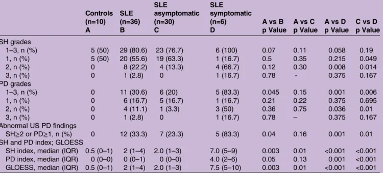
Table 3 Prevalence of US PD findings and global US PD scores in each group Controls SLE SLE asymptomatic SLE symptomatic A vs B p Value A vs C p Value A vs D p Value C vs D p Value (n=10) (n=36) (n=30) (n=6) A B C D SH grades 1–3, n (%) 5 (50) 29 (80.6) 23 (76.7) 6 (100) 0.07 0.11 0.058 0.19 1, n (%) 5 (50) 20 (55.6) 19 (63.3) 1 (16.7) 0.5 0.35 0.215 0.049 2, n (%) 0 8 (22.2) 4 (13.3) 4 (66.7) 0.12 0.30 0.008 0.014 3, n (%) 0 1 (2.8) 0 1 (16.7) 0.78 - 0.375 0.167 PD grades 1–3, n (%) 0 11 (30.6) 6 (20) 5 (83.3) 0.045 0.15 0.001 0.006 1, n (%) 0 6 (16.7) 5 (16.7) 1 (16.7) 0.21 0.22 0.375 0.695 2, n (%) 0 4 (11.1) 1 (3.3) 3 (50) 0.36 0.75 0.036 0.01 3, n (%) 0 1 (2.8) 0 1 (16.7) 0.78 – 0.375 0.167 Abnormal US PD findings SH≥2 or PD≥1, n (%) 0 12 (33.3) 7 (23.3) 5 (83.3) 0.04 0.16 0.001 0.01
SH and PD index; GLOESS
SH index, median (IQR) 0.5 (0–1) 2 (1–4) 2.0 (1–3) 7.0 (5–9) 0.003 0.01 <0.001 <0.001 PD index, median (IQR) 0 (0–0) 0 (0–1) 0 (0–0) 4.0 (2–6) 0.05 0.13 0.001 <0.001 GLOESS, median (IQR) 0.5 (0–1) 2 (1–4) 2.0 (1–3) 7.5 (5–10) 0.003 0.01 <0.001 <0.001 Abnormal US PD findings were defined as presence of SH≥2 or PD signal≥1. Statistical tests: Wilcoxon rank sum test, χ2test, Fisher
’s exact test.
GLOESS, Global OMERACT–EULAR Synovitis Score; PD, power Doppler; SH, synovial hypertrophy; SLE, systemic lupus erythematosus; US, ultrasound.
Author affiliations
1Radiology Department, Hospital de Santo António dos Capuchos, Centro Hospitalar de Lisboa Central (CHLC), Lisbon, Portugal
2Autoimmune Disease Unit, Unidade de Doenças Auto-imunes/Serviço Medicina 3, Hospital de Santo António dos Capuchos, CHLC, Lisbon, Portugal 3Núcleo de Estudos de Doenças Auto-imunes da Sociedade Portuguesa de Medicina Interna (NEDAI/SPMI), Lisbon, Portugal
4Autoimmune Disease Unit, Unidade de Doenças Auto-imunes/Serviço Medicina 7.2, Hospital Curry Cabral, CHLC, Lisbon, Portugal
Contributors All persons who meet authorship criteria are listed as authors, and all authors certify that they have made substantial contributions to the work to take public responsibility for the content. Each author agrees to be accountable for all aspects of the work in ensuring that questions related to the accuracy or integrity of any part of the work are appropriately investigated and resolved. Furthermore, each author certifies that this material or similar material has not been and will not be submitted to or published in any other publication. Specific contributions made by each author: Category 1– Conception and design of study: CAR, SP, LSV, MFMF: Acquisition of data: CAR, RMo, JFO, SP, LSV, MFMF: Analysis and/or interpretation of data: CAR, MFM-F. Category 2– Drafting the manuscript: CAR MFMF – Revising the manuscript critically for important intellectual content: CAR, RM, JFO, SP, LSV, MFM-F. Category 3– Approval of the version of the manuscript to be published: CAR, RM, JFO, SP, LSV, MFM-F.
Competing interests None declared.
Patient consent Obtained.
Ethics approval All procedures performed were in accordance with the ethical standards of the Institutional Ethics Committee ((Comissão de Ética do Centro Hospitalar de Lisboa Central; approval number 1115 (2014)) and with the 1964 Helsinki declaration and its later amendments or comparable ethical standards.
Provenance and peer review Not commissioned; externally peer reviewed.
Data sharing statement No additional data are available.
Open Access This is an Open Access article distributed in accordance with the Creative Commons Attribution Non Commercial (CC BY-NC 4.0) license, which permits others to distribute, remix, adapt, build upon this work non-commercially, and license their derivative works on different terms, provided the original work is properly cited and the use is non-commercial. See: http:// creativecommons.org/licenses/by-nc/4.0/
REFERENCES
1. Grossman JM. Lupus arthritis.Best Pr Res Clin Rheumatol
2009;23:495–506.
2. Malcus Johnsson P, Sandqvist G, Bengtsson A, et al. Hand function and performance of daily activities in systemic lupus erythematosus.
Arthritis Rheum2008;59:1432–8.
3. Ball EM, Bell AL. Lupus arthritis-do we have a clinically useful classification?Rheumatology (Oxford)2012;51:771–9.
4. van Vugt RM, Derksen RH, Kater L, et al. Deforming arthropathy or lupus and rhupus hands in systemic lupus erythematosus.
Ann Rheum Dis1998;57:540–4.
5. Mosca M, Tani C, Carli L, et al. The role of imaging in the evaluation of joint involvement in 102 consecutive patients with systemic lupus erythematosus.Autoimmun Rev2015;14:10–15.
6. Kane D, Grassi W, Sturrock R, et al. Musculoskeletal ultrasound—A state of the art review in rheumatology. Part 2: Clinical indications for musculoskeletal ultrasound in rheumatology.Rheumatology (Oxford)
2004;43:829–38.
7. Szkudlarek M, Court-Payen M, Strandberg C, et al. Power Doppler ultrasonography for assessment of synovitis in the
metacarpophalangeal joints of patients with rheumatoid arthritis: a comparison with dynamic magnetic resonance imaging.Arthritis Rheum2001;44:2018–23.
8. Terslev L, Torp-Pedersen S, Savnik A, et al. Doppler ultrasound and magnetic resonance imaging of synovial inflammation of the hand in rheumatoid arthritis: a comparative study.Arthritis Rheum
2003;48:2434–41.
9. Iagnocco A, Epis O, Delle Sedie A, et al. Ultrasound imaging for the rheumatologist XVII. Role of colour Doppler and power Doppler. Clin Exp Rheumatol 2008;26:759–62.
10. Marcusa DP, Mandl LA. Challenges in imaging in preclinical rheumatoid arthritis.Rheum Dis Clin North Am2014;40:727–34. 11. Bresnihan B, Kane D. Sonography and subclinical synovitis.
Ann Rheum Dis2004;63:333–4.
12. Colebatch AN, Edwards CJ, Østergaard M, et al. EULAR recommendations for the use of imaging of the joints in the clinical management of rheumatoid arthritis.Ann Rheum Dis
2013;72:804–14.
13. Zayat AS, Md Yusof MY, Wakefield RJ, et al. The role of ultrasound in assessing musculoskeletal symptoms of systemic lupus erythematosus: a systematic literature review.Rheumatology (Oxford)2016;55:485–94.
14. Yoon HS, Kim KJ, Baek IW, et al. Ultrasonography is useful to detect subclinical synovitis in SLE patients without musculoskeletal involvement before symptoms appear.Clin Rheumatol
2014;33:341–8.
15. Ejbjerg B, Narvestad E, Rostrup E, et al. Magnetic resonance imaging of wrist and finger joints in healthy subjects occasionally shows changes resembling erosions and synovitis as seen in rheumatoid arthritis.Arthritis Rheum2004;50:1097–106.
16. Parodi M, Silvestri E, Garlaschi G, et al. How normal are the hands of normal controls? A study with dedicated magnetic resonance imaging. Clin Exp Rheumatol 2006;24:134–41.
17. Mangnus L, Schoones JW, van der Helm-van Mil AH. What is the prevalence of MRI-detected inflammation and erosions in small joints in the general population? A collation and analysis of published data.RMD Open2015;1:e000005.
18. Padovano I, Costantino F, Breban M, et al. Prevalence of ultrasound synovial inflammatory findings in healthy subjects.Ann Rheum Dis
2016;75:1819–23.
19. Tan EM, Cohen AS, Fries JF, et al. The 1982 revised criteria for the classification of systemic lupus erythrematosus.Arthritis Rheum
1982;25:1271–7.
20. Moraes-Fontes MF, Lúcio I, Santos C, et al. Neuropsychiatric features of a cohort of patients with systemic lupus erythematosus.
ISRN Rheumatol2012;2012:989218.
21. Panush RS, Lawrence N, Longley S, et al.‘Rhupus’ Syndrome.
Arch Intern Med1988;148:1633–6.
22. Alarcon-Segovia D, Abud-Mendoza C, Diaz-Jouanen E, et al. Deforming arthropathy of the hands in systemic lupus erythematosus. J Rheumatol 1988;15:65–9.
23. Grunke M, Antoni CE, Kavanaugh A, et al. Standardization of joint examination technique leads to a significant decrease in variability among different examiners.J Rheumatol2010;37:860–4.
24. Romero-Diaz J, Isenberg D, Ramsey-Goldman R. Measures of adult systemic lupus erythematosus: updated version of British Isles Lupus Assessment Group (BILAG 2004), European Consensus Lupus Activity Measurements (ECLAM), Systemic Lupus Activity Measure, Revised (SLAM-R), Systemic Lupus Activity Questionnaire for Population Studies (SLAQ), Systemic Lupus Erythematosus Disease Activity Index 2000 (SLEDAI-2 K), and Systemic Lupus International Collaborating Clinics/American College of Rheumatology Damage Index (SDI).Arthritis Care Res2011;63 (Suppl. 11):S37–46.
25. Gladman D, Ginzler E, Goldsmith C, et al. The development and initial validation of the systemic lupus international
collaborating clinics/American College of Rheumatology Damage Index for Systemic Lupus Erythematosus.Arthritis Rheum
1996;39:363–9.
26. Yee CS, Farewell VT, Isenberg DA, et al. The use of systemic lupus erythematosus disease activity index-2000 to define active disease and minimal clinically meaningful change based on data from a large cohort of systemic lupus erythematosus patients.Rheumatology (Oxford)2011;50:982–8.
27. Torp-Pedersen ST, Terslev L. Settings and artefacts relevant in colour/power Doppler ultrasound in rheumatology.Ann Rheum Dis
2008;67:143–9.
28. Backhaus M, Burmester GR, Gerber T, et al. Guidelines for musculoskeletal ultrasound in rheumatology.Ann Rheum Dis
2001;60:641–9.
29. Wakefield RJ, Balint PV, Szkudlarek M, et al. Musculoskeletal ultrasound including definitions for ultrasonographic pathology. J Rheumatol 2005;32:2485–7.
30. Szkudlarek M, Court-Payen M, Jacobsen S, et al. Interobserver agreement in ultrasonography of the finger and toe joints in rheumatoid arthritis.Arthritis Rheum2003;48:955–62.
31. D’Agostino MA, Wakefield RJ, Berner-Hammer H, et al. Value of ultrasonography as a marker of early response to abatacept in patients with rheumatoid arthritis and an inadequate response to methotrexate: results from the APPRAISE study.Ann Rheum Dis
32. Hammer HB, Bolton-King P, Bakkeheim V, et al. Examination of intra and interrater reliability with a new ultrasonographic reference atlas for scoring of synovitis in patients with rheumatoid arthritis.
Ann Rheum Dis2011;70:1995–8.
33. Machado FS, Furtado RNV, Takahashi RD, et al. Sonographic cutoff values for detection of abnormalities in small, medium and large joints: A comparative study between patients with rheumatoid arthritis and healthy volunteers.Ultrasound Med Biol2015;41:989–98. 34. Filer A, de Pablo P, Allen G, et al. Utility of ultrasound joint counts in
the prediction of rheumatoid arthritis in patients with very early synovitis.Ann Rheum Dis2011;70:500–7.
35. Delle Sedie A, Riente L, Scirè CA, et al. Ultrasound imaging for the rheumatologist XXIV. Sonographic evaluation of wrist and hand joint and tendon involvement in systemic lupus erythematosus. Clin Exp Rheumatol 2009;27:897–901.
36. Gabba A, Piga M, Vacca A, et al. Joint and tendon involvement in systemic lupus erythematosus: An ultrasound study of
hands and wrists in 108 patients.Rheumatol2012; 51:2278–85.
37. Torrente-Segarra V, Lisbona MP, Rotés-Sala D, et al. Hand and wrist arthralgia in systemic lupus erythematosus is associated to ultrasonographic abnormalities.Jt Bone Spine
2013;80:402–6.
38. Dreyer L, Jacobsen S, Juul L, et al. Ultrasonographic abnormalities and inter-reader reliability in Danish patients with systemic lupus erythematosus—a comparison with clinical examination of wrist and metacarpophalangeal joints.Lupus
2015;24:712–19.
39. Iagnocco A, Ceccarelli F, Rizzo C, et al. Ultrasound evaluation of hand, wrist and foot joint synovitis in systemic lupus erythematosus.
Rheumatol2014;53:465–72.
40. Lins CF, Santiago MB. Ultrasound evaluation of joints in systemic lupus erythematosus: a systematic review.Eur Radiol
without musculoskeletal symptoms
patients with systemic lupus erythematosus
inflammation in the hands and wrists of
Ultrasound detects subclinical joint
Vieira and Maria Francisca Moraes-Fontes
Carina A Ruano, Rui Malheiro, João F Oliveira, Sofia Pinheiro, Luís S
doi: 10.1136/lupus-2016-000184
2017 4:Lupus Sci Med
http://lupus.bmj.com/content/4/1/e000184 Updated information and services can be found at:
These include:
References
#BIBL
http://lupus.bmj.com/content/4/1/e000184
This article cites 40 articles, 19 of which you can access for free at:
Open Access
http://creativecommons.org/licenses/by-nc/4.0/
non-commercial. See:
provided the original work is properly cited and the use is
non-commercially, and license their derivative works on different terms, permits others to distribute, remix, adapt, build upon this work
Commons Attribution Non Commercial (CC BY-NC 4.0) license, which This is an Open Access article distributed in accordance with the Creative
service
Email alerting
box at the top right corner of the online article.
Receive free email alerts when new articles cite this article. Sign up in the
Collections
Topic
Articles on similar topics can be found in the following collections (6)Immunology and Inflammation
Notes
http://group.bmj.com/group/rights-licensing/permissions To request permissions go to:
http://journals.bmj.com/cgi/reprintform To order reprints go to:
http://group.bmj.com/subscribe/ To subscribe to BMJ go to: