FACULDADE DE FARMÁCIA
SOMATIC EXPRESSION OF THE TESTIS-SPECIFIC
PDHA2 GENE: MECHANISMS OF
ACTIVATION/SILENCING
Ana Sofia da Costa Pinheiro
DOUTORAMENTO EM FARMÁCIA
Biologia Celular e Molecular
FACULDADE DE FARMÁCIA
SOMATIC EXPRESSION OF THE TESTIS-SPECIFIC
PDHA2 GENE: MECHANISMS OF
ACTIVATION/SILENCING
b
EXPRESSÃO SOMÁTICA DO GENE PDHA2:
MECANISMOS DE ACTIVAÇÃO E SILENCIAMENTO
Research Advisor: Professora Doutora Isabel Antolin Rivera Co-advisor: Professora Doutora Maria João Ferreira da Silva
Ana Sofia da Costa Pinheiro
Lisboa
2012
Dissertação apresentada à Faculdade de Farmácia da Universidade de Lisboa para obtenção do grau de Doutor em Farmácia (Biologia Celular e Molecular)
De acordo com o disposto no ponto 1 do artigo nº41 do Regulamento de Estudos Pós-Graduados da Universidade de Lisboa, deliberação nº93/2006, publicada em Diário da República – II série nº153 – 5 de Julho de 2003, a autora desta dissertação declara que participou na concepção e execução do trabalho experimental, interpretação dos resultados obtidos e redacção dos manuscritos.
The studies presented in this thesis were performed at Research Institute for Medicines and Pharmaceutical Sciences (iMed.UL), Faculty of Pharmacy, University of Lisbon - Portugal, under the scientific supervision of Professor Isabel Antolin Rivera and Professor Maria João Ferreira da Silva.
Ana Sofia da Costa Pinheiro is the recipient of a PhD fellowship (SFRH / BD / 31264 / 2006) from Fundação para a Ciência e Tecnologia (FCT), Lisbon, Portugal. This work was supported in part by POCI / SAU-MMO / 57052 / 2004 from FCT.
ACKNOWLEDGEMENTS
Em primeiro lugar, gostaria de expressar a minha gratidão e reconhecimento a todos que, directa ou indirectamente, contribuiram para a realização desta tese.
As minhas primeiras palavras de agradecimento dirigem-se à Professora Doutora Isabel Tavares de Almeida, por me ter aceite no seu grupo de investigação e por ter reunido todas as condições que permitiram conduzir este trabalho a bom porto.
E às minhas orientadoras, Professoras Doutoras Isabel Antolin Rivera e Maria João Silva, a quem dedico esta tese... Muito mais que orientadoras, colegas e amigas, a palavra “obrigada” não consegue tão pouco reflectir o que sinto.
Professora Isabel: obrigada por TUDO... Em particular agradeço-lhe todos os ensinamentos, a inestimável ajuda na realização da tese e a mais que permanente disponibilidade. Acima de tudo, obrigada por ter acreditado!
Professora João: obrigada por me ajudar nos importantes primeiros passos, por me ter deixado entrar no mundo da PDH e da AVB, e por me ter permitido continuar o seu trabalho. Agradeço-lhe também o apoio, interesse e preocupação constantes!
Professora Doutora Elsa Rodrigues: obrigada por ser orientadora sem o ser! A si lhe agradeço as inúmeras sugestões e discussões de trabalho prático, a importante revisão da escrita e, claro, o seu constante optimismo!
Aos Professores Doutores Mário de Sousa e Alberto Barros manifesto o meu mais sentido agradecimento pela disponibilização do material biológico que possibilitou a maioria do trabalho realizado. Em especial, agradeço ao Professor Mário os preciosos ensinamentos quando me acolheu no seu laboratório, e a revisão cuidada dos artigos.
Ao Professor Doutor Domingos Henrique agradeço o período que passei no IMM e todo o conhecimento que o seu grupo me transmitiu.
I deeply acknowledge Professor Mulchand Patel for being a gentle collaborator and sharing his knowledge and experience with our group.
É com a maior satisfação que expresso uma palavra de reconhecimento a todos os que contribuíram para a obtenção dos resultados... À Inês Faustino pela implementação do MS-PCR, pela amizade e admirável competência; à Madalena e à Ângela pelo apoio, persistência e incansável dedicação ao NB; à Cristina pelos resultados da 2D e pela organização do laboratório. Em especial, agradeço à Inês Milagre e à Maria os inúmeros e cruciais ensinamentos práticos, e claro, a paciência e
disponibilidade que sempre demonstraram. Ao Miguel agradeço também o “template” da figura 1.8.
A todos os outros colegas e amigos que estiveram presentes durante este tempo: Mónica, Paula, Cátia, Sandra, Andreia, Ruben R., Ana Isabel, Marisa, Ruben E., Sara, Israel, Marco, João, Ana Serrão, Conceição e tantos outros. Obrigada por me terem ouvido e encorajado sempre! Este percurso não seria o mesmo sem a vossa presença!
Em geral expresso uma palavra de gratidão a todos os Professores, Colegas, Amigos e Técnicos da FFUL, IMM e ICBAS.
Por fim, não poderia deixar de agradecer à minha família... Aos meus Pais e Irmão, a quem eu devo o que sou hoje! E à família do Tiago que me “adoptou”.
A ti Tiago, agradeço por estares ao meu lado estes anos todos, e por tudo o que me deste e que só nós sabemos... Obrigada pelo que percebes, e especialmente pelo que não percebes!
TABLE OF CONTENTS
Abbreviations ... xvii
Abstract ... xxi
Resumo ... xxiii
CHAPTER 1: 1. General Introduction and Objectives ... 1
1.1. Pyruvate Dehydrogenase Complex (PDC) ... 3
1.1.1. Biological importance ... 3
1.1.2. Structural organization ... 5
1.1.3. Biochemical and molecular structure ... 6
1.1.4. Regulation ... 9
1.1.5. Pyruvate Dehydrogenase Complex Deficiency (PDCD) ... 11
1.2. PDHA2 gene ... 15
1.2.1. Structure and evolution ... 15
1.2.2. Tissue- and cell-specific expression ... 17
1.2.3. Transcriptional regulation ... 19
1.2.3.1. Promoter structure ... 19
1.2.3.2. Methylation and tissue specificity ... 20
1.3. Transcriptional regulation of gene expression ... 22
1.3.1. Core promoter elements and general transcription factors ... 23
1.3.2. Other regulatory elements ... 24
1.3.3. Chromatin remodelling ... 25
1.3.3.1 Histone variants and histone modifications ... 26
1.3.3.2 DNA methylation ... 28
1.4 Objectives ... 33
CHAPTER 2: 2. The unique case of a family expressing the testis-specific PDHA2 gene in somatic tissues ... 35
2.1. Abstract ... 37
2.2. Introduction ... 37
2.3.1. Case report ... 39
2.3.2. Sample preparation ... 40
2.3.3. Western immunoblotting ... 40
2.3.4. Two-dimensional electrophoresis ... 41
2.3.5. Preparation of genomic DNA, total RNA and cDNA ... 41
2.3.6. PCR of genomic DNA and cDNA ... 42
2.3.7. Single strand conformational polymorphism analysis (SSCP) ... 42
2.3.8. qPCR for PDHA1 and PDHA2 expression analysis and PDHA1 gene dosage ... 44
2.3.9. Sequence analysis of PDHA1 and PDHA2 promoter regions ... 44
2.3.10. Sodium bisulfite genomic DNA sequencing of the CpG islands in PDHA2 gene ... 45
2.4. Results ... 45
2.4.1. Identification of a 5’-truncated PDHA1 transcript and a full-length PDHA2 transcript in somatic cells ... 46
2.4.2. Protein expression analysis reveals the co-existence of both PDHA proteins in somatic tissues ... 50
2.4.3. Trying to explain the truncated PDHA1 transcript ... 51
2.4.4. Potential mechanism underlying PDHA2 expression in somatic cells ... 52
2.5. Discussion ... 53
CHAPTER 3: 3. Pyruvate dehydrogenase complex: mRNA and protein expression patterns of PDHα subunit genes in human spermatogenesis ... 59
3.1. Abstract ... 61
3.2. Introduction ... 61
3.3. Materials and methods ... 63
3.3.1. Human samples ... 63
3.3.2. Cell isolation ... 64
3.3.3. Western immunoblotting ... 65
3.3.4. Two-dimensional electrophoresis ... 65
3.3.5. Immunohistochemistry ... 66
3.3.6. RNA isolation and qRT-PCR ... 66
3.4.1. Expression pattern of PDHA proteins in testis and somatic tissues ... 67
3.4.2. Specific detection of testis and somatic PDHA mRNAs ... 71
3.5. Discussion ... 73
CHAPTER 4: 4. Human testis-specific PDHA2 gene: methylation status of a CpG island in the open reading frame correlates with transcriptional activity ... 77
4.1. Abstract ... 79
4.2. Introduction ... 79
4.3. Material and methods ... 82
4.3.1. Source of human samples ... 82
4.3.2. Cell isolation ... 82
4.3.3. Nucleic acids extraction ... 83
4.3.4. Sodium bisulfite genomic DNA sequencing ... 83
4.3.5. Reverse transcription-polymerase chain reaction (RT-PCR) ... 84
4.3.6. CpG island prediction analysis ... 84
4.4. Results ... 85
4.5. Discussion ... 87
CHAPTER 5: 5. Demethylation of the coding region triggers the activation of the human testis-specific PDHA2 gene in somatic tissues ... 91
5.1. Abstract ... 93
5.2. Introduction ... 93
5.3. Materials and methods ... 95
5.3.1. Cloning of the human PDHA2 proximal promoter ... 95
5.3.2. PDHA2 promoter reporter constructs ... 96
5.3.3. Transactivation studies ... 96
5.3.4. Cell cultures and treatments ... 96
5.3.5. RNA isolation and qRT-PCR for PDHA2 expression analysis ... 97
5.3.6. Genomic DNA isolation and bisulfite sequencing for PDHA2 methylation analysis ... 98
5.3.7. Chromatin immunoprecipitation assays ... 98
5.4. Results ... 99
5.4.1. PDHA2 promoter constructs display basal activity in human somatic cell lines ... 99 5.4.2. Activation of PDHA2 expression in somatic cells after treatment with
epigenetic drugs ... 101 5.4.3. DAC induces demethylation of PDHA2 coding region ... 103
5.5. Discussion ... 103
CHAPTER 6:
6. Concluding Remarks ... 109
REFERENCES ... 117
FIGURES
Figure 1.1. PDC relationships with energy metabolism …….….…...………... 3
Figure 1.2. Sequential reactions catalyzed by PDC ………...… 4
Figure 1.3. Schematic representation of eukaryotic PDC structure ………... 5
Figure 1.4. Schematic representation of PDC global regulation ….……….. 10
Figure 1.5. Illustration of mutations prevalence in PDCD .………... 12
Figure 1.6. Schematic representation of human spermatogenesis ..………... 17
Figure 1.7. Proximal promoter sequence of the human PDHA2 gene ..………. 19
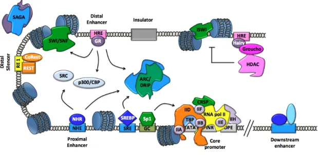
Figure 1.8. Elements of eukaryotic gene transcriptional regulation ..……… 22
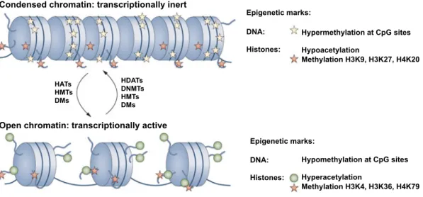
Figure 1.9. Epigenetic marks in chromatin remodelling ..……….. 26
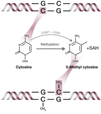
Figure 1.10. Methylation modification of DNA ………...………. 29
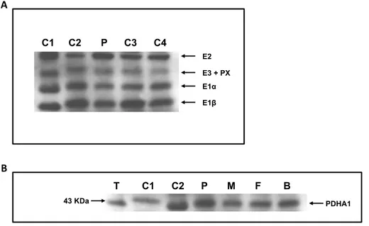
Figure 2.1. Western blot analysis of lymphocyte and testis homogenates .………... 46
Figure 2.2. RT-PCR analysis of PDH E1α transcripts .………. 48
Figure 2.3. Schematic representation of the PDHA1 mRNA sequence …….……… 48
Figure 2.4. qRT-PCR analysis to evaluate PDHA1 and PDHA2 gene expression … 50 Figure 2.5. Bidimensional analysis of PDHA1 in several cellular homogenates ….. 51
Figure 2.6. DNA methylation status of PDHA2 gene coding region ……….. 53
Figure 2.7. Segregation of the X chromosome in the family under study …….…… 55
Figure 3.1. Western blot analysis of E1α in several human tissues …..………… 68
Figure 3.2. Immunoblot analysis of human tissues using anti-PDHA1 antibody …. 69 Figure 3.3. Immunostaining of E1α protein in a human testis section …………. 70
Figure 3.4. qRT-PCR analysis of PDH genes expression in different cells ………. 71
Figure 3.5. qRT-PCR analysis of PDH genes expression during spermatogenesis ... 72
Figure 4.1. Schematic representation of CpG sites distribution in PDHA2 gene ….. 85
Figure 4.2. DNA sequencing analysis of CpG island II in PDHA2 gene ………….. 86
Figure 4.3. Methylation profiles of the two CpG islands present in PDHA2 gene … 86 Figure 4.4. Transcription profiles of PDHA genes in different tissues ………. 87
Figure 5.1. Functional deletion analysis of the human PDHA2 gene promoter …… 100
Figure 5.2. DAC increases PDHA2 mRNA levels in SH-SY5Y cells ……….. 102
Figure 5.3. Recruitment of RNA pol II to the human PDHA2 gene promoter. …… 102
TABLES
Table 1.1. Summary of the genes encoding PDC components ……….. 7 Table 2.1. List of primers used in the family´s study……….. 43 Table 2.2. Calculations for determining the copy number of PDHA1 gene …...… 52 Table 3.1. Tissue-specific profile of human PDHA genes ... 73 Table 3.2. Cell-specific profile of human PDHA genes during spermatogenesis .. 73 Table 5.1. List of the primers used in the human PDHA2 promoter cloning ...….. 95 Table 5.2. Statistical analysis of PDHA2 gene methylation results …...………… 105
ABBREVIATIONS
5hmC 5-hydroxymethylcytosine
5mC 5-methylcytosine
A adenosine
aa amino acids
ADP adenosine diphosphate
ALF TFIIAα/β-life factor
ARC/DRIP activator-recruited cofactor/ vitamin D receptor interacting protein ATF/CREB activating transcription factor/ cAMP-response element-binding protein ATP adenosine triphosphate
BORIS brother of the regulator of imprinted sites
bp base pairs
C cytidine
Ca2+
calcium ion
cAMP cyclic adenosine monophosphate
cDNA complementary DNA
ChIP chromatin immunoprecipitation
Chr chromosome
CNS central nervous system
CoA coenzyme A
CpG dinucleotide of cytosine and guanine separated by one phosphate group
CREMτ cAMP-responsive element modulator τ
CRSP co-factor required for Sp1 activation
Da Dalton
DAC 5’-aza-2’-deoxycytidine
DMEM Dulbecco’s modified Eagle’s medium
DNA deoxyribonucleic acid
DNMT DNA methyltransferase
DPE downstream promoter element
E1 pyruvate dehydrogenase
E2 dihydrolipoyllysine-residue acetyltransferase
E3 dihydrolipoyl dehydrogenase
Ets E-twenty six
G guanosine
GR glucocorticoid receptor
GTFs general transcription factors
HAT histone acetyl transferase
HDAC histone deacetylase
HRE hormone responsive element
Inr initiator
ISWI imitation switch
kb kilo base pairs
kDa kilo Daltons
MBD methyl-CpG binding domain
MEP mouse E1α promoter transcription factor
Mg2+
magnesium ion
mRNA messenger RNA
NAD+
nicotinamide adenine dinucleotide
NADH nicotinamide adenine dinucleotide (reduced form)
NF-κB nuclear factor κ B
NHE nuclease hypersensitive element
NHR nuclear hormone receptor
NT2 Ntera2/clone D1
p300/CBP E1A binding protein p300 / CREB-binding protein
PDC pyruvate dehydrogenase complex
PDCD pyruvate dehydrogenase complex deficiency
PDK pyruvate dehydrogenase kinase
PDP pyruvate dehydrogenase phosphatase
Pi inorganic phosphate
REST RE 1-silencing transcription factor
RNA ribonucleic acid
RNA pol II RNA polymerase II
SAGA Spt-Ada-Gcn5-Acetyltransferase
SAM S-adenosyl methionine
Sp1 specificity protein 1
SRE sterol-regulatory element
SREBP sterol-regulatory element binding protein
SSCP single-strand conformation polymorphism
SWI/SNF SWItch/Sucrose NonFermentable
T thymidine
TAFs TBP-associated factors
TBP TATA binding protein
TFs transcription factors
TPP thiamine pyrophosphate
TSA trichostatin A
U uridine
ABSTRACT
Pyruvate Dehydrogenase Complex Deficiency (PDCD) is a severe inborn error of metabolism that causes cell energy impairment, and predominantly results from mutations in PDHA1 gene that encodes the crucial E1α subunit. Albeit, there is a second isoform encoded by PDHA2, a testis-specific gene, whose potential activation in somatic tissues constitutes a conceptual therapy for PDCD.
Since the human PDHA2 gene regulation has remained elusive over the time, the present work was designed to disclose the regulatory mechanisms involved in this gene tissue-specific expression, namely its activation in spermatogenic cells and its silencing in somatic ones.
The first part of this thesis (chapter 1) comprises a general introduction with a review of the literature concerning Pyruvate Dehydrogenase Complex (PDC), PDHA2 gene and transcriptional regulation, later ending with the objectives of the present work.
Our studies commenced by exploring an interesting case that was found during our routine laboratory diagnostics (chapter 2). We reported a family, whose PDC molecular analysis revealed the presence in circulating lymphocytes of an incomplete
PDHA1 transcript together with a normal PDHA2 transcript. Moreover, both transcripts
were translated into full-length proteins, thus leading to the co-existence of both PDHA isoforms in somatic cells. This was the first time that the expression of the PDHA2 gene was observed in somatic cells and, accordingly, it represented a unique opportunity to study PDHA2 regulatory mechanisms. Our findings led us to speculate that the explanation for the somatic activation of PDHA2 gene expression should rely on epigenetic mechanisms, namely DNA methylation.
Before any meaningful attempt to elucidate PDHA2 regulation, it was required to fully characterize the expression pattern of this gene. Therefore, in chapter 3 we described for the first time the transcriptional and translational patterns of human PDHA genes during spermatogenesis. We confirmed the ubiquitous expression of PDHA1 gene in all somatic cells, whereas the expression of PDHA2 gene was restricted to germ cells. Moreover, we could establish that the switch from the X-linked to the autosomic gene expression occurs in spermatocytes, before the first meiotic division of spermatogenesis.
Subsequently, and consistently with the knowledge that DNA methylation is a mechanism involved in tissue-specific regulation of PDHA2 gene in mouse, our first
approach was to analyze the transcriptional profile and DNA methylation status of
PDHA2 gene (chapter 4). Indeed, we revealed that PDHA2 methylation status was
perfectly correlated with transcriptional activity, although, and surprisingly, it was the methylation status of the gene-coding region that underlied this correlation.
In the following studies, we further explored the regulatory mechanisms that govern PDHA2 gene expression, including the role of DNA methylation and histone modifications (chapter 5). Functional deletion studies revealed that the -122 to -6 promoter region is indispensable for basal expression of this TATA-less promoter, and suggested that an epigenetic program was controlling PDHA2 gene expression. Indeed, we found that 5’-aza-2’-deoxycytidine (a hypomethylating agent) induced a relevant demethylation of the coding region and the concomitant PDHA2 gene derepression in a somatic cell line.
Taken together, our results proved the involvement of DNA methylation as a primary regulatory mechanism of PDHA2 gene expression. Moreover, we disclosed the possibility to activate PDHA2 gene expression in somatic cells, which may open new therapeutic avenues for PDCD caused by PDHA1 mutations.
Keywords: Pyruvate Dehydrogenase Complex Deficiency; PDHA2 gene;
RESUMO
O Complexo do Piruvato Desidrogenase (cPDH) desempenha um papel central no metabolismo celular por estabelecer a ligação entre a glicólise e o ciclo dos ácidos tricarboxílicos, realizando a conversão de piruvato em acetil-CoA, fundamental para a obtenção de energia. As deficiências associadas a este complexo constituem uma das doenças genéticas mais comuns do metabolismo energético mitocondrial.
O fenótipo clínico das deficiências do cPDH é caracterizado principalmente por grave hiperlactacidémia e neurodegenerescência crónica, sendo no entanto observado um largo espectro de características clínicas. Dada esta heterogeneidade, o fenótipo é normalmente classificado em três categorias distintas de acordo com a gravidade apresentada: neonatal, infantil e benigno.
As terapias actualmente empregues visam estimular a actividade do complexo e melhorar o fenótipo clínico, e consistem, por exemplo, na implementação de uma dieta cetogénica, na suplementação com cofactores (tiamina, carnitina e ácido lipóico) e/ou na administração de dicloroacetato. Contudo, estas terapias revelam-se claramente insuficientes, pois raramente alteram a degenerescência neurológica associada à patologia, o que torna deste modo pertinente a procura de novas abordagens terapêuticas, nomeadamente ao nível genotípico.
O cPDH é um sistema multienzimático constituído por múltiplas cópias de diferentes componentes: E1 (piruvato desidrogenase), um heterotetrâmero de duas subunidades α e duas β; E2 (di-hidrolipoíl-lisina acetiltransferase); E3 (di-hidrolipoíl desidrogenase); E3BP (proteína de ligação a E3); PDK (piruvato desidrogenase cinase) e PDP (piruvato desidrogenase fosfatase).
Apesar deste complexo ser codificado por 13 genes diferentes, a maioria dos casos reportados de deficiência em cPDH resulta de mutações no gene PDHA1, que codifica a subunidade α do primeiro componente catalítico do complexo, E1. Esta subunidade apresenta-se sob duas isoformas distintas: uma codificada pelo gene
PDHA1 e expressa em todos os tecidos somáticos; e uma segunda, codificada pelo gene PDHA2 e exclusivamente expressa nas células espermatogénicas. A existência desta
segunda isoforma, e a sua potencial activação nos tecidos somáticos, foi apontada por alguns autores como a terapia ideal para os casos de deficiência em cPDH que resultam de mutações no gene PDHA1. No entanto, o conhecimento dos mecanismos de regulação da expressão do gene PDHA2 humano é extremamente diminuto e,
consequentemente, o principal objectivo do presente trabalho consistiu em investigar os mecanismos que modulam a especificidade tecidular deste gene, nomeadamente a sua activação nas células espermatogénicas e a sua repressão nos tecidos somáticos.
O primeiro capítulo da tese apresenta uma revisão geral da literatura sobre o cPDH, o gene PDHA2 e a regulação da transcrição génica, terminando com a descrição dos objectivos delineados para o presente trabalho.
O trabalho iniciou-se com um interessante caso clínico que surgiu na nossa rotina laboratorial de diagnóstico (capítulo 2), o qual constituiu o ponto de partida para os estudos subsequentes. Neste capítulo reportamos o estudo de uma família, cuja análise molecular ao nível dos linfócitos revelou a presença simultânea de um transcrito
PDHA1 incompleto e de um transcrito PDHA2 íntegro. Segundo o nosso conhecimento,
esta é a primeira vez que a expressão do gene PDHA2 foi observada em células somáticas, o que representa uma oportunidade única para estudar os mecanismos de regulação deste gene. A análise deste caso permitiu-nos postular que a activação somática do gene PDHA2 se encontra, muito provavelmente, relacionada com mecanismos epigenéticos de regulação, nomeadamente com o estado de metilação do DNA.
Antes de qualquer tentativa de elucidar os mecanismos envolvidos na regulação da expressão do gene PDHA2, revelou-se necessário obter algum conhecimento relativamente ao padrão de expressão deste gene em tecidos humanos, dada a ausência de dados na literatura. Desse modo, no capítulo subsequente (capítulo 3) descrevemos pela primeira vez o padrão de expressão dos genes PDHA1 e PDHA2, ao nível de expressão quer das proteínas quer dos transcritos, em diversos tecidos e células, e em particular durante a espermatogénese. Os resultados permitiram-nos confirmar a expressão somática do gene PDHA1, bem como a expressão exclusiva do gene PDHA2 em células espermatogénicas.
Para além disso, a análise do padrão de expressão destes genes durante a espermatogénese levou-nos a concluir que a forma somática PDHA1 é substituída pelo forma testicular PDHA2 ao nível do espermatócito, antes da primeira divisão meiótica das células germinativas. Observámos também que, quer ao nível do homogeneizado testicular, quer ao nível do espermatozóide maduro, a proteína PDHA2 é a única isoforma encontrada, embora com uma massa molecular inferior à esperada, e concluímos que a activação do gene PDHA2 é uma maneira de assegurar a expressão da
subunidade E1α durante a espermatogénese e garantir a viabilidade e funcionalidade destas células germinativas.
Numa primeira abordagem para compreender a regulação da expressão do gene
PDHA2, e de acordo com o conhecimento sobre a metilação do DNA como um
mecanismo importante na regulação da especificidade tecidular deste gene no murganho, começámos por avaliar os seus padrões de metilação e de transcrição em tecidos somáticos e testiculares humanos (capítulo 4). A análise da sequência do gene
PDHA2 revelou a presença de duas ilhas CpG: uma no promotor próximo que se
apresenta metilada em todos os tipos de tecidos estudados, e outra localizada na região codificante que se apresenta metilada nos tecidos somáticos e completamente desmetilada nas células espermatogénicas, onde o transcrito PDHA2 é normalmente expresso. É importante salientar que este padrão de metilação revelou uma correlação perfeita com a actividade transcricional, mas que neste caso particular é a desmetilação da ilha localizada na região codificante, e não no promotor do gene, que se correlaciona com a sua expressão.
Nos estudos posteriores, pretendemos estudar os mecanismos de regulação e clarificar o papel da metilação na expressão do gene PDHA2 (capítulo 5). Começámos por clonar diferentes porções do promotor do gene PDHA2 e procedemos à análise das respectivas actividades transcricionais após transfecção em diversas linhas de células somáticas: SH-SY5Y (neuroblastoma humano), HeLa (adenocarcinoma cervical humano) e Ntera2/Clone D1 (teratocarcinoma humano). Os resultados revelaram que a transcrição dirigida pelo promotor ocorre nestas linhas celulares, as quais não expressam normalmente o gene PDHA2, e que esta actividade transcricional não apresentou diferenças óbvias entre as diversas linhas. Estes resultados sugerem que, após introdução transiente em células somáticas de plasmídeos contendo a região 5’ adjacente do gene PDHA2, o promotor não responde aos mecanismos de regulação que normalmente operam nestas células para inibir a expressão do gene in vivo. Para além disso, estes dados sugerem também que a expressão específica de tecido do gene
PDHA2 não deverá envolver factores de transcrição específicos celulares, quer
activadores ou repressores, mas possivelmente outro nível de modulação, como por exemplo mecanismos epigenéticos de regulação.
Numa abordagem para avaliar o envolvimento de modificações epigenéticas na regulação da expressão deste gene, nomeadamente uma possível interacção entre a metilação do DNA e a acetilação das histonas, procedemos ao tratamento de uma linha
celular somática (SH-SY5Y) com o fármaco hipometilante 5’-aza-2’-desoxicitidina (DAC) e com tricostatina A (TSA) que inibe as desacetilases de histonas.
Os resultados da avaliação dos níveis de mRNA por qRT-PCR mostraram que o DAC induz a activação do gene PDHA2 em linhas celulares somáticas (SH-SY5Y), tendo-se observado, após estudos de imunoprecipitação da cromatina, o concomitante recrutamento da RNA polimerase II para o promotor próximo. Paralelamente, observou-se que o DAC induz uma desmetilação relevante da ilha CpG localizada na região codificante, o que constitui assim a prova de que a metilação do DNA na região codificante desempenha uma função importante na regulação da expressão do gene
PDHA2.
Para terminar a presente tese, é apresentada no capítulo 6 uma discussão dos resultados obtidos, incluindo uma análise integrada de todo o trabalho e respectivas conclusões, bem como algumas perspectivas de trabalho futuro.
Em suma, este trabalho contribuiu para o esclarecimento dos mecanismos de regulação da especificidade tecidular do gene PDHA2, demonstrando o envolvimento da metilação do DNA como mecanismo primário de regulação da expressão deste gene. Para além disso, demonstrou-se a possibilidade de activação do gene em células somáticas, o que constitui um avanço relevante na direcção de novas abordagens terapêuticas para as deficiências do cPDH causadas por mutações no gene PDHA1.
Palavras-Chave: Deficiência do Complexo Piruvato Desidrogenase; gene PDHA2; espermatogénese; expressão e regulação génicas; metilação do DNA
CHAPTER 1
1.
GENERAL INTRODUCTION AND OBJECTIVES
1.1. Pyruvate Dehydrogenase Complex (PDC) 1.1.1. Biological importance
Pyruvate dehydrogenase complex (PDC) is a multienzyme system located in the mitochondrial matrix that fulfills a major role in aerobic energy metabolism (Robinson, 2001). This system represents the critical link between glycolysis and the tricarboxylic acid cycle by catalyzing the conversion of pyruvate, mainly derived from dietary glucose, to acetyl-CoA, which is then further oxidized for energy production or serves as substrate for biosynthetic processes (Patel and Harris, 1995) (Figure 1.1.).
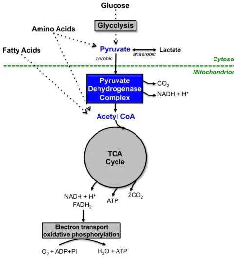
Figure 1.1. Cellular localization of pyruvate dehydrogenase complex (PDC) and its relationships with energy metabolism. PDC links glycolysis to tricarboxylic acid (TCA) cycle by converting pyruvate derived from carbohydrates (mainly glucose) into acetyl-CoA. Subsequently, acetyl-CoA is metabolized in the TCA cycle, which presents a central role in the chemical conversion of carbohydrates, fatty acids and amino acids into carbon dioxide, water and usable energy (ATP) by cooperation with the electron transport chain (adapted from Patel and Harris, 1995 and http://www.wiley.com/college/ boyer/ 0470003790/animations/pdc/pdc.htm). anaerobic Glucose Pyruvate Acetyl CoA aerobic Lactate TCA Cycle Pyruvate Dehydrogenase Complex Electron transport oxidative phosphorylation ATP 2CO2 NADH + H+ FADH2 H2O + ATP O2 + ADP+Pi Amino Acids Fatty Acids Cytosol Mitochondrion NADH + H+ CO2 Glycolysis
The irreversible oxidative decarboxylation of pyruvate into acetyl-CoA occurs
via a postulated “flip-flop” mechanism, through a series of sequential reactions
catalyzed by PDC components and requiring diverse cofactors (Figure 1.2.).
The level of PDC activity regulates the balance between carbohydrates and alternative substrates, such as fatty acids and amino acids, to be used for energy production. Because approximately 50% of daily energy production derives from carbohydrates intake, PDC is a key regulatory step in the central pathways of energy metabolism (Patel and Korotchkina, 2006). Although PDC plays an important role in all metabolically active tissues, its function is particularly critical in the brain. Actually, the brain cannot metabolize fatty acids and, in a normal state, is almost exclusively dependent on the aerobic oxidation of glucose for energy production (Brown et al., 1994; De Vivo, 1998).
A reduction in PDC activity leads to the accumulation of pyruvic and lactic acids and, consequently, to a severe energy impairment. The biological importance of PDC is particularly evident in the pathological condition known as Pyruvate Dehydrogenase Complex Deficiency (PDCD), a neurometabolic disorder that will be further characterized in section 1.1.5.
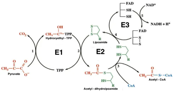
Figure 1.2. Sequential reactions catalyzed by PDC. E1, a TPP-requiring enzyme, catalyzes the first two partial reactions (oxidative decarboxylation and reductive acetylation), resulting in acetylation of the lipoyl group covalently linked to lysyl residue(s) of E2 component. E2 then catalyzes the transfer of the acetyl group to coenzyme A, forming acetyl-CoA and leaving reduced the dithionate ring of the lipoyl moiety. E3, a flavoprotein, reoxidizes the lipoyl moiety and transfers the electrons to NAD+ in a two-step
1.1.2. Structural organization
Pyruvate dehydrogenase complex is one of the largest and more intricate multienzyme structures ever known. The nuclear encoded mammalian PDC, with a molecular mass of 9.5 million Da, is composed by multiple copies of three catalytic, one structural and two regulatory components: pyruvate dehydrogenase (E1), dihydrolipoyllysine-residue acetyltransferase (E2), dihydrolipoyl dehydrogenase (E3); E3 binding protein (E3BP); pyruvate dehydrogenase kinase (PDK) and pyruvate dehydrogenase phosphatase (PDP).
The highly organized PDC structure relies on a central core, which plays a crucial role in the organization, integration of chemical reactions and regulation of the complex activity. In mammals, the central core consists of 48 E2 subunits and 12 E3BP subunits, arranged in a pentagonal dodecahedron structure (Hiromasa et al., 2004; Ciszak et al., 2006). All other components bind noncovalently to this central core: 30 E1 subunits bind to E2-binding domain, and 6 E3 subunits bind to E3BP-binding domain (Sanderson et al., 1996; Zhou et al., 2001). This organization, in which E1/E2 and E3/E3BP form sub-complexes with a 1:2 stoichiometry, implies the existence of a network of “cross-bridges” across the surface of PDC assembly (Smolle et al., 2006).
Figure 1.3. Schematic representation of eukaryotic PDC structure. The central core is formed by E2 (green) and E3BP (pink) subunits. The E2 and E3BP subunits display binding domains (BD) that bind E1 (orange) and E3 (blue), respectively. Additionally, E2 present lipoyl domains (LD) that bind PDK/PDP (purple). This representation puts in evidence the E1/E2 and E3/E3BP “cross-bridges” (adapted from Smolle et al., 2006). E2 BD E2 BD E2 LD E2 LD E2 LD E2 LD E3B P LD E3B P LD E3B P BD E3B P BD E3 E3 E1 E1 P D K P D K P D P P D P
The regulatory enzymes of the complex (PDK and PDP) bind to lipoyl domains of E2 and E3BP, which are extremely flexible domains that move between the active sites of E1, E2 and E3, transferring substrates, products and reducing equivalents (Perham, 2000; Patel and Korotchkina, 2006) (Figure 1.3.).
Therefore, the assembly of this multienzyme complex favors substrate channelling and active site coupling; this allows catalytic reactions to proceed sequentially with the rapid transfer of intermediates between PDC individual components without diffusion into the bulk medium.
1.1.3. Biochemical and molecular structure
The complexity of PDC structure is even more astonishing if we consider that is encoded by 13 different nuclear genes. The precursor proteins are synthesized in cytoplasmic ribosomes and are further transported into the mitochondrial matrix, where the perfect assembly of a fully functional complex occurs after the proteolytic cleavage of their leader sequences. In this section it will be presented a short resume of PDC components and their encoding genes.
Pyruvate dehydrogenase or E1 (EC 1.2.4.1) is the first component of PDC and performs the rate limiting reaction, the decarboxylation of pyruvate and the reductive acetylation of lipoic acid in the E2 subunit. Mammalian E1 is a heterotetramer composed of two α and two β subunits (43 and 36 kDa, respectively) (Ciszak et al., 2001). This enzyme presents two catalytic sites, each requiring a thiamine pyrophosphate (TPP) molecule and a magnesium ion as cofactors. Each α subunit binds magnesium ion and pyrophosphate fragment, while each β subunit binds the pyrimidine fragment of TPP, thus originating one catalytic site at the interface of each α/β dimer (Ciszak et al., 2003). E1 subunit is also responsible for global PDC regulation, which relies on a mechanism of dephosphorylation (activation) / phosphorylation (inactivation) acting upon 3 specific serine residues in the E1α subunit (Korotchkina and Patel, 2001).
The E1α subunit exists as two isoforms encoded by two different genes, PDHA1 and PDHA2, whose sequences display 87% identity and 93% homology. The PDHA1 gene is located on region p22.1 of the human X chromosome, contains 11 exons separated by introns, spans approximately 17 kb of genomic DNA and is selectively expressed in somatic tissues. Additionally, an autosomal locus, PDHA2, lies on
chromosome 4 (4q22-23), completely lacks introns, spans 1.4 kb of genomic DNA and displays characteristics of a functional processed gene; it is expressed only in testis after the onset of spermatogenesis (Dahl et al., 1990).
The E1β subunit is in turn coded for by PDHB gene. The main characteristics of these genes are displayed in Table 1.1.
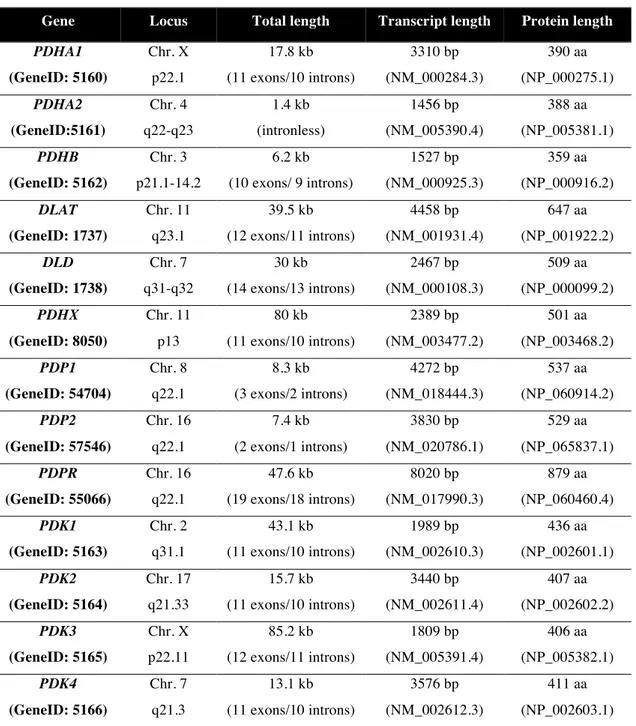
Table 1.1. Summary of the genes encoding PDC components. Characteristics of the genes and respective transcripts and proteins (Source: http://www.ncbi.nlm.nih.gov/gene)
Gene Locus Total length Transcript length Protein length
PDHA1 (GeneID: 5160) Chr. X p22.1 17.8 kb (11 exons/10 introns) 3310 bp (NM_000284.3) 390 aa (NP_000275.1) PDHA2 (GeneID:5161) Chr. 4 q22-q23 1.4 kb (intronless) 1456 bp (NM_005390.4) 388 aa (NP_005381.1) PDHB (GeneID: 5162) Chr. 3 p21.1-14.2 6.2 kb (10 exons/ 9 introns) 1527 bp (NM_000925.3) 359 aa (NP_000916.2) DLAT (GeneID: 1737) Chr. 11 q23.1 39.5 kb (12 exons/11 introns) 4458 bp (NM_001931.4) 647 aa (NP_001922.2) DLD (GeneID: 1738) Chr. 7 q31-q32 30 kb (14 exons/13 introns) 2467 bp (NM_000108.3) 509 aa (NP_000099.2) PDHX (GeneID: 8050) Chr. 11 p13 80 kb (11 exons/10 introns) 2389 bp (NM_003477.2) 501 aa (NP_003468.2) PDP1 (GeneID: 54704) Chr. 8 q22.1 8.3 kb (3 exons/2 introns) 4272 bp (NM_018444.3) 537 aa (NP_060914.2) PDP2 (GeneID: 57546) Chr. 16 q22.1 7.4 kb (2 exons/1 introns) 3830 bp (NM_020786.1) 529 aa (NP_065837.1) PDPR (GeneID: 55066) Chr. 16 q22.1 47.6 kb (19 exons/18 introns) 8020 bp (NM_017990.3) 879 aa (NP_060460.4) PDK1 (GeneID: 5163) Chr. 2 q31.1 43.1 kb (11 exons/10 introns) 1989 bp (NM_002610.3) 436 aa (NP_002601.1) PDK2 (GeneID: 5164) Chr. 17 q21.33 15.7 kb (11 exons/10 introns) 3440 bp (NM_002611.4) 407 aa (NP_002602.2) PDK3 (GeneID: 5165) Chr. X p22.11 85.2 kb (12 exons/11 introns) 1809 bp (NM_005391.4) 406 aa (NP_005382.1) PDK4 (GeneID: 5166) Chr. 7 q21.3 13.1 kb (11 exons/10 introns) 3576 bp (NM_002612.3) 411 aa (NP_002603.1)
The dihydrolipoyllysine-residue acetyltransferase or E2 (EC 2.3.1.12), the second enzyme participating in pyruvate metabolisation, is an acetyltransferase responsible for the transfer of acetyl groups from the lipoyl domains to CoA (Patel and Roche, 1990). The mammalian E2 is a monomer of 59.5 kDa and its structure comprises different domains: an inner domain, a subunit-binding domain and two lipoyl domains. This structure allows E2 to be the core of the complex to which all the other components bind. Moreover, the lipoyl domains are extremely flexible and allow the transfer of the reaction intermediates between the active sites of E1, E2 and E3 (Thekkumkara et al., 1989). Therefore, E2 is crucial for PDC organization, integrative reaction and regulation. In humans, E2 is encoded by the DLAT gene, located in the long arm of chromosome 11 (see Table 1.1.)
The dihydrolipoyl dehydrogenase or E3 (EC 1.8.1.4) finishes the PDC reaction by reoxidizing the lipoamide bound to E2 subunit. This reaction is dependent on two cofactors, FAD and NAD+
, acting as electron acceptors. The active E3 component is a homodimer of 51 kDa and encompasses four structural domains: one catalytic domain, one binding domain to E3BP and two other binding domains to each of the cofactors, FAD and NAD+
(Reed, 2001). This enzyme, encoded by the DLD gene (see Table 1.1.), is also a component of other enzyme systems, namely the α-ketoglutarate dehydrogenase complex, the branched-chain α-keto acid dehydrogenase complex, and the glycine cleavage system (Patel and Roche, 1990).
The E3-binding protein (E3BP), previously called Protein X, is an accessory protein present in eukaryotes that displays the structural function of binding E3 subunit to the complex core; moreover, it was also suggested a possible role on the transfer of acetyl groups (Harris et al., 1997). E3BP is a monomer of 50 kDa showing a similar structure to E2 subunit, once it has an inner domain, a binding domain (to E3 subunit), but only one lipoyl domain (Harris et al., 1997). The PDHX gene codes for this structural element and, curiously, is also located in chromosome 11 (see Table 1.1.), like the DLAT gene encoding the E2 subunit.
PDP (EC 3.1.3.43) is a specific phosphatase that removes Pi from phosphorylated serine residues of E1 (Site 1: 264; Site 2: 271 and Site 3: Ser-203), thus reactivating the component and the whole PDC on a Mg2+
dependent reaction. Mammalian PDP is a heterodimer composed by two different subunits: a catalytic subunit (PDPc) of 52 kDa and a regulatory one (PDPr) of 96 kDa (Chen et al., 1996; Lawson et al., 1997). The catalytic subunits of PDP exist as two isoforms (PDP1
and PDP2), which display some differences concerning their activity, regulation, and tissue distribution (Karpova et al., 2003). It is known that binding of PDP1 to the mammalian PDC occurs through the lipoyl domain of E2 subunit and requires the presence of Ca2+
which plays a bridging role (Roche et al., 2003). The catalytic subunits of PDP are encoded by PDP1 and PDP2 genes, whose loci are non-syntenic, while the regulatory subunit is coded for by PDPR gene that is located near PDP2 gene in chromosome 16 (see Table 1.1.).
PDK (EC 2.7.11.2) is a specific kinase that inactivates PDC by phosphorylating the same previously referred three serine residues in E1α subunit, using ATP as donor of phosphate groups (Kolobova et al., 2001). Mammalian PDK has been shown to exist in four isoforms. Each PDK isoform is a homodimer composed by subunits of molecular masses between 39 and 48 kDa. PDK binds mainly to the mobile lipoyl domains of E2 subunit, and only the PDK4 isoform prefers the lipoyl domain of the E3BP subunit (Roche et al., 2003). Such localization of PDKs on the “swinging arms”, and their transfer from one lipoyl domain to another, gives the possibility of reaching almost all phosphorylation sites in the E1α subunits (Patel and Korotchkina, 2006). It is known that these four mammalian isoenzymes are differently distributed among tissues and also that they present different activities towards the three phosphorylation sites (Korotchkina and Patel, 2001); this adds another control level underlying the regulation of the complex. The genes coding for the different PDKs isoforms are PDK1, PDK2,
PDK3 and PDK4 and their characteristics are displayed in Table 1.1.
1.1.4. Regulation
The central role of PDC in the maintenance of glucose homeostasis relies on a meticulous control of its activity by elaborated mechanisms (Patel and Korochkina, 2003) (Figure 1.4.). This regulation can be divided in two different types: a short-term control that involves a post-translational regulation and a long-term control that occurs at a transcriptional level.
The short-term regulation involves a phosphorylation/dephosphorylation mechanism, that relies on specific kinases (PDK) and phosphatases (PDP), which bind to PDC mainly via the E2-lipoyl domains (Harris et al., 2002) and catalyze the phosphorylation and dephosphorylation of three specific serine residues in E1α (Patel and Korotchkina, 2006), as previously referred. The phosphorylation and concomitant
inactivation of E1 enzyme is catalyzed by PDKs and renders the entire complex inactive. On the other hand, the dephosphorylation and reactivation of E1 enzyme by PDPs reinstates the complex activity (Linn et al., 1969).
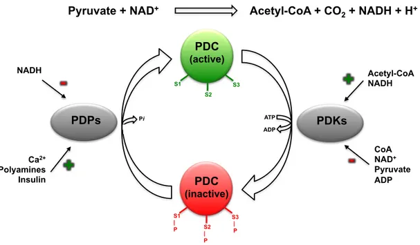
Figure 1.4. Schematic representation of PDC global regulation. Regulation of PDC activity by interconversion between active (unphosphorylated) and inactive (phosphorylated) forms catalyzed by PDPs and PDKs. The (de)phosphorylation (P) occurs in three specific serine residues (S1, S2, S3) of E1α subunit (adapted from Patel and Korotchkina, 2003).
The enzymes of this complex are not only dependent on the interactions among the catalytic and regulatory components of the complex, but also sensitive to the intramitochondrial redox state and metabolite levels, as indicators of energy status. The activities of PDKs are regulated by the concentrations of the physiological ligands pyruvate, ADP and Pi, as well as by NADH/NAD+
and acetyl-CoA/CoA ratios (Holness and Sugden, 2003). PDK activity is inhibited by high levels of pyruvate and a low energy state (high levels of ADP, NAD+
, CoA and Pi) thus increasing PDC activity. NADH and acetyl-CoA, the end-products of the PDC reaction, stimulate PDK activity through reduction and acetylation of the E2 lipoyl moieties, which in turn allosterically affect PDK bound to the lipoyl domain (Patel and Korotchkina, 2006). The activity of PDPs is mainly regulated by the interaction of Mg2+
and Ca2+
, by polyamines (spermine, spermidine and putrescine) and by the NADH/NAD+
ratio (Patel and Korotchkina, 2006).
Pyruvate + NAD+ Acetyl-CoA + CO
2 + NADH + H+ PDC (active) PDC (inactive) PDKs PDPs Acetyl-CoA NADH CoA NAD+ Pyruvate ADP NADH Ca2+ Polyamines Insulin
+
++
S1 S2 S3 S1 | P S2 | P S3 | P Pi ATP ADPBesides the short-term regulation by PDK and PDP, there is also a long-term control of the amount of these same enzymes at the transcriptional level, which will further allow the regulation of PDC activity in response to different nutritional and disease states. For example, PDK4, and to a lesser extent PDK2, were shown to be up-regulated in several tissues under conditions such as starvation, diabetes and hyperthyroidism; on the contrary, PDP1 and PDP2 were shown to be down-regulated in starvation and diabetes (Harris et al., 2002; Holness and Sugden, 2003; Patel and Korotchkina, 2006).
1.1.5. Pyruvate Dehydrogenase Complex Deficiency (PDCD)
Pyruvate Dehydrogenase Complex Deficiency (PDCD) is one of the most common genetic disorders associated with an abnormal mitochondrial metabolism. PDCD, first described by Blass and co-workers, results from a reduction in the enzymatic activity of Pyruvate Dehydrogenase Complex (PDC) and leads to energy deprivation, especially in the central nervous system (Blass et al., 1970).
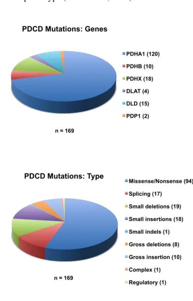
PDCD can be caused by mutations in any of the genes encoding the complex subunits. Though defects may occur in all of its proteins, the E1α subunit is predominantly the culprit. Concerning all PDCD reported mutations, around 70% are assigned to PDHA1 gene (OMIM #312170), and more than a half are of missense type (Figure 1.5.).
As the majority of mutations occur in the X-linked PDHA1 gene, an X-linked pattern of inheritance is observed, but the number of affected males and females is similar (Dahl et al., 1992). PDCD is a rare disorder being most of mutations sporadic ones and the recurrence rate very low.
Nevertheless, the true occurrence of this disorder is unknown because mild mutations may be asymptomatic, especially in females. The variability of the phenotype in heterozygous females appears to be largely determined by differences in X-inactivation patterns (Brown et al., 1994; Willemsen et al., 2006). Moreover, it has been observed that male patients who survived the neonatal period and early infancy generally have a milder phenotype, which seems inconsistent with a general rule of most X-linked dominant diseases (i.e. hemizygous male patients usually have a more severe phenotype than heterozygous female patients). A possible explanation for this apparent discrepancy may be that males carrying a mutant PDHA1 allele that leads to
12
severe deficiency of the enzyme are selected antenatally and, consequently, only male patients who harbor a milder mutant PDHA1 allele survive the neonatal period. In females, on the other hand, possession of a mutant allele leading to severe enzyme deficiency could still be consistent with a live birth, depending on the pattern of X-inactivation; such individuals would then present a more severe phenotype than male patients who have the mild phenotype (Wada et al., 2004).
Figure 1.5. Illustration of mutations prevalence in PDCD. PDCD reported mutations are represented
by gene type and mutation type (Source: The Human Gene Mutation Database
http://www.hgmd.cf.ac.uk/ac/index.php in 2011-03-21).
Not only PDCD results in severe energy impairment, but it is also one of the major causes of lactic acidosis in children. The clinical presentation is extremely heterogeneous, with a spectrum ranging from fatal lactic acidosis in the newborn period to a chronic neurodegenerative condition. The primary phenotypic manifestation is the
PDHA1 (120) PDHB (10) PDHX (18) DLAT (4) DLD (15) PDP1 (2) Missense/Nonsense (94) Splicing (17) Small deletions (19) Small insertions (18) Small indels (1) Gross deletions (8) Gross insertion (10) Complex (1) Regulatory (1)
PDCD Mutations: Genes PDCD Mutations: Type
!!!!
n = 169 n = 169PDHA1 (120)
PDHB (10)
PDHX (18)
DLAT (4)
DLD (15)
PDP1 (2)
Missense/Nonsense (94)
Splicing (17)
Small deletions (19)
Small insertions (18)
Small indels (1)
Gross deletions (8)
Gross insertion (10)
Complex (1)
Regulatory (1)
PDHA1 (120) PDHB (10) PDHX (18) DLAT (4) DLD (15) PDP1 (2) Missense/Nonsense (94) Splicing (17) Small deletions (19) Small insertions (18) Small indels (1) Gross deletions (8) Gross insertion (10) Complex (1) Regulatory (1)PDCD Mutations: Genes PDCD Mutations: Type
!!!!
n = 169 n = 169PDHA1 (120)
PDHB (10)
PDHX (18)
DLAT (4)
DLD (15)
PDP1 (2)
Missense/Nonsense (94)
Splicing (17)
Small deletions (19)
Small insertions (18)
Small indels (1)
Gross deletions (8)
Gross insertion (10)
Complex (1)
impairment of neurological function and/or development, but a wide range of clinical characteristics may be observed: hypotonia, seizures, CNS degeneration and malformations, ataxia, respiratory alterations (apnea, hypoventilation), facial dysmorphic features (narrow head, frontal bossing, wide narrow bridge), peripheral and skeletal/cardiac neuropathy. Several authors classify the phenotype in three major categories, according to the observed severity: neonatal, infantile and benign (Brown et
al., 1994; Wexler et al., 1997).
The current available therapy for PDCD relies on a palliative treatment aiming to stimulate PDC activity, to provide alternative fuels or to correct acidosis. However, it rarely influences the course of the disease and merely prevents the acute worsening of the syndrome. Ketogenic diets, that minimize carbohydrate and maximize fat daily intake, have been used to control lactic acidosis with minimal success. Although the ketogenic diet may reduce the blood levels of lactic acid and extend lifespan, CNS metabolic abnormalities persist, as evidenced by high lactic acid levels in the cerebrospinal fluid and progressive neurological degeneration. Moreover, due to the dependence of the brain on glucose as a fuel, this type of diet increases CNS vulnerability (Wexler et al., 1997; Weber et al., 2001).
In order to optimize PDC function, the standard care option is diet supplementation with cofactors, such as thiamine, carnitine, and lipoic acid. In selected PDCD cases, caused by the so-called thiamine-responsive mutations, high doses of thiamine are particularly effective and a more favorable outcome can be expected for these extremely rare patients (Robinson et al., 1996). There is some evidence that dichloroacetate, which inhibits PDC kinases and thereby activates any residual functioning complex, will also reduce the metabolic disturbance in some patients but, again, this is rarely accompanied by any objective improvement in neurological performance (Berendzen et al., 2006).
Despite these therapeutic approaches, all directed to correction of the phenotype, it is clear they are insufficient and that new avenues must be open, namely at the genotype level.
As previously mentioned, the majority of mutations underlying PDCD are missense ones. Recently, many lines of evidence have suggested that this kind of mutations result in disturbances of protein folding, either at translational or post-translational levels. Disorders based on this pathogenic mechanism are known as conformational diseases (Gregersen et al., 2006) and, accordingly, most cases of PDCD
may fit this denomination. Moreover, it was also shown that missense mutations affect the maturation of polypeptides in different cellular compartments and that chemical or molecular chaperones may modulate the disease phenotype (Baynes et al., 2005). Actually, our own group has recently described a male patient with mild neurological involvement, who displayed low PDC activity and absence of E1α subunit on immunoblotting analysis (Silva et al., 2009); molecular studies revealed a novel mutation, R253G. Surprisingly, following a period of arginine aspartate (Asparten®
) intake, clinical signs and symptoms clearly reversed, fact confirmed by complete normalization of global PDC activity and E1α expression levels. Moreover, when stopping treatment, clinical signs and symptoms quickly return. This case may depict a new therapeutic approach, the use of chemical or molecular chaperones, which represents a more personalized therapy, once is genotype-specific.
However, and specifically for most PDCD cases which are caused by mutations in the PDHA1 gene, the long dreamed therapy referred by several authors (Robinson et
al., 1996; Datta et al., 1999) would be the somatic activation of the autosomic PDHA2
gene that encodes the E1α subunit in spermatogenic cells. The activation of PDHA2 gene in somatic cells would allow circumventing the absence or inactivation of the altered PDHA1 subunit.
1.2. PDHA2 gene
Pyruvate dehydrogenase complex (PDC) plays a crucial role in cell energy metabolism, particularly in those tissues that obtain almost all their energy from carbohydrates. This is the case of the brain, as previously mentioned, but also of spermatogenic cells, which are therefore likely to be more dependent on PDC function.
During spermatogenesis, spermatogenic cells go through meiotic divisions and become haploid, so half the cells will not contain an X chromosome. In addition, the X chromosome is inactivated early in spermatogenesis, so even those cells containing an X chromosome appear not to express the X-linked genes.
Accordingly, the X chromosome location of the normally expressed E1α gene (PDHA1) could represent a serious setback for energy production in sperm. Notably, this apparent problem to express the essential E1α subunit could be overcome in two different ways: by pre-making E1α subunit (or a precursor) when the X chromosome is still functional and storing it for later use; or, by generating E1α subunit from an autosomally located gene, once autosomes, unlike sex chromosomes, are functional in the haploid spermatogenic cells. Indeed, there is a second isoform of E1α in spermatogenic cells, encoded by the PDHA2 gene located on chromosome 4, which thus allows the accomplishment of energy requirements in these cells.
In this way, PDHA genes quite resemble the extensively studied phosphoglycerate kinase (PGK) genes regarding their organization and expression patterns. In placental mammals both have two variants: a somatic isoform (PGK1 and
PDHA1) encoded by an X chromosome-located gene and a testis-specific isoform
(PGK2 and PDHA2) that are expressed only in spermatogenic cells and are encoded by autosome-located and intronless genes (Fitzgerald et al., 1996).
1.2.1. Structure and evolution
PDHA2 gene, first identified by Dahl and co-workers (1990), was mapped to
region q22-q23 on chromosome 4. Unlike PDHA1 gene, that contains 10 introns and spans ∼17 kb of genomic DNA, PDHA2 is an intronless gene spanning only 1.4 kb (Maragos et al., 1989; Dahl et al., 1990). The PDHA1 and PDHA2 gene sequences have 84% and 86% similarity at the coding nucleotide and amino acid sequence levels, respectively (Dahl et al., 1990). The majority of the changes are conservative ones, with sequences surrounding important functional domains remaining highly conserved, such
as the three phosphorylation sites and the thiamine pyrophosphate binding site (Young
et al., 1998).
Mouse orthologs for PDHA1 and PDHA2 genes, Pdha1 and Pdha2, respectively, have been extensively described. These genes also show different chromosomal locations: Pdha1 is X-linked with a similar structure to PDHA1, whereas
Pdha2 is intronless and located in chromosome 19. A comparative analysis of PDHA1
and Pdha1 sequences indicate that both are highly homologous, 86% at transcript level and 98% at protein level. However, the sequences of testis genes, PDHA2 and Pdha2, show less similarity with each other presenting 78% and 75% at the coding nucleotide and amino acid sequence levels, respectively (Brown et al., 1990; Dahl et al., 1990; Fitzgerald et al., 1992).
The genetic derivation of both human and mouse intronless autosomal genes is not clear; however, comparative mapping of eutherian and marsupial PDHA homologs has shed some light over this matter. In contrast to eutherians, marsupials only have one
PDHA gene, which is autosomic, contains introns and is expressed in both testis and
somatic tissues. This led to the conclusion that a common ancestor of marsupials and eutherians also had a single autosomal PDHA gene and that PDHA1 and PDHA2 arose after marsupials and eutherians diverged (Fitzgerald et al., 1993). The X-linked PDHA1 is presumed to have arisen following translocation of the autosomal gene to the short arm of the X chromosome and the testis-specific autosomal intronless PDHA2 is believed to have become selected over time to overcome the absence or apparent inactivation of X chromosome in the haploid spermatogenic cells (Dahl et al., 1996).
Several proposals have been suggested to account for the appearance of these testis-specific variants. One hypothesis suggests that such genes represent functional retroposons, meaning that they are functional processed genes generated by reverse transcription of mRNAs, which were subsequently reinserted into the genome. It is probable that these retroposons were inserted into multiple sites, with selective pressure favoring the promulgation of functional genes that were only active during spermatogenesis, when the X chromosome is either absent or inactive (McCarrey and Thomas, 1987).
Nevertheless, although both human and mouse testis-specific genes appear to be retroposons, it is possible that the human promoter region of PDHA2 gene did not evolve from the same retroposon as the mouse Pdha2 promoter, but rather from an independent retroposon-mediated insertion of the coding region of PDHA1 (Datta et al.,
1999). If this is the case, then the mechanisms underlying regulation of human PDHA2 may be significantly different from that of the mouse species.
1.2.2. Tissue- and cell-specific expression
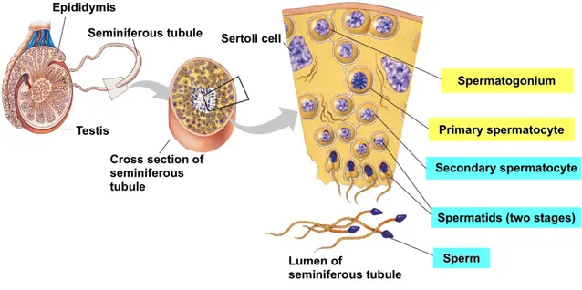
Spermatogenesis is a complex biological process of cellular transformation that produces male haploid germ cells from diploid spermatogonial stem cells. This cell differentiation occurs within seminiferous tubule boundaries of the testis and involves different stages such as mitosis, meiosis and spermiogenesis. Briefly, spermatogenesis begins when primitive type A spermatogonia proliferate by mitosis into type A and type B cells. Type B cells then sequentially differentiate into primary spermatocytes, which after two stages of meiosis result in haploid round spermatids. Finally, round spermatids undergo marked morphological differentiation to become mature sperm (Figure 1.6.).
Figure 1.6. Schematic representation of human spermatogenesis. Spermatogenesis is the process of male germ cell differentiation from spermatogonia to fully functional spermatozoa, which occurs in testis
seminiferous tubules (adapted from http://www.esu.edu/~milewski/intro_biol_one_lect/web
links/oogenesis-spermatogenesis/1-Spermatogenesis.jpg).
This complex process requires the coordinated expression of a number of testis-specific genes. The understanding of the regulatory mechanisms underlying these genes’ expression is crucial and the testis-specific gene that codes for the E1α subunit
was soon pointed out as a reliable model to elucidate gene regulation during spermatogenesis (Iannello et al., 1995).
The first approach to understand E1α testis-specific gene was performed in mouse, rather than in human. The reason underlying this choice was not only due to the relatively easy access to mouse tissues, but also because germ cell differentiation in mouse development is synchronized as the mouse proceeds towards sexual maturity, which provides an excellent opportunity to examine the temporal expression of genes in mouse testis. As a consequence, the transcriptional and translational expression patterns of the testis-specific and somatic forms of E1α subunit genes have already been studied in detail in mouse testis (Takakubo and Dahl, 1992).
Briefly, Pdha2 transcripts were found in all spermatocytes and spermatids stages with a particular increased level in pachytene spermatocytes. In contrast, the expression of Pdha1 was only detected in those cells where Pdha2 expression was not detected (spermatogonia, Leydig and Sertoli cells). Immunostaining with a specific anti-E1α antibody showed that the synthesis of E1α protein was dramatically increased in primary spermatocytes, reaching the highest abundance in pachytene spermatocytes. The amount of protein remained at high levels throughout spermiogenesis; however, it remarkably declined in epididymal spermatozoa. Also, spermatogonia, Leydig and Sertoli cells displayed low levels of E1α protein. These results suggest that the transcriptional switch from the somatic Pdha1 to the testis-specific Pdha2 gene occurs during the first meiotic prophase of spermatogenesis, and also that the E1α protein involved in the development of spermatogenic cells is coded for by the Pdha2 gene (Takakubo and Dahl, 1992).
Moreover, two transcripts for Pdha2 have been identified. In the early stages of
Pdha2 expression, a 2.0 kb mRNA is the only form evident; however, as
spermatogenesis proceeds and the mouse enters sexual maturity, a second smaller transcript becomes apparent. This smaller 1.7 kb transcript is produced in haploid cells and results from the use of an alternative polyadenylation signal (Fitzgerald et al., 1992; Iannello and Dahl, 1992). Although the functional implications of transcript shortening are not clearly understood, it was suggested it might facilitate long-term stability of the mRNA (Young et al., 1998).
1.2.3. Transcriptional regulation
1.2.3.1. Promoter structure
In an attempt to elucidate the mechanisms leading to the human PDHA2 repression in the somatic tissues, Datta and co-workers (1999) isolated and characterized the proximal portion of the human promoter, including the location of the transcription start site (Figure 1.7.). According to these authors, the transcription start site is located 71 nucleotides upstream the start methionine, 10 bp further upstream the transcription start site of mouse Pdha2 (Iannello et al., 1993). Similar to the mouse
Pdha2, the human PDHA2 promoter lacks the classic TATA or CAAT boxes and the
transcription initiation may rely on a Sp1-binding dependent mechanism.
Figure 1.7. Proximal promoter sequence of the human PDHA2 gene. Sequence of the 5´-regulatory region and localization of the potential regulatory elements (Ets, Sp1 and MEP-2) identified by Datta and co-workers (1999). (*) Indicates the transcription start site and the long arrow bellow ATG marks the start methionine (adapted from Datta et al., 1999).
DNAse I footprinting analysis, using both testis and liver extracts, allowed the identification of two sites protected by nuclear proteins: considering the A from the start ATG codon as +1, one site was located between -177 to -144 which may correspond to the putative Sp1 binding site (located around -175), and the second one located between