Universidade de Lisboa
Faculdade de Farmácia
Research Institute for Medicines and Pharmaceutical Sciences
(iMed.UL)
Neuron Glia Biology in Health and Disease Group
Profiling new histological biomarkers for
oligodendrogliomas
Sandra Cristina Lourenço de Macedo
Dissertação
Mestrado em Ciências Biofarmacêuticas
Universidade de Lisboa
Faculdade de Farmácia
Research Institute for Medicines and Pharmaceutical Sciences
(iMed.UL)
Neuron Glia Biology in Health and Disease Group
Profiling new histological biomarkers for
oligodendrogliomas
Sandra Cristina Lourenço de Macedo
Dissertação orientada pela Professora Doutora Dora Brites e pela
Doutora Ana Sofia Falcão
Mestrado em Ciências Biofarmacêuticas
Part of the results discussed in this thesis were presented in the following publications/ communications
Macedo S, Falcão AS, Cardoso F, Silvestre AR, Pereira P, Pimentel J, Brites D. Assessment of new biomarkers for anaplastic oligodendrogliomas. VIII Congresso Nacional da APLF, Leiria, 17-18 November, 2012 [Best Communication Award].
Macedo S, Falcão AS, Cardoso F, Silvestre AR, Pereira P, Pimentel J, Brites D. Profiling new histological biomarkers for oligodendrogliomas grade III. Research Institute for Medicines and Pharmaceutical Sciences, Lisbon, 20th December, 2012 [Poster comunication].
Macedo S, Falcão AS, Cardoso F, Silvestre AR, Pereira P, Cabeçada J, Pimentel J, Brites D. Exploring new molecular signatures for oligodendrogliomas. Research Institute for Medicines and Pharmaceutical Sciences, Lisbon, 18th July, 2013 [Poster comunication].
Acknowledgments
Agradecer a todos os que me ajudaram a construir este projecto é sem dúvida muito difícil e seguramente não conseguirei dar o meu agradecimento como devia a todos os que contribuíram para esta importante etapa da minha vida. Assim, ficam apenas algumas palavras que podendo ser poucas, são sinceras.
Em primeiro lugar, agradeço-Te a Ti meu grande Amigo! Agradeço-Te pelas pessoas que passaram e que partilharam das minhas angústias e alegrias. Contudo, só conTigo dividi todas as dificuldades destes últimos anos. Assim, estas breves palavras apenas pretendem agradecer os momentos que ficaram por dizer “Obrigada!”
Seguidamente, como não poderia deixar de ser, agradeço à professora Doutora Dora Brites pela disponibilidade de me acolher no seu grupo. Foi sem dúvida uma experiência enriquecedora e o realizar de um projecto pelo qual ansiava há muito. A sua sinceridade e honestidade permitiram um crescimento constante e deram incentivo para alcançar sempre o melhor. O seu espírito crítico e enorme saber foram fulcrais na progressão deste trabalho. Foi para mim uma honra poder fazer parte do seu grupo.
Sofia, sei que não foi fácil co-orientar este projecto e que ambas gostaríamos que as oportunidades tivessem sido outras. Estou-te muito grata por toda a disponibilidade e atenção que tiveste comigo e pelo tempo de dedicação a este trabalho. Nesta fase final, o teu contributo foi determinante e o rigor e qualidade nas tuas sugestões foram características marcantes neste projecto. A tua dedicação é para mim um exemplo e incentivo de crescimento pessoal.
Filipa! A ti nem sei como agradecer. Foste sem dúvida a “madrinha do meu mestrado”. Senão fosses tu, o caminho que percorri tinha sido muito mais difícil. Deste-me a mão em todas as dificuldades e foste sempre compreensiva. Tinhas sempre a palavra certa para “aquele” momento, mesmo que não fosse a que mais gostava de ouvir. Muito mais que uma colega de grupo, encontrei em ti uma amiga em quem confiar. Obrigada pela paciência de aturares esta “Filipete” que não te deixava descansar nem no fim-de-semana ☺. Muito mais poderia agradecer-te, mas sei que sabes o carinho que tenho por ti
e o quão importante é para mim conhecer-te.
Quero também agradecer ao Professor Dr. José Pimentel e à Drª Ana Rita Silvestre do Hospital de Santa Maria bem como ao Dr. José Cabeçadas e Drª Gabriela Gasparinho do Instituto Português de Oncologia de Lisboa pela dádiva de amostras de tecidos humanos que permitiram a elaboração deste projecto nos moldes definidos. Neste âmbito,
gostaria também de agradecer à Técnica Teresa Ferreira pela disponibilidade, acompanhamento e aconselhamento quando ainda tudo era uma ideia.
Seguidamente, agradeço aos restantes elementos do grupo Neuron Glia Biology in Health & Disease por partilharem comigo o laboratório e pelos dias passados nas minhas “férias” do trabalho. Um particular agradecimento para a Cátia, a Inês e a Gisela por partilharem comigo conhecimentos relevantes para este trabalho e também pela disponibilidade, atenção e simpatia.
Duarte, não poderia deixar passar o apoio e carinho com que me recebeste no CPM. As nossas conversas, mesmo no “vão da escada”, permitiram momentos mais descontraídos e uma rajada de incentivo para a etapa seguinte. Obrigada!
A todos os meus colegas de trabalho do Hospital Beatriz Ângelo quero agradecer a vossa paciência nos dias de rabugice, a disponibilidade, o apoio e a partilha de conhecimento que me transmitem todos os dias. É uma honra poder fazer parte desta equipa. Obrigada por proporcionarem um ambiente onde “Há Bué Alegria”! ☺ Drª Claúdia,
agradeço a confiança e a oportunidade de fazer parte da sua equipa. “Chefa” Rosana e Pedro, a vocês que estiveram comigo desde o início aliás, ainda antes do início! Foi uma honra fazer parte da equipa de abertura do Hospital ao vosso lado, não poderia ter melhores colegas! ☺ A ti Pedro, ainda tenho de te agradecer as minhas dores de cabeça e
alterações de humor pois sem dúvida foste tu quem me incentivou a atirar de cabeça nesta aventura. Por fim, mas com igual carinho, agradeço aos amigos que ficaram do Hospital da Luz com quem partilhei também bons momentos!
A todos os meus amigos que me apoiaram e fizeram caminho comigo. Pelas constantes demonstrações de interesse e encorajamento. Agradeço os cafés que tão bem me fizeram e incentivaram, e a vossa compreensão para as minhas constantes ausências. Prometo compensar! ☺
Bianca, Gonçalo, Lara e Martim, confesso que muitas vezes fui egoísta e procurei em vocês um abrigo. Vocês enchem-me o coração! “Fofuras”, gosto de vocês daqui até à Lua!” ☺
À minha família, em especial aos meus pais e à minha irmã. O vosso apoio incondicional, compreensão, amor e constante incentivo foram fundamentais para o desenvolvimento deste projecto. A vocês agradeço-vos tudo. Sou o que sou (o bom e o mau ☺) porque vocês existem! Obrigada por estarem sempre presentes e partilharem tão bons momentos comigo. Adoro-vos! ☺
Tiago, agradeço com um carinho muito especial a tua presença, partilha, compreensão e incentivo. De entre sapos, ovelhas, baleias e koalas, parece ser difícil mas vamos conseguindo que todas as espécies se dêem bem! ☺ Obrigada por todos os momentos passados ao teu lado e o conforto que me deste durante estes anos! Adoro-te!!
Contents
FIGURE INDEX ... 8 TABLE INDEX ... 10 ABBREVIATIONS ... 11 RESUMO ... 12 ABSTRACT ... 14 I. INTRODUCTION ... 161. ORGANIZATION OF THE BRAIN ... 16
1.1. Brain anatomy ... 16
1.2. Cellular components of the brain ... 19
1.2.1. Neuronal cells ... 20 1.2.2. Glial cells ... 21 1.2.2.1. Astrocytes ... 21 1.2.2.2. Microglial cells ... 22 1.2.2.3. Oligodendrocytes ... 23 1.2.2.4. Ependymal cells... 25
1.2.3. Other non-neuronal cells ... 25
1.2.3.1. Pericytes ... 25
2. BRAIN TUMORS: GLIOMAS ... 27
2.1. WHO classification ... 28
2.1.1. Classification based on cell types ... 28
2.1.2. Classification based on malignancy grade ... 29
2.1.3. Classification based on tumor location ... 30
2.3. Symptoms and diagnosis ... 32
2.4. Treatment ... 34
3. GLIOMA BIOMARKERS ... 36
3.1. Characteristics of a biomarker ... 36
3.2. Classical biomarkers ... 37
3.3. New glioma biomarkers: exploring cellular and molecular changes in gliomas . 39 3.3.1. Proliferation and Differentiation ... 39
3.3.2. Adherens junctions ... 42
3.3.3. Transcellular pathway across endothelial cells ... 43
3.3.4. Autophagy ... 44
3.3.5. Neuronal cytoskeleton ... 46
3.3.6. Microvasculature changes ... 47
3.3.7. Drug resistance... 48
4. AIMS ... 50
II. MATERIAL AND METHODS ... 51
5. ETHICS STATEMENT ... 51
6. REAGENTS AND ANTIBODIES ... 51
7. CLINICAL DATA COLLECTION ... 51
8. IMMUNOFLUORESCENT STAINING METHOD ... 52
9. EVALUATION OF CELLULAR BIOMARKERS ... 53
10. STATISTICAL ANALYSIS ... 55
III. RESULTS ... 56
IV. DISCUSSION ... 69
V. FINAL CONSIDERATIONS AND PERSPECTIVES ... 75
Figure Index
I.
Introduction………..………..16
Fig. I.1 – Drawing of the human brain from a top view………... 17
Fig. I.2 – Simplified view of the gray and white mater location in a transversal section of the human brain……… 17
Fig. I.3 – Human brain diagram: lateral view (a) and midsagittal view (b)……….. 18
Fig. I.4 - Schematic view of the brain parenchymal cells involved in the blood brain barrier………... 19
Fig. I.5 - Drawing of a neuron... 20
Fig. I.6 - Drawing of astrocytes ... 22
Fig. I.7 - Oligodendrocytes are myelinating cells that surround the neurons ... 24
Fig. I.8 - Pericytes are contractile cells that wrap around the endothelial vessels 26 Fig. I.9 - Glial cell types and associated tumors of CNS. ... 28
Fig. I.10 - Oligodendroglioma grades……….... 30
Fig. I.11 - Histopathological classification of the oligodendroglioma according to WHO ... 31
Fig. I.12 - NSC differentiation into oligodendrocytes... 41
Fig. I.13 - β-catenin intracellular localization... 43
Fig. I.14 - Schematic diagram of the role of caveolin-1... 44
Fig. I.15 - Schematic diagram of the role of LC3 in autophagy... 46
Fig. I.16 - Glioma cells and their relationship and interdependence with cerebral vasculature... 48
II.
Material and methods………..………51
Fig. II.1 - Simple preparation scheme ... 53III.
Results……….……..…………..………56
Fig. III.1 – Expression of Sox-2, a neural stem cell marker, in healthy (control) and neoplastic samples (oligodendroglioma grade I, II and III) ... 57 Fig.III.2 – Expression of NG-2, an oligodendrocyte progenitor cell marker, in
healthy (control) and neoplastic samples (oligodendroglioma grade I, II and III) ... 58 Fig. III.3 – Expression of β-catenin, a regulator of cellular adhesion, in healthy
(control) and neoplastic samples (oligodendroglioma grade I, II and III)...60 Fig. III.4 – Expression of caveolin-1, a major component of caveolae, in healthy
(control) and neoplastic samples (oligodendroglioma grade I, II and III).. 61 Fig. III.5 – Expression of LC3, an autophagic cell marker, in healthy (control)
and neoplastic samples (oligodendroglioma grade I, II and III). ... 64 Fig. III.6 – Expression of NF, a neuronal cytoskeleton marker, in healthy
(control) and neoplastic samples (oligodendroglioma grade I, II and III).. 65 Fig. III.7 – Expression of CD31, a marker for microvasculature, in healthy
(control) and neoplastic samples (oligodendroglioma grade I, II and III).. 66 Fig. III.8 – Expression of α-actin, a pericyte cell marker, in healthy (control) and
neoplastic samples (oligodendroglioma grade I, II and III). ... 67 Fig. III.9 – Expression of P-gp, a marker for multidrug resistance, in healthy
(control) and neoplastic samples (oligodendroglioma grade I, II and III).. 68
V.
Final considerations and perspectives…….………...………75
Fig. V.1 – Variation in promising biomarkers for oligodendroglioma I (A) and oligodendroglioma III (B) ... 76
Table Index
I.
Introduction………..…..………16
Table I.1- Molecular markers used on diagnosis, prognosis and predictive behavior of oligodendrogliomas in patients. ... 33
Table I.2- Scheme therapy for the different types of oligodendrogliomas. ... 34
II.
Material and methods…..……..………..………51
Table II.1 – Clinical data of control and neoplastic samples ... 52
Table II.2 – List of antibodies used for immunohistochemistry and their dilutions. 53 Table II.3 – Biomarkers used to evaluate specific cellular molecular changes and respective quantification method. ... 54
Abbreviations
AJ Adherens junctions
BBB Blood-brain barrier
CD31 Cluster differentiation
CNS Central nervous system
CSC Cancer stem cells
GFAP Glial fibrillary acidic protein
Iba-1 Calcium-binding protein
IDH Isocitrate dehydrogenase
IF Intermediate filaments
LC3 Light chain 3
MDR Multidrug resistance
MGMT O–methylguanylmethyltransferase
NF Neurofilaments
NFTPs Neurofilament triplet proteins
NG-2 Chondroitin sulfate proteoglycan neuron-glial 2
NPC Neural percursor cells
NSC Neural stem cells
Olig 1 Oligodendrocyte transcription factor 1
Olig2 Oligodendrocyte transcription factor 2
OMS Organização mundial da saúde
OPC Oligodendrocyte precursor cells
p53 Protein 53
PDGFRA Platelet-derived growth factor receptor alpha
PECAM-1 Platelet endothelial cell adhesion molecule 1
ROS Reactive oxygen species
SGZ Subgranular zones
Sox-2 Sex determining region Y-box 2
SVZ Subventricular zone
VE-cadherin Vascular endothelial-cadherin
WHO Worl health organization
Wnt Wingless
α-actin Alpha-actin
Resumo
Os tumores cerebrais são a segunda neoplasia mais comum em crianças e a sua incidência é igualmente elevada na população adulta.
Actualmente a classificação dos gliomas é baseada em características histológicas definidas pela Organização Mundial da Saúde (OMS) que os distingue em astrocitomas, oligodendrogliomas e ependimomas. Os oligodendrogliomas representam cerca de 10-15% do total dos gliomas sendo o seu prognóstico mais favorável comparativamente aos restantes. Uma outra classificação, utilizada de forma complementar, baseia-se no grau de malignidade dos gliomas, permitindo a sua classificação entre o estadios I a IV de acordo com a sua malignidade, sendo os de nível IV os mais agressivos. Embora pareça que a classificação dos gliomas está bem estabelecida, a mesma padece de determinada subjectividade, sendo também limitadamente reprodutível uma vez que o desenvolvimento neoplásico pode ser impulsionado por diversas vias de sinalização que podem ser activadas ou silenciadas em paralelo, ou mesmo convergir para complexas interacções. Desta forma, a identificação de características biológicas para cada tipo de glioma é de extrema importância. Esta identificação pode ser realizada através do perfil de proteínas presentes nas células tumorais ou nos fluidos circundantes. Os perfis definidos podem não só fornecer indicações sobre a classificação do tumor, como também identificar biomarcadores clínicos e patológicos que levem ao desenvolvimento de tratamentos personalizados.
Desta forma, com este trabalho pretendemos identificar novos potenciais marcadores para a classificação de oligodendrogliomas. Para alcançar o nosso objectivo, foram usadas amostras humanas de tecido não neoplásico e amostras de tecidos de pacientes com oligodendrogliomas de graus variáveis entre I e III. As avaliações destes marcadores foram realizadas por imunocitoquímica com o fim de quantificar a expressão de algumas moléculas associadas a processos como a proliferação e diferenciação (Sox-2 e NG2, respectivamente), autofagia (LC3), endocitose (Caveolina-1), transdução de sinal (β-catenina) e multi-resistência a fármacos (P-glicoproteina), e ainda relativamente ao citosqueleto das células neuronais (neurofilamentos, NF) e a alterações na microvasculatura (CD31, α-actina).
Pretendemos também com este trabalho comparar a expressão das moléculas referidas entre os diferentes graus de oligodendrogliomas e avaliar a possibilidade de classificar os diferentes estadios através da quantificação de novos marcadores.
Apesar dos resultados apresentados serem preliminares, acreditamos que poderão dar origem a conclusões bastante promissoras. Dos resultados obtidos, observou-se que os oligodendrogliomas, em comparação com amostras de controlo, apresentam níveis de expressão mais baixos de β-catenina e de α-actina e níveis mais elevados dos outros biomarcadores avaliados. Em particular, distinguimos como mais importantes pelo seu potencial como marcador específico para o diagnóstico de oligodendrogliomas de grau I, os marcadores Sox-2, NG2, P-gp, caveolina-1 e α-actina e para oligodendrogliomas de grau III, a β-catenina, LC3, NF e o CD31.
Assim podemos concluir que, através da realização deste trabalho, a maioria dos marcadores testados podem ser potencialmente usados para o diagnóstico, prognóstico e evolução dos oligodendrogliomas, fornecendo pistas que possam servir de auxílio ao tratamento deste tipo de tumor para o qual as opções terapêuticas são ainda bastante limitadas.
Abstract
Brain tumors are the second most common neoplasm in children and its incidence is also relatively high in the adult population, with gliomas accounting for the majority of cases. The World Health Organization (WHO) classification distinguishes astrocytomas, oligodendrogliomas and ependymomas, based on the histological characteristics of tumor cells. Oligodendrogliomas accomplish about 10-15% of total gliomas and are the ones with a better prognostic. A complementary classification based on glioma malignancy, allows the grading of these tumors from grade I to IV (the most aggressive ones). Although it seems that the classification of gliomas is well established, these classifications have subjective criteria and lack reproducibility since the neoplastic process is driven by too many signalling pathways that can be activated or silenced in parallel, or even converge to complex interactions. Thus, identifying biological signatures of each glioma subtype through protein biomarker profiling of tumor or tumor-proximal fluids is therefore of high priority. Such profiling not only may provide clues regarding tumor classification but may identify as well clinical biomarkers and pathologic targets for the development of personalized treatments.
Therefore, with this work we aim to identify potential new biomarkers for the classification of oligodendrogliomas. To accomplish our goal, human samples from normal tissue and from patients with oligodendrogliomas (grades I to III) will be studied by immunocytochemistry in order to quantify the expression of some molecules associated with proliferation and differentiation processes (Sox-2 and NG2), autophagy (LC3), endocytosis (caveolin-1), signal transduction (β-catenin), neuronal cells cytoskeleton (neurofilaments, NF), microvasculature changes (CD31, α-actin) and multidrug resistance protein (P-glycoprotein). We will evaluate how the expression of these proteins varies between the different oligodendroglioma grades and we will look on whether it is possible to classify them accordingly to the presence of these new biomarkers for gliomas.
Although preliminary, we believe that our results give rise to very promising conclusions. We have observed that, as compared to control samples, oligodendrogliomas show a lower expression of β-catenin and α-actin, but a higher expression of other biomarkers. In particular, Sox-2, NG2, P-gp, caveolin-1 and α-actin revealed to be potential biomarkers to be used for the diagnosis of oligodendrogliomas type I, while β-catenin, LC3, NF and CD31 revealed to be potential biomarkers for the diagnosis of oligodendrogliomas type III. Thus, we may conclude that most of the tested biomarkers can be potentially used
for oligodendroglioma diagnosis, prognosis and surveillance, providing clues to assist therapeutic decisions of this still untreatable brain cancer.
I.
Introduction
1. Organization of the Brain
1.1. Brain anatomy
The central nervous system (CNS) is the most amazing structure in human being considered as the major controlling, regulatory and communicating system, able to receive and interpret millions of informations from the inside or outside of the body. An introductory description of the brain anatomy is a basic, although essential topic of this thesis, due to the frequent references to various structures of the brain. Thus, this brief summary on the major regions of the CNS intends to help the reader to further understand all the used terminology.
CNS is composed by two main parts: brain and spinal cord which are continuous at the foramen magnum. Due to their vital importance, these structures are protected from injury by a bony skull. The brain is located in the cranial vault and the spinal cord is placed in the vertebral column (1-3).
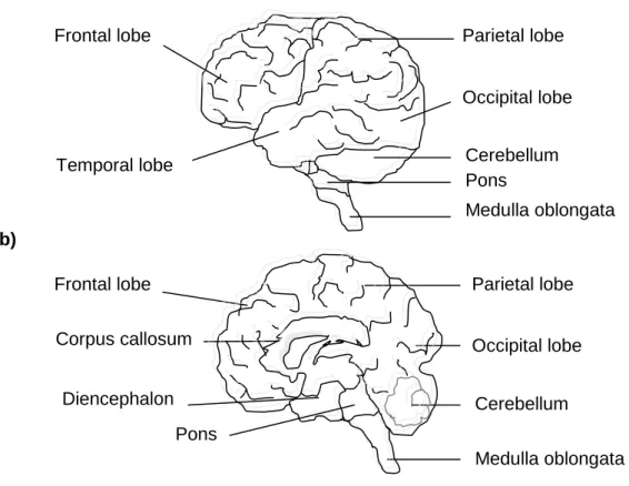
Considering brain, this organ can be divided into three main structures: forebrain, midbrain and hindbrain. The forebrain is the largest portion of the brain since it contains the cerebrum, the corpus callosum and the diencephalon. The midbrain (or mesencephalon) connects the pons and the cerebellum with the cerebral hemispheres. The hindbrain is the region consisting of the medulla oblongata, pons and cerebellum (1). All the structures of the brain are surrounded by connective tissue membranes, called meninges, and fluid-filled spaces named ventricles (2, 3).
The cerebrum is divided into two hemispheres by a deep longitudinal fissure (interhemispheric fissure) and connected by the corpus callosum. The outer surfaces of the hemispheres (cerebral cortex) are composed by many gyri or convolutions caused by infolding of the cortex, separated by sulcus or fissures (Fig.I.1) (1-3). The hemispheres are identically divided into four lobes, which names derived from the cranial bones that overlie them and the location that they assume in the brain. The four lobes, occipital, temporal, parietal and frontal were originally a mere anatomical classification (1, 2). Currently they have been related with the capacity to process and initiate distinct functions such as motor activities, vision and perception of auditory and olfactory stimuli, memory and speech (2, 3).
The diencephalon is composed by the thalamus and hypothalamus and is located between corpus callosum and brainstem. Thalamus is important to process sensorial information and the hypothalamus controls emotions, autonomic and endocrine functions (1-3).
Fig. I.1 – Drawing of the human brain from a top view. The paired cerebral hemispheres are
divided by a deep longitudinal fissure (interhemispheric fissure). The outer surfaces of these hemispheres display many gyri or convolutions separated by sulcus or fissures.
Within the brain, there are areas dominated by grayish appearance while others appear as white mater (Fig. I.2).
The gray matter is located on the surface of the hemispheres and is composed of neuronal cell bodies, glial cells and blood vessels. The white matter is located in deeper nuclei and it mainly consists of nerve fibers that cover the axons (2).
Fig. I.2 –Simplified view of the gray and white mater location in a transversal section of the human brain. The gray matter is situated on the surface of the hemispheres and made up by cell
bodies, while the white matter is located in deeper nuclei and composed by nerve fibers. Sulcus
Gyri
Cerebellum Interhemispheric fissure
Left cerebral hemisphere
Right cerebral hemisphere
Anterior Posterior White matter Gray matter White matter Gray matter Ventricles
The cerebellum is in the posterior part of the brain and it is connected to the pons and medulla oblongata. Cerebellum is essential for the coordination of movements and learning of motor tasks. Pons is related to somatic and visceral motor control while medulla oblongata controls autonomic functions (Fig. I.3) (1-3).
a)
b)
Fig. I.3 –Human brain diagram: lateral view (a) and midsagittal view (b). In the lateral view it
can be visualized the four lobes of the brain, with cerebellum and corpus callosum visualized in the midsagittal view.
Medulla oblongata is continuous with the spinal cord which extends from the foramen magnum at the base of the skull to the level of the first lumbar vertebra. The spinal cord receives and processes sensory information and controls movement of the limbs and the trunk (1, 2). Parietal lobe Occipital lobe Cerebellum Medulla oblongata Frontal lobe Corpus callosum Diencephalon Pons Parietal lobe Occipital lobe Cerebellum Medulla oblongata Frontal lobe Pons Temporal lobe Anterior Posterior
1.2. Cellular components of the brain
The CNS being such a complex and essential structure requires a highly specialized system to maintain a stable environment. With this purpose, CNS is separated from circulatory system by a dynamic interface called blood
by Lewandowski in 1900. This barrier limits the entry of several components such as red blood cells, leukocytes and plasma components into the brain
barrier to many components
structure and function of the BBB depends on the interaction between several different cell types (Fig. I.4).
Despite the complexity of the nervous system and the tremendous number of cells, there are only two main types of cells in
(microglia, astrocytes, and oligodendrocytes)
the brain, assisting in nervous system repair and maintenance, in synapse elimination, and providing metabolic functions to
system and the responsible to transmit impulses. Glial cells are structural filler, since they highly
contributing to maintain the inte
Fig. I.4 - Schematic view of the
The “neurovascular unit” that is essential for the health and function of CNS cerebral microvascular endothelium,
oligodendrocytes and astrocytes), together with the extracellular matrix
Cellular components of the brain
The CNS being such a complex and essential structure requires a highly specialized system to maintain a stable environment. With this purpose, CNS is separated from circulatory system by a dynamic interface called blood-brain barrier (BBB), first introduce
in 1900. This barrier limits the entry of several components such as red blood cells, leukocytes and plasma components into the brain (4). It serves also as a
mponents located inside the brain, preventing them from leaving it structure and function of the BBB depends on the interaction between several different cell
Despite the complexity of the nervous system and the tremendous number of cells, there are only two main types of cells in the nervous tissue: neurons and glial cells
microglia, astrocytes, and oligodendrocytes) with several functions that include
the brain, assisting in nervous system repair and maintenance, in synapse elimination, and roviding metabolic functions to neurons. Neurons are the structural unit of the nervous system and the responsible to transmit impulses. Glial cells are much m
since they highly contribute to keep the functional homeostasis of the brain maintain the integrity of the neurons (5).
Schematic view of the brain parenchymal cells involved in the blood brain barrier
The “neurovascular unit” that is essential for the health and function of CNS
cerebral microvascular endothelium, pericytes, neurons and the surrounding glial cells (microglia and astrocytes), together with the extracellular matrix.
The CNS being such a complex and essential structure requires a highly specialized system to maintain a stable environment. With this purpose, CNS is separated from brain barrier (BBB), first introduced in 1900. This barrier limits the entry of several components such as red . It serves also as a from leaving it. The structure and function of the BBB depends on the interaction between several different cell
Despite the complexity of the nervous system and the tremendous number of cells, tissue: neurons and glial cells with several functions that include support for the brain, assisting in nervous system repair and maintenance, in synapse elimination, and Neurons are the structural unit of the nervous much more than a mere contribute to keep the functional homeostasis of the brain
lved in the blood brain barrier.
The “neurovascular unit” that is essential for the health and function of CNS comprehends the and the surrounding glial cells (microglia,
1.2.1. Neuronal cells
The neuron is an electrically excitable cell
and transmit information. These cells are highly specialized and the structure of a typical neuron.
Fig. I.5 - Drawing of a neuron
axon.
The neuron is composed by three basic parts: cell body or soma, one or more dendrites and a unique axon.
to other types of cells. It has a nucleus with at least one nucleolus and contains many of the typical cytoplasmic organelles that provide energy and synthesize organic materials, especially neurotransmitters
The lack of centrioles is consistent to their amitotic nature, since the function of centrioles is related with cell division. Additionally, the cytoplasm contains clusters of roug endoplasmic reticulum and free ribosomes, called Nissl bodies, which account for the dark color of the gray matter (3, 6
The dendrites and axons are cytoplasmic extensions that Dendrites are usually short and branched (rarely longer than 1
receiving and processing, from other neurons, the majority of the excitatory synaptic inputs. Due to their actin cytoskeleton, microtubules and
dendrites are stable extensions
neurons have hundreds of small protrusions
(3). Some authors refer that abnormalities in the number, size and morphology of dendrites and their respective dendritic spines, can be associated with several diseases
Neuronal cells
The neuron is an electrically excitable cell of the CNS, conceived to receive, integrate and transmit information. These cells are highly specialized and amitotic.
the structure of a typical neuron.
euron. A typical neuron is constituted by cell body, dendrites and a
The neuron is composed by three basic parts: cell body or soma, one or more dendrites and a unique axon. The cell body is the central part of the neuron and is similar to other types of cells. It has a nucleus with at least one nucleolus and contains many of the typical cytoplasmic organelles that provide energy and synthesize organic materials, urotransmitters that are important molecules in cell-to-cell communication. The lack of centrioles is consistent to their amitotic nature, since the function of centrioles is related with cell division. Additionally, the cytoplasm contains clusters of roug endoplasmic reticulum and free ribosomes, called Nissl bodies, which account for the dark
6).
The dendrites and axons are cytoplasmic extensions that project from the cell body. Dendrites are usually short and branched (rarely longer than 1-2 mm) specialized for receiving and processing, from other neurons, the majority of the excitatory synaptic inputs. Due to their actin cytoskeleton, microtubules and microtubule-associated proteins, dendrites are stable extensions that serve to increase the surface area of the cell
neurons have hundreds of small protrusions along the dendrites known as dendritic spines ome authors refer that abnormalities in the number, size and morphology of dendrites
respective dendritic spines, can be associated with several diseases Cell body Dendrites
Axon
to receive, integrate amitotic. Fig.I.5 illustrates
A typical neuron is constituted by cell body, dendrites and a single
The neuron is composed by three basic parts: cell body or soma, one or more The cell body is the central part of the neuron and is similar to other types of cells. It has a nucleus with at least one nucleolus and contains many of the typical cytoplasmic organelles that provide energy and synthesize organic materials, cell communication. The lack of centrioles is consistent to their amitotic nature, since the function of centrioles is related with cell division. Additionally, the cytoplasm contains clusters of rough endoplasmic reticulum and free ribosomes, called Nissl bodies, which account for the dark
project from the cell body. mm) specialized for receiving and processing, from other neurons, the majority of the excitatory synaptic inputs. associated proteins, surface area of the cell. Some known as dendritic spines ome authors refer that abnormalities in the number, size and morphology of dendrites
respective dendritic spines, can be associated with several diseases. Cell body Dendrites
The axon is usually elongated and it carries impulses away from the cell body up to a meter or more away. An axon may have infrequent branches called axon collaterals. Axons and axon collaterals terminate in many short branches or telodendria. The distal ends of the telodendria are slightly enlarged to form synaptic bulbs. Many axons are surrounded by a segmented, white and fatty called myelin or myelin sheath. Myelinated fibers make up the white matter of the CNS. The unmyelinated regions between the myelin segments are called the nodes of Ranvier (2).
1.2.2. Glial cells
The name glial (meaning "glue") reflects the original concept that glial cells were merely supportive cells for neurons. Currently it has become apparent that glial cells are not only supportive cells but instead they play a number of important active functions (7). They comprise 90% of the brain's cells and are capable of mitosis, but they do not conduct nerve impulses. These cells perform a plethora of important functions, as they can provide support for the brain, assist in nervous system repair and maintenance, produce myelin for neurons and also supply physic, nutritional and oxygen support for neuronal cells (7, 8).
Four types of glial cells are usually considered on CNS: macroglial cells (which consist of astrocytes and oligodendrocytes), microglia cells, NG2 cells and ependymal cells (7, 8).
1.2.2.1. Astrocytes
Astrocytes, also known by astroglia, are star-shaped cells (Fig. I.6) that make up the largest glial population cells (9).
They exert structural functions, are required for neuronal survival, neurite formation and angiogenesis, and maintain CNS homeostasis by regulating pH, ionic concentrations and osmolarity (10). Astrocytes also provide metabolic support to neurons by fueling their activity with energy and substrates, while removing excess neurotransmitter molecules from the extracellular space (11). These cells also form the neurovascular unit of the BBB since their end-feet wrap around the capillary surface (12). These cells can induce tight junctions in endothelial cells, thus participating in the regulation of BBB permeability (13).
Fig. I.6 - Drawing of astrocyt
be classified as protoplasmic (a) or fibrous (b).
Astrocytes are usually
astrocytes. The location and morphology of these two types of astrocytes are different. While protoplasmic astrocytes are distributed in grey matter and have a large, flat and round cell body, wide processes and few branches
in white matter and are typically
Astrocytes can be identified histologically by their expression protein (GFAP), a specific intermediate filament
cells. This is a useful tool to evaluate the presence of these cells in brain samples
1.2.2.2. Microglial cells
Microglial cells are believed to originate from myeloid cells and comprise approximately 5 to 12% of the brain
macrophages in CNS as they are described to be part of the innate immune response in the brain. Thus, microglial cells can phagoc
(18, 20-23).
Microglial cells develop in the brain and spinal cord and are distributed througho the gray and white matter. It is known that these cells develop a branched form with intimate interaction with neurons and other glial cells. Historically, descriptions of microglia report remarkable morphological transformations in response to tissue i
including CNS infections, neuroin diseases (8, 20).
Although the exact role of microglia cells in CNS development, homeostasis, and disease remain almost unknown, resting microglial cells are described to exhibit a
a)
astrocytes. Astrocytes are glial cells with a star shape
be classified as protoplasmic (a) or fibrous (b).
are usually classified in two different types: protoplasmic an
tion and morphology of these two types of astrocytes are different. While protoplasmic astrocytes are distributed in grey matter and have a large, flat and
de processes and few branches, fibrous astrocytes are are typically slim and small with thinner branches (14
Astrocytes can be identified histologically by their expression of glial fibrillary acidic (GFAP), a specific intermediate filament that allows the cytoplasm
tool to evaluate the presence of these cells in brain samples
Microglial cells
are believed to originate from myeloid cells and comprise approximately 5 to 12% of the brain (18-21). These cells are mostly compared to macrophages in CNS as they are described to be part of the innate immune response in the brain. Thus, microglial cells can phagocyte cell debris, waste products and pathogens
Microglial cells develop in the brain and spinal cord and are distributed througho the gray and white matter. It is known that these cells develop a branched form with intimate interaction with neurons and other glial cells. Historically, descriptions of microglia report remarkable morphological transformations in response to tissue i
including CNS infections, neuroinflammatory lesions, brain tumors, and neurodegenerative
Although the exact role of microglia cells in CNS development, homeostasis, and disease remain almost unknown, resting microglial cells are described to exhibit a
Extensions Cell body
b)
star shape morphology that can
classified in two different types: protoplasmic and fibrous tion and morphology of these two types of astrocytes are different. While protoplasmic astrocytes are distributed in grey matter and have a large, flat and are usually localized 14, 15).
glial fibrillary acidic cytoplasm staining of these tool to evaluate the presence of these cells in brain samples (16, 17).
are believed to originate from myeloid cells and comprise . These cells are mostly compared to macrophages in CNS as they are described to be part of the innate immune response in yte cell debris, waste products and pathogens
Microglial cells develop in the brain and spinal cord and are distributed throughout the gray and white matter. It is known that these cells develop a branched form with intimate interaction with neurons and other glial cells. Historically, descriptions of microglia report remarkable morphological transformations in response to tissue injury or disease flammatory lesions, brain tumors, and neurodegenerative
Although the exact role of microglia cells in CNS development, homeostasis, and disease remain almost unknown, resting microglial cells are described to exhibit a
characteristic ramified morphology and are responsible for immune surveillance. However, in response to brain injury or to immunological stimuli they become rapidly activated and undergo dramatic morphologic alterations to an amoeboid state (8, 18, 20, 23). An unregulated response or overactivation of microglia cells can have devastating neurotoxic consequences. Thus, depending on the progression of the disease and the type of stimulus, microglia activated cells can have neurotoxic or neuroprotective properties (20).
Concerning the phenotype markers of these cells, calcium-binding protein (Iba-1) stands out from others markers, being often used as a marker of microglia. Nevertheless, since Iba-1 it is also expressed by most tissue macrophages, it cannot be used to differentiate between tissue resident microglia and other tissue macrophages (8).
1.2.2.3. Oligodendrocytes
Oligodendrocytes are the myelinating cells of the CNS and constitute about 5 to 10% of the total glial population. Oligodendrocytes have compact and uniform dark nuclei, a small round body and about four to six branching processes (24).
These cells wrap themselves around part of the surrounding axons forming myelin, an insulating lipid-rich membrane sheath that speeds the conduction of electrical impulses (Fig. I.7) and they are able to myelinate more than sixty axons (24, 25). The major components of myelin are lipids, which represent about 70% of the total composition. The remaining 30% are proteins, such as the myelin basic protein (MBP), proteolipid protein (PLP) and 2′,3′-cyclic nucleotide 3′-phosphodiesterase (CNP). These three proteins together comprise the main protein components of myelin (24). The principal physiologic function of myelin is to conduct, in a high velocity, the potential action in the axon. Moreover, periodic gaps (node of ranvier) in the insulating sheath on the axon of certain neurons serve to facilitate the rapid conduction of nerve impulses. The conduction performed by saltatory mode is much faster comparing to the unmyelinated axon of the same diameter (24).
Fig. I.7 - Oligodendrocytes are myelinating cells that surround the neurons. These cells
provide support to neurons and produce large amounts of specialized membrane (myelin) that form multiple wraps around the contracted axons.
Two of the most important functions of oligodendrocytes are the promotion in axonal conduction and the regulation of neuronal properties by maintaining axonal integrity (24, 25). A large number of brain´s diseases have been reported to be related with modifications in oligodendrocytes. These modifications are associated with the decrease in oligodendrocyte number and density, as well as with an abnormal myelin morphology. In some disorders, imaging studies using tensor imaging revealed a decrease in white matter integrity that may subsist prior to disease onset (24, 25). Thus, many studies have been made in order to promote myelin repair, delaying the disease progression, and to recover the loss of neurological functions.
The origin of oligodendrocytes is still disputed, however, recent studies indicate that in spinal cord most oligodendrocytes derive from a specific domain in ventricular zone, which give rise to oligodendrocyte precursor cells (OPC) that migrate to their final location and then differentiate (24, 26, 27). During the progression along oligodendroglial lineage, several markers are expressed in specific stages of cell maturation. In this context, these makers, as well as the morphologic complexity and the migratory capacity, can contribute to distinguish the different stages of oligodendrocyte maturation. Initially, OPC express some specific markers like chondroitin sulfate proteoglycan NG2, ganglioside A2B5 and platelet-derived growth factor receptor alpha (PDGFRA). During the differentiation process these cells give rise to pre-oligodendrocytes that express, in addition to OPC markers, the sulfatide recognized by the O4 antibody. After loss of A2B5 and NG2 markers, the immature oligodendrocytes continue to express O4 and start the expression of galactocerebroside C (GalC). Finally, mature oligodendrocytes are able to produce the myelin proteins, such as MBP, proteolipid protein PLP, myelin associated glycoprotein
Oligodendrocyte
Axons
(MAG) and myelin oligodendrocyte glycoprotein (MOG) (24, 26-28). In addition, many transcription factors have been implicated in the specification of different stages of oligodendrocyte maturation. Regarding that, one of the best known transcription factors are the Olig genes, Olig1 and Olig2. Expression of Olig2 occurs during all stages of oligodendrocyte maturation and is required for the development of NG2 positive progenitor cells (29, 30). On the other hand, Olig1 appears to be mostly implicated in oligodendrocytes maturation, being involved in the final stages of myelin production (30, 31).
1.2.2.4. Ependymal cells
Ependymal cells are ciliated cells coating the ventricular walls of the brain and the central canal in the spinal cord. These cells are important for the propulsion of cerebrospinal fluid (CSF), provide a barrier to the parenchyma and have recently been related to neuroendocrine functions (32).
Other type of cells, choroid ependymocytes, occupy an important position interposed between fenestrated blood capillaries of the choroid plexus and the CSF, where they constitute an active blood-CSF barrier, also playing a role in the production of this ventricular fluid (2, 3).
1.2.3. Other non-neuronal cells
Related to cells in the CNS, it has been describe that other type of cells, pericytes, in spite of not being neural cells, are imperative for the CNS integrity and maintenance. For this reason we will add to the introduction section, a brief topic concerning these cells.
1.2.3.1. Pericytes
Pericytes play an integral role in the maintenance of the BBB as well as in several other homeostatic and hemostatic functions of the brain (33). These cells are also a key component of the neurovascular unit, which further includes endothelial cells, the basement membrane, astrocytes, microglia and neurons (22) as previously indicated.
Pericytes exhibit multipolar primary processes extending along the longitudinal axis of capillaries and secondary branches wrapping around the vessels enveloped in the basement membrane and in direct contact with endothelial cells (Fig. I.8) (34). Thus, these
cells are responsible for a variety of functions such as capillary blood flow regulation, modulation of BBB permeability
can also stabilize and monitor the maturation of endothelial cells by direct co across the basement membrane, as well
shown, in recent studies, that the lack of pericytes in the CNS can cause the breakdo the BBB and lead to other degenerative changes in the brain
Fig. I.8 - Pericytes are contractile cells that
have a close physical association with endothelial cells.
Pericytes are known to express multiple muscle actin, NG2, PDGFR
and II, and also desmin (34
marker expression suggesting the existence of pericyte heterogeneity. For instance, there are some pericyte subpopulations that lack
Indeed, only those located near arterioles in the brain are positive negatively stained in fresh
cells are responsible for a variety of functions such as capillary blood flow regulation, permeability and clearance and phagocytosis of cellular debris
can also stabilize and monitor the maturation of endothelial cells by direct co the basement membrane, as well as by paracrine signaling (35
that the lack of pericytes in the CNS can cause the breakdo the BBB and lead to other degenerative changes in the brain (33).
are contractile cells that wrap around the endothelial
have a close physical association with the endothelium, sharing the basement membrane with the
s are known to express multiple immunologic markers such as
muscle actin, NG2, PDGFR-α, PDGFR-β, major histocompatibility complex (MHC) class I 34). However, not all pericyte subpopulations display the same sting the existence of pericyte heterogeneity. For instance, there are some pericyte subpopulations that lack α-smooth muscle actin expression Indeed, only those located near arterioles in the brain are positive. M
negatively stained in fresh primary cultures (36).
Endothelial cell Pericyte
Vessel
cells are responsible for a variety of functions such as capillary blood flow regulation, of cellular debris. They can also stabilize and monitor the maturation of endothelial cells by direct communication 35). It has also been that the lack of pericytes in the CNS can cause the breakdown of
endothelial vessels. Pericytes
the endothelium, sharing the basement membrane with the
immunologic markers such as α-smooth , major histocompatibility complex (MHC) class I . However, not all pericyte subpopulations display the same sting the existence of pericyte heterogeneity. For instance, there smooth muscle actin expression (34). More than 95% are
Endothelial cell Pericyte
2. Brain tumors: Gliomas
In general, a brain tumor is characterized by an abnormal mass of cells growing in the brain. It develops from abnormal cells that have the ability to multiply uncontrollably for multiple or unknown reasons. This mass of cells can be divided into two main groups: benign and malignant. In clinical practice, a benign tumor is not considered cancerous. Their capacity to grow is slow and they can be usually removed. Recidives in benign tumor are not recurrent and cells do not spread or invade other tissues. In contrast, malignant tumors are cancerous, grow fast and can invade and damage nearby tissues or organs also having the capacity to metastasize (37). However, this classification is not trivial since a tumor with benign characteristics may be considered malignant if it is located in a critical area or its size is life-threatening.
The ability of cancer cells to disconnect from the malignant mass, enter the bloodstream or lymphatic system to form tumors in other parts of the body is called metastasis. For this reason, a tumor can be primary or secondary. A primary brain tumor arise “de novo” in brain. Secondary brain tumor is originated from cells which arise from other tissue or organ and spread into healthy areas (37).
Brain tumors have an incidence of 12.8 per 100,000 people (38, 39) and a median survival of 14 months (40).They are usually caused by a change in genetic structure, such as mutated or missing genes resulting in abnormal cells. Gliomas are the most common primary brain tumor accounting for more than 70% of total primary malignant intracranial tumor (41, 42). Some controversy still exists in turn of the real origin of gliomas (41). Nevertheless, some studies have demonstrated that these tumors can consist of a heterogenous mixture of several glial phenotypes that goes from immature cell types, through poorly differentiated cells to mature cells (43). This heterogeneity leads to vary different courses of gliomas compromising prognosis and therapy.
Due to the diversity of gliomas, it was required to establish a unique classification in order to be followed by the clinical community worldwilde. In this context the WHO proposed a universal classification of CNS tumors. The present thesis is focus in WHO classification of gliomas. So, the following section intends to provide some concepts to be taking into account when considering the sample profiling used in the present work.
2.1. WHO classification
In 1993, the WHO created their first guideline for comprehensive classification of neoplasms affecting the CNS. Recently, in 2007, the WHO published a review of their classification of tumors where several neoplasms were included. This classification is based mainly on three parameters
2.1.1. Classification based on cell types
Despite the knowledge regarding the origin of gliomas the cell type from which gliomas are
heterogeneity. This classification is based on histological similarities involving gliomas and normal glial cells such as astrocytes, oligodendrocytes and ependymal cells. Thereb gliomas can be classified as astrocytomas, oligodendrogliomas and ependymomas, respectively (Fig. I.9). Tumors displaying a mixture of different cells are designated by oligoastrocytomas (or mixed gliomas)
oligodendrogliomas are the goal of this work.
Fig. I.9 - Glial cell types and associated tumors of CNS.
categories based on their cell origin, in accordance to World Health Organization Oligodendroglioma
Oligoastrocytoma
WHO classification
In 1993, the WHO created their first guideline for comprehensive classification of neoplasms affecting the CNS. Recently, in 2007, the WHO published a review of their classification of tumors where several neoplasms were included. This classification is
parameters: cell type, malignancy grade and tumor location
Classification based on cell types
knowledge regarding the origin of gliomas still remain
cell type from which gliomas are initiated seems to have influence in glioma heterogeneity. This classification is based on histological similarities involving gliomas and normal glial cells such as astrocytes, oligodendrocytes and ependymal cells. Thereb gliomas can be classified as astrocytomas, oligodendrogliomas and ependymomas,
. Tumors displaying a mixture of different cells are designated by oligoastrocytomas (or mixed gliomas) (38, 45). Among all these types of tumors, oligodendrogliomas are the goal of this work.
Glial cell types and associated tumors of CNS. Gliomas can be divided in different
categories based on their cell origin, in accordance to World Health Organization Oligodendroglioma Astrocytoma Ependymoma
Oligoastrocytoma
In 1993, the WHO created their first guideline for comprehensive classification of neoplasms affecting the CNS. Recently, in 2007, the WHO published a review of their classification of tumors where several neoplasms were included. This classification is
and tumor location (44).
remain overshadowed; ems to have influence in glioma heterogeneity. This classification is based on histological similarities involving gliomas and normal glial cells such as astrocytes, oligodendrocytes and ependymal cells. Thereby, gliomas can be classified as astrocytomas, oligodendrogliomas and ependymomas, . Tumors displaying a mixture of different cells are designated by . Among all these types of tumors,
Gliomas can be divided in different categories based on their cell origin, in accordance to World Health Organization.
2.1.2. Classification based on malignancy grade
The WHO grading scheme for glial neoplasms assigns four distinct stages (from Grade I to Grade IV). Accurate scaling of glioma facilitates the treatment and the prediction of their outcome. The grade designates its degree of malignancy and it is acquired based on the tumor’s histopathology using the following features: similarity to normal cells (atypia), rate of growth (mitotic index), indication of uncontrolled growth, dead cancer cells in the center of the tumor (necrosis), potential for invasion (infiltration) and blood supply (vascularisation) (42).
Grade I gliomas are the less malignant tumors, sometimes considered benign and are typically related with long-term survival (46, 47). The median survival for these patients is between 5 and 10 years compared with a survival of about 14 months in patients with malignant glioma (48, 49). These tumors exhibit a slow grow, a limited cell proliferation potential and have an almost normal appearance by microscope observation (47, 50). These tumors are not likely to spread. Grade II tumors have a relatively slow growing rate, a low-level of proliferative activity and a slightly abnormal microscopic appearance. Some of these tumors are infiltrative and can either recur or progress to a higher-grade glioma (44, 50). Grade III tumors are, by definition, malignant. The cells of a grade III tumor are actively reproducing abnormal cells, which grow into nearby normal brain tissue. These tumors tend to recur, often as the higher grade IV, which are the most malignant. When gliomas contain several grades, the malignancy is established by the highest malignant grade of the cells, even if most of the tumor is of a lower grade kind.
Regarding oligodendrogliomas, the main focus of the present work, they can be classified in low-grade/WHO Grades I and II (Oligodendroglioma I and II, respectively) or high-grade/WHO Grade III (Anaplastic Oligodendrogliomas). Concerning low-grade oligodendrogliomas, grade I is almost inexistent and there are few studies regarding these tumors. Oligodendrogliomas are also found in oligoastrocytomas, generally related to the grade IV, but not exclusive of this degree (51, 52). Typically, oligodendrogliomas progress from low grade lesions into high grade but this natural progression may not be seen in some patients (53). Fig.I.10 shows the classical progression of oligodendrogliomas.
Fig. I.10 - Oligodendroglioma grades. Oligodendrogliomas can be divided into three categories
based on their malignant grade and general tumour characteristics, in accordance to World Health Organization.
2.1.3. Classification based on tumor location
Gliomas can also be classified according to their location, whether above or below the tentorium, a membrane that separates the cerebrum (above) from the cerebellum (bellow). Hence, they are defined as supratentorial, which develop above the tentorium, and as infratentorial, which develop below the tentorium (44).
2.2. Oligodendrogliomas: Definition and Grade
As described above, oligodrendrogliomas are usually divided into low-grade tumors (grade I and II) and high-grade tumors (grade III) (51) since, in general, grade IV also consider the presence of astrocytomas.
Currently, clinical evaluation is made by routine hematoxylin- eosin. When observed in a microscope, grades I and II show tumour cells arranged in a solid or honeycomb appearance. The cells are usually small and rounded with a well-defined nucleus surrounded by a perinuclear clear halo. Grade II can also exhibit calcifications (52, 54). Histological examination of anaplastic oligodendrogliomas shows an aggressive form of oligodendrogliomas often associated with increased cellularity, nuclear atypia and widespread mitotic activity and apoptosis. Haemorrhage, tumor necrosis, calcification and microvascular proliferation may be present, but are variable in severity and extent (52, 55).
Oligoastrocytomas show a bizarre appearance, have a high mitotic activity and can easily spread into the surrounding normal brain tissue. These tumors have also an enormous ability to form new blood vessels (angiogenesis) so they can maintain their rapid growth. Necrotic zones are usually associated with a rapid evolution and fatal outcome (44,
Malignancy Oligodendrocyte Oligodendroglioma I Oligodendroglioma II Anaplastic Oligodendroglioma
well-defined oligodendroglial and astrocytic morphology; oligoastrocytomas this may occur. Fig.I
classification of oligodendrogliomas and oligoastrocytomas.
Fig. I.11 - Histopathological
Oligodendrocytes in normal brain white matter (A),
and delicate chromatin pattern and a perinuclear halo. Low
some calcifications (white arrows). Anaplastic oligodendroglioma (C) is characterized by a higher number of cells and blood vessel
and oligodendrogliomas (left). D which show magnification x100
Oligodendrogliomas comprise about 4% of primary intracranial tumor, representing about 10-15% of the gliomas
between 30-50 years with a higher incidence in male than tumors an age and gender
relatively rare form of cancer, their location in the brain is commonly problematic accounting to high morbidity and mortality
and 63.6% for 10 years, which also depend
6% of these tumors are found in infants and children which are survival rates (93% for 5 years)
Oligodendrogliomas have more susceptibility to invade cerebral hemispheres in the white matter of the frontal lobe but they can also occur in other locations such as
A
C
glial and astrocytic morphology; however rocytomas this may occur. Fig.I.11 shows a general view of the classification of oligodendrogliomas and oligoastrocytomas.
al classification of the oligodendroglioma according to WHO.
normal brain white matter (A), present round nucleus with a relatively dense and delicate chromatin pattern and a perinuclear halo. Low-grade oligodendroglioma (B) contains calcifications (white arrows). Anaplastic oligodendroglioma (C) is characterized by a higher s and blood vessel density. (D) Proliferation biphasic pattern of astrocytomas (right) and oligodendrogliomas (left). Haematoxylin-eosin staining; sections magnification x
100 Adapted from Wesseling (2011) and Portelli (2012)
Oligodendrogliomas comprise about 4% of primary intracranial tumor, representing 15% of the gliomas (52, 57). The peak of the onset of oligodendrogliomas occurs 50 years with a higher incidence in male than in female, which makes these tumors an age and gender-related disease (52, 55). Although oligodendrogliomas are a relatively rare form of cancer, their location in the brain is commonly problematic morbidity and mortality rates, ranging from 94.2% for 1 year; 79.5% for 5 6% for 10 years, which also depends on the severity of the malignancy
6% of these tumors are found in infants and children which are associated with better 5 years) (59) comparing to older individuals (58).
Oligodendrogliomas have more susceptibility to invade cerebral hemispheres in the white matter of the frontal lobe but they can also occur in other locations such as
A B
C D
however, in some the histopathological
ligodendroglioma according to WHO.
present round nucleus with a relatively dense grade oligodendroglioma (B) contains calcifications (white arrows). Anaplastic oligodendroglioma (C) is characterized by a higher of astrocytomas (right) magnification x200, except for (2011) and Portelli (2012) (42, 54).
Oligodendrogliomas comprise about 4% of primary intracranial tumor, representing . The peak of the onset of oligodendrogliomas occurs female, which makes these oligodendrogliomas are a relatively rare form of cancer, their location in the brain is commonly problematic , ranging from 94.2% for 1 year; 79.5% for 5 of the malignancy (58). Only associated with better
Oligodendrogliomas have more susceptibility to invade cerebral hemispheres in the white matter of the frontal lobe but they can also occur in other locations such as
cerebellum, brainstem and spinal cord (60, 61). Even though oligodendrogliomas rarely metastasize outside the brain, they are capable to spread extensively into surrounding normal brain tissue (42).
Diverse studies had shown that oligodendrogliomas have better prognosis and are more sensitive to chemotherapy than other gliomas with the same WHO grade. In this way, it is very important an accurate diagnosis in order to predict the survival rate (52, 62).
2.3. Symptoms and diagnosis
Brain tumors may have a variety of warning signs ranging from pain to stroke. Different parts of the brain control different functions, so symptoms will vary depending on the tumor's location, type and size of tumor. However, possible symptoms of a brain tumor can be headaches, seizures, hearing and vision loss, sensory and motor loss, changes in behavior, weakness and difficulty with short-term memory (63). Usually, gliomas (in most cases oligodendrogliomas) that affect the temporal lobe are silent causing few symptoms and sometimes, seizures and language problems (39).
For a complete diagnosis, it is useful to perform an entire physical and neurological examination. Depending on the results of the physical and neurological examinations, other strategies can be applied such as computerized tomography or computerized axial tomography, magnetic resonance imaging, angiogram or arteriogram and brain scan. Recently, these tests are being replaced by positron emission tomography scanning due to its superior resolution and accuracy. Surgical biopsies of brain can also be used (62, 64).
An accurate distinction between the different glioma types is crucial for prognostic and therapeutic implications. As seen before, histopathology represents the gold standard for typing and grading gliomas and relies largely on particular architectural similarities of tumor cells with non neoplastic glial cells (47, 65, 66). However, this type of classification is not trivial and is strongly associated with interobserver variability (47, 67) presenting discrepancies ranging from 23 to 43% (66). This clearly impacts on treatment decision and on the outcome and interpretation of clinical studies. Thus, the availability of molecular biomarkers or signature protein patterns identified from plasma or tumor biopsies, may have potential to improve routine diagnosis (66).
Some authors claim that the knowledge of the genetic alterations that can occur in the various types and malignancy grades of glioma can be useful to classify tumors in clinical practice. In this regard, various studies have been done to understand genetic mutations.
Recently, three molecular markers stand out from others assuming an important role in the diagnosis and/or prognosis of gliomas: 1p/19q co-deletion, isocitrate
methylation (60, 63, 68). The detection of these mutations is usually predictive of a favorable prognosis as it is associated with longer progression-free time and longer median survival time (47, 57, 62). These mutations can be detected by different techniques including polymerase chain reaction, fluorescent in situ hybridization and array-based comparative genomic hybridization (52). Other mutations also found in oligodendrogliomas are loss of chromosome 9p, loss of the tumor suppressor genes CDKN2A and CDKN2B, mutation/deletion of RB1 and loss of chromosome 10q. Experts in the investigation of gliomas claimed that some genetic mutations are linked to tumor locations. Table I.1 illustrates the most important genetic mutations in oligodendrogliomas.
Table I.1- Molecular markers used on diagnosis, prognosis and predictive behavior of oligodendrogliomas in patients. O li g o d e n d ro g li o m a
Chromosomal Type of mutation Tumor Location Clinical Application
1p/19q (47) Co-deletion Frontal, parietal and occipital lobes Diagnosis and Prognosis 9q and/or 10q (46)
Loss Inconsistent data
Prognosis
Genomic
MGMT (47) Hypermethylation Prognosis and Predictive
IDH (47) Somatic
mutations Frontal lobe
Diagnosis and Prognosis
CDKN2A/B (69) Hypermethylation
and Deletion Inconsistent data Predictive RB1 (70) Hypermethylation Inconsistent data Predictive
MGMT - O–methylguanylmethyltransferase; IDH - Isocitrate dehydrogenase; CDKN2A - cyclin-dependent kinase inhibitor 2A; CDKN2B - cyclin-dependent kinase inhibitor 2B; RB1 - retinoblastoma-associated protein.