ORIGINAL ARTICLE
642
Proteomics analysis of tissue samples from patients with
squamous cell carcinoma of the penis and positive to human
papillomavirus
_______________________________________________
Leandro Koifman
1,2,3, Paulo Ornellas
3,4, Antonio Augusto Ornellas
2,5, Denise de Abreu Pereira
3,6,7,
Benedeta Russolina Zingali
7, Silvia Maria Baeta Cavalcanti
8, Larissa Alves Afonso
8, Vanessa Sandim
3,7,
Gilda Alves
31Serviço de Urologia, Hospital Municipal Souza Aguiar, Rio de Janeiro, Rio de Janeiro, Brasil; 2Serviço
de Urologia, Hospital Mário Kröeff, Rio de Janeiro, Rio de Janeiro, Brasil; 3Serviço de Hematologia, Instituto Nacional de Câncer - Laboratório de Genética Aplicada, Rio de Janeiro, Rio de Janeiro, Brasil; 4Programa de Pós-Graduação em Ciências Médicas (PGCM), Universidade Estadual do Rio de Janeiro,
Rio de Janeiro, Brasil; 5Departmento de Urologia, Instituto Nacional de Câncer, Rio de Janeiro, Brasil; 6Instituto Nacional de Câncer - Programa de Carcinogênese Molecular, Coordenação Geral de Ensino
e Pesquisa, Rio de Janeiro, Brasil; 7Universidade Federal do Rio de Janeiro - Instituto de Bioquímica Médica, Unidade de Espectrometria de Massas e Proteômica, Instituto Nacional de Biologia Estrutural e Bioimagem (INBEB), Rio de Janeiro, Brasil; 8Universidade Federal Fluminense - Laboratório de Diagnóstico Virológico, Departamento de Microbiologia e Parasitologia, Instituto Biomédico, Rio de Janeiro, Brasil
ABSTRACT
ARTICLE
INFO
______________________________________________________________ ______________________
Purpose: The aim of this study was to identify possible protein biomarkers and/or candidates for therapeutic targets in tissues of patients with SCCP, infected by HPV, applying one dimensional electrophoresis (1DE), followed by direct mass spectrometry (MS) analysis.
Materials and Methods: Tissues from 10 HPV positive patients with SCCP and from 10 patients with HPV negative non-tumorous penile foreskins were analyzed applying 1D electrophoresis, followed by analysis with direct mass spectrometry (MS).
Results: Sixty-three different proteins were identified in the first group and 50 in the second group. Recognition was possible for 28 proteins exclusively detected in Group 1 and 21 proteins presented only in Group 2.
Conclusion: Some proteins in the first group are directly involved in the development of other types of cancer, and therefore, suitable for analysis. Complement C3 protein is a strong candidate for evaluating SCCP patients.
Key words:
Carcinoma, Squamous Cell; Penis; Human papillomavirus
Int Braz J Urol. 2014; 41: 642-54
_____________________
Submitted for publication: January 31, 2014
_____________________
Accepted after revision: June 25, 2014
INTRODUCTION
Cancer of the penis is a rare neoplasm with a high incidence in developing countries. This fact clearly indicates the disease’s association with local
economic conditions (1). Penile cancer has a low overall incidence, representing approximately 0.4% of malignancies in the Unites States. In Brazil, des-pite the high incidence in some regions, this disease accounts for about 2.1% of malignancies (2, 3). A
IBJU| PROTEOMICS ANALYSIS OF TISSUE SAMPLES FROM PATIENTS WITH PENILE CARCINOMA AND POSITIVE TO HPV
643
recent Brazilian epidemiologic study on penile car-cinoma revealed the profile of these patients (4).The etiology of penile cancer has not been fully elucidated. However, its incidence va-ries according to the practice of circumcision, personal hygiene, presence of phimosis, human papillomavirus (HPV) infection, and tobacco use (5-9). The mechanism of tumor induction and promotion related to HPV infection is not com-pletely understood. It is believed that the incor-poration of viral DNA to the human genome lea-ds to hyper-expression of viral genes E6 and E7 and inactivates the host cell’s tumor suppressor gene products p53 and pRb (10).
The presence and extent of inguinal me-tastases are the most important prognostic fac-tor related to the survival of patients with penile
carcinoma. At the time of its initial
presenta-tion, 50% of patients with SCCP have inguinal lymphadenopathy; however, only half of these actually show metastatic lymph node involve-ment. Furthermore, 20% of patients with clini-cally negative inguinal lymph nodes have mi-cro-metastases that will only be diagnosed by histopathologic examination of surgical speci-mens obtained from lymphadenectomy, a pro-cedure associated with a significant morbidity (1, 4). Therefore, SCCP remains a challenge for the urologist, because there is no consensus for an appropriate therapy for all forms of disea-se predisea-sentation. The possibility of using reliable biomarkers to predict disease prognosis and to establish procedures less aggressive for patients at low risk for metastasis becomes necessary. In this sense, the development of more accurate molecular diagnostic methods and prognostic value tumor markers is essential.
Proteomics is the large-scale identifica-tion of proteins. Proteomics technologies are currently under development and several metho-dological approaches can be applied depending on the objectives. The great advantage of proteo-mics over genoproteo-mics or transcriptoproteo-mics studies is that the real functional molecules of the cell are being studied. Therefore, in this study, the aim was to identify possible protein biomarkers and/ or candidates for therapeutic targets in tissues of patients with SCCP, infected by HPV, applying
one dimensional electrophoresis (1DE), followed by direct mass spectrometry (MS) analysis.
MATERIALS AND METHODS
Patients and controls
Between January 2009 and December 2011, 20 patients treated at three health institu-tions in the state of Rio de Janeiro were recrui-ted and divided into two groups for prospective tissue proteomic analysis. Group 1 was composed of 10 patients with positive HPV malignant SCCP treated at the Brazilian National Cancer Institute (INCA) and Mario Kröeff Hospital. Group 2 (con-trol group) was composed of 10 patients with HPV negative non-tumorous penile foreskins collected at Santa Veronica Hospital after circumcision pro-cedures. HPV typing was performed as previously published (11, 12) and reported (13).
IBJU| PROTEOMICS ANALYSIS OF TISSUE SAMPLES FROM PATIENTS WITH PENILE CARCINOMA AND POSITIVE TO HPV
644
informed consent. This study was approved by the Brazilian National Cancer Institute Ethical Board (re-gistrations # 38/05 and 67/07). Because this was a pilot study and unprecedented in literature the num-ber of patients was pre-established in both groups in the design of work, aiming preliminary results for further investigation. The only exclusion crite-rion was positivity for HPV in the control group. Our study aimed qualitative detection of proteins in the 2 groups not being our objective to quantify the identified proteins. All tests in tumor samples from patients revealed the presence of HPV. Because of the rarity of HPV-negative patients, a second study with HPV-negative patients will be necessary.Tissue protein extraction and quantification Tissues were macerated in 200µL of lysis buffer (7 M urea, 2 M Thiourea, 4% CHAPS and
1% DTT) with the addition of 0.2-mM PMSF. This mixture was stirred for 1 hour at room temperatu-re and then centrifuged at 14.000g for 15 minutes. The supernatant was collected and stored at -80ºC (14) until experimentation.
The protein extracts were quantified by 2D Quant Kit (GE Healthcare, Cat #. 80-6483-56), ac-cording to the manufacturer’s instructions. Mea-surement was performed at 650 nm in Elisa Spec-tra Max 190 device from Molecular Devices. The analysis of quantification was performed by the program SOFT® Pro 4.3 max, Life Sciences Edition.
Gel 1D
After quantification, two protein pools were formed with 10 SCCP tissues and with 10 control tissues, separately. Each pool contained 3.3µg of proteins from each sample, a total of Table 1 - Histopathologic findings, pathologic staging and treatment option for patients from group 1.
Pts Histology Grade Stage TNM HPV type Surgery
1 Squamous cell carcinoma
G2 T4N2Mx MY-/16+ Total Amputation + Bilateral RIL
2 Squamous cell carcinoma
G1 T2N3Mx MY-/18+ Partial Amputation + Bilateral RIL
3 Squamous cell carcinoma
G2 T2N0Mx 16+;45+ Partial Amputation + Bilateral RIL
4 Squamous cell carcinoma
G2 T2N0Mx MY-/18+ Partial Amputation + Bilateral RIL
5 Squamous cell carcinoma
G1 T1N1Mx MY-/45+ Partial Amputation + Bilateral RIL
6 Squamous cell carcinoma
G2 T1N0Mx 45+ Partial Amputation + Bilateral RIL
7 Squamous cell carcinoma
G1 T2N0Mx MY-/16+ 45+ Partial Amputation + Bilateral RIL
8 Squamous cell carcinoma
G2 T2N1Mx MY+/45+ Partial Amputation + Bilateral RIL
9 Squamous cell carcinoma
G2 T2N1Mx MY+/16+ Partial Amputation + Bilateral RIL
10 Squamous cell carcinoma
G2 T2N0Mx MY-/45+ Partial Amputation + Bilateral RIL
IBJU| PROTEOMICS ANALYSIS OF TISSUE SAMPLES FROM PATIENTS WITH PENILE CARCINOMA AND POSITIVE TO HPV
645
33µg. The SCCP and control pools were applied on a 12% SDS-PAGE gel. Proteins were separated in Tris-Glycine buffer (25-mM Tris and 250-mM Glycine pH 8,3) and 0.1% SDS at 80 V and 50 mA (15). The proteins were visualized with Coomas-sie blue G-250. The gels were scanned on Image ScannerTM (GE Healthcare) using the program La-bscan™ (GE Healthcare) for protein lanes reading.Mass spectrometry analysis
The lanes were fractioned in approxima-tely 2-5 mm slices. The bands in the slices were destained in a solution of 25-mM ammonium
bi-carbonate (NH4HCO3) pH 8.8/50% and acetonitrile
(ACN) overnight on a shaker, at room temperature. To reduce proteins, the gel was incubated with
10-mM DTT in 25-10-mM NH4HCO3 at 56ºC for 1 hour.
The supernatant was discarded and the gel was
washed in a solution of 25-mM NH4HCO3 twice.
After protein disulfide bonds were reduced, cystei-nes were alkylated with iodoacetamide 55 mM for 45 minutes at room temperature in the dark. The supernatant was discarded and the gel was
wa-shed with 25-mM NH4HCO3 solution in 50% ACN.
The supernatant was removed again and gel slices were dehydrated with 100% ACN for 5 minutes and posteriorly in a vacuum centrifuge. Proteins were digested with trypsin (Promega) 10ng/µL dilution, overnight, at 37ºC. After digestion with trypsin, peptides were extracted from gels by adding a so-lution containing 0.1% formic acid/50% ACN for 30 minutes. This solution was transferred to ano-ther tube and the procedure was repeated twice. The samples were completely dried in a vacuum centrifuge. The pellets were resuspended in wa-ter and purified through Ziptip Perfect Pure C18 (Eppendorf, cat # 0030.008.405) and then dried in a vacuum centrifuge.
For mass spectrometry analysis, the pepti-des were resuspended in 20µL of acetonitrile 3% and acid formic 0.1% solution. The peptides were analyzed by mass spectrometer ESI-Q/TOF Micro (Waters) linked to a nanoACQUITYUPLC® (Waters). The peptides was loaded on symmetric C18 trap column (Waters) followed by fraction in a nanoE-ase BEH 130 C18 100 mm × 100µm column (Wa-ters) at a flow rate of 0.5µL/min and eluted with a linear acetonitrile gradient (from 10 to 50%) of
0.1% formic acid. Spectrometer analysis was per-formed on positive mode. Acquisition parameters on mass spectrometer was: cone voltage 30 V, ca-pillary voltage 3500 V, source temperature 80ºC, scanning a mass-to-charge ratio (m/z) MS mode 400-2000 and MS/MS mode 50-2000. The three ions with more intensity with charge states of +2, +3, or +4 were selected for MS/MS fragmentation. The reference ion used was the monocharged ion m/z 588.8692 of phosphoric acid. The data acqui-sition was performed by MassLynx 4.0 software (Micromass/Waters) and the process data by pro-teinLynx Global Service (PLGS 2.4, Waters).
Proteins were identified by correlation of tandem mass spectra to the NCBInr proteins data-base, using Mascot on-line (Matrix Science, Lon-don, UK - http://www.matrixscience.com/cgi/se-arch_form.pl?FORMVER=2&SEARCH=MIS) with restricted taxonomy Homo sapiens. The NCBI (National Center for Biotechnology Information) protein database is an on line collection of se-quences from several sources, including transla-tions from annotated coding regions in GenBanK, RefSeq and TPA, as well as records from Swiss-Prot, PIR, PRF, and PDB; “nr” refers to non-re-dundant protein sequences. The NCBI is a divi-sion of the National Library of Medicine (NLM) at the National Institutes of Health (NIH), USA. The parameters were as follows: MS and MS/MS tolerance of 0.1 Da, tryptic specificity allowing for one missed cleavage, fixed modification of carbamidomethylation of cysteine residues, and variable modification of oxidation of methionine, phosphorylation of tyrosine, serine and threonine residues and propionamide. Positive protein iden-tification was accepted with at least two peptides
with a Mascot peptide score ≥35.
RESULTS
The pathological features of the primary tumor and inguinal lymph nodes, the type of tre-atment instituted, and the HPV type (13) for each patient from Group 1 are described in Table-1.
The protein extracts obtained from Groups 1 and 2 were separated by electrophoresis in a
12% SDS-PAGE gel.The protein bands of each
differen-IBJU| PROTEOMICS ANALYSIS OF TISSUE SAMPLES FROM PATIENTS WITH PENILE CARCINOMA AND POSITIVE TO HPV
646
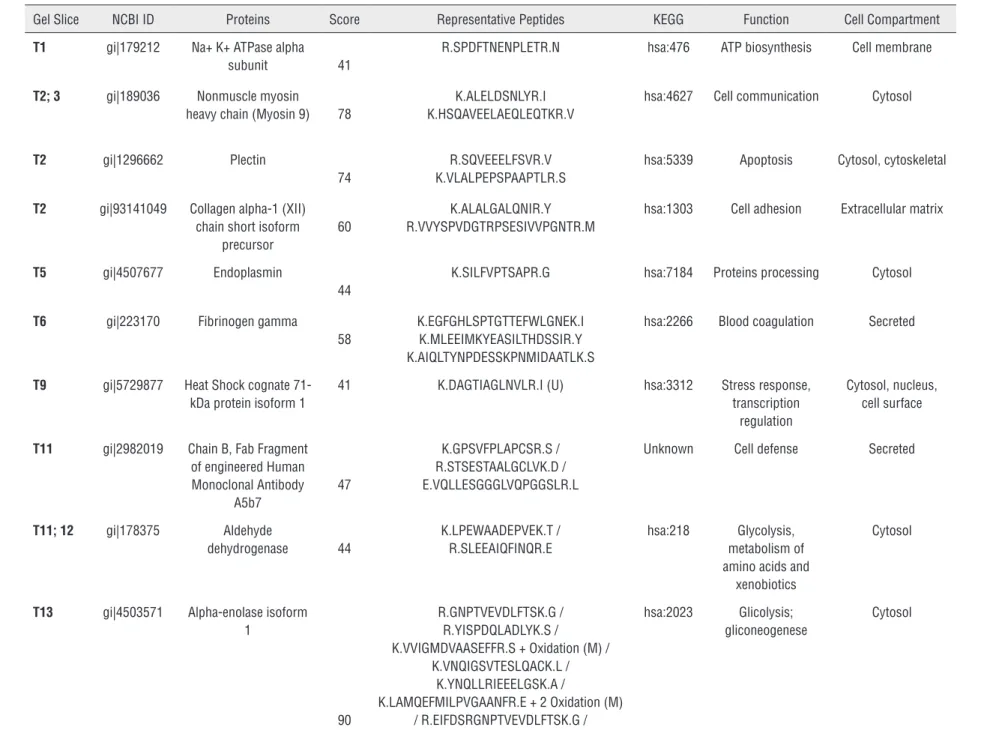
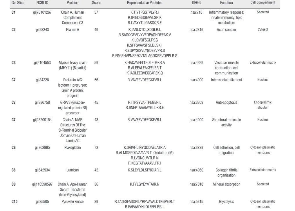
ces. Twenty-six protein spots from Group 1 and 21 from Group 2 were identified, sliced out from the gel and analyzed through mass spectrometry (Fi-gure-1). Sixty-three different proteins were iden-tified in Group 1 and 50 in Group 2. After a com-parative analysis of both groups, it was possible to recognize 28 proteins exclusively detected in Group 1 and 21 proteins presented only in Group 2 (Tables 2 and 3).DISCUSSION
A large number of proteins were identified in both Groups 1 and 2. Some of these proteins
found in Group 1 are also directly involved in the development of other types of cancers and there-fore, suitable for analysis.
The major stress-inducible heat shock pro-tein, Hsp70, that is a chaperone protein abundan-tly and preferentially expressed in tumors, was detected in Group 1. Owing to the ability of Hsp70 to protect cells from a wide range of apoptotic and necrotic stimuli, it has been assumed that Hsp70 may confer survival advantage to tumor cell li-nes. Nylandsted et al. (16) demonstrated that the depletion of Hsp70 by an adenovirus expressing antisense Hsp70 resulted in a massive cell death of tumorigenic cell lines of breast, colon,
prosta-Figure 1 - 1DE analysis of tissue samples from SCCP HPV patients and control group. Each pool contained 3.3µg of proteins from each sample, a total of 33µg. The SCCP and control pools were applied on a 12% SDS-PAGE gel. The gel was stained with Coomassie blue G. The markers and numbers in gel represent the sections that were excised for mass spectrometry analysis.
T=penile cancer SCCP; C=control group; SMW=Standard Molecular Weight
IBJU
|
PRO
TEOMICS ANAL
YSIS OF TISSUE SAMPLES FROM P
A
TIENTS WITH PENILE CARCINOMA AND POSITIVE T
O HPV
647
Table 2 - Proteins identified in pool of patients with SCCP (Group 1)
Gel Slice NCBI ID Proteins Score Representative Peptides KEGG Function Cell Compartment
T1 gi|179212 Na+ K+ ATPase alpha subunit 41
R.SPDFTNENPLETR.N hsa:476 ATP biosynthesis Cell membrane
T2; 3 gi|189036 Nonmuscle myosin
heavy chain (Myosin 9) 78
K.ALELDSNLYR.I K.HSQAVEELAEQLEQTKR.V
hsa:4627 Cell communication Cytosol
T2 gi|1296662 Plectin
74
R.SQVEEELFSVR.V K.VLALPEPSPAAPTLR.S
hsa:5339 Apoptosis Cytosol, cytoskeletal
T2 gi|93141049 Collagen alpha-1 (XII) chain short isoform
precursor
60
K.ALALGALQNIR.Y R.VVYSPVDGTRPSESIVVPGNTR.M
hsa:1303 Cell adhesion Extracellular matrix
T5 gi|4507677 Endoplasmin
44
K.SILFVPTSAPR.G hsa:7184 Proteins processing Cytosol
T6 gi|223170 Fibrinogen gamma
58
K.EGFGHLSPTGTTEFWLGNEK.I K.MLEEIMKYEASILTHDSSIR.Y K.AIQLTYNPDESSKPNMIDAATLK.S
hsa:2266 Blood coagulation Secreted
T9 gi|5729877 Heat Shock cognate 71-kDa protein isoform 1
41 K.DAGTIAGLNVLR.I (U) hsa:3312 Stress response, transcription
regulation
Cytosol, nucleus, cell surface
T11 gi|2982019 Chain B, Fab Fragment of engineered Human Monoclonal Antibody
A5b7
47
K.GPSVFPLAPCSR.S / R.STSESTAALGCLVK.D / E.VQLLESGGGLVQPGGSLR.L
Unknown Cell defense Secreted
T11; 12 gi|178375 Aldehyde
dehydrogenase 44
K.LPEWAADEPVEK.T / R.SLEEAIQFINQR.E
hsa:218 Glycolysis, metabolism of amino acids and
xenobiotics
Cytosol
T13 gi|4503571 Alpha-enolase isoform 1
90
R.GNPTVEVDLFTSK.G / R.YISPDQLADLYK.S /
K.VVIGMDVAASEFFR.S + Oxidation (M) / K.VNQIGSVTESLQACK.L /
K.YNQLLRIEEELGSK.A /
K.LAMQEFMILPVGAANFR.E + 2 Oxidation (M) / R.EIFDSRGNPTVEVDLFTSK.G / K.DATNVGDEGGFAPNILENKEGLELLK.T
hsa:2023 Glicolysis; gliconeogenese
IBJU
|
PRO
TEOMICS ANAL
YSIS OF TISSUE SAMPLES FROM P
A
TIENTS WITH PENILE CARCINOMA AND POSITIVE T
O HPV
648
T13 gi|1710248 Protein disulfide isomerase-related
protein 5
55
R.TGEAIVDAALSALR.Q / K.LAAVDATVNQVLASR.Y
hsa:10130 Proteins processing Endoplasmatic reticulum
T13 gi|31170 Chain A, Crystal Structure Of Human
Beta Enolase Enob
44
K.VNQIGSVTESIQACK.L hsa:2027 Glicolysis Cytosol,fosfopiruvat hydratase complex
T13 gi|40068518 6-phosphogluconate dehydrogenase, decarboxylating
42
K.IISYAQGFMLLR.Q + Oxidation (M) / K.GILFVGSGVSGGEEGAR.Y
hsa:5226 Pentose pathways Cytosol
T14 gi|4505763 Phosphoglycerate
kinase 1 39
K.ITLPVDFVTADKFDENAK.T hsa:5230 Glicolysis Cytosol
T14 gi|306882 Haptoglobin precursor 36
K.VTSIQDWVQK.T /
K.SPVGVQPILNEHTFCAGMSK.Y + Oxidation (M)
hsa:3240 Defense Secreted
T15 gi|35222 70-kDa heat shock protein
43 R.TTPSYVAFTDTER.L (U) hsa:3312 Regulation of cell cycle; cellular
membrane organization
Cytosol; plasma membrane
T16 gi|63252913 Macrophage-capping protein 44
R.QAALQVAEGFISR.M hsa:822 Actin filament organization
Cytosol
T17 gi|5174391 Alcohol dehydrogenase
[NADP+] 44
K.GLVQALGLSNFNSR.Q / R.GLEVTAYSPLGSSDR.A
hsa:10327 Glicolysis, glicerolipids metabolism
Cytosol
T17 gi|31397 Fibronectin precursor 40
R.VPGTSTSATLTGLTR.G hsa:2335 Angiogenesis, cell adhesion, platelet activation and degranulation
Secreted, extracellular matrix
T18 gi|31645 Glyceraldehyde-3-phosphate dehydrogenase
46 R.GALQNIIPASTGAAK.A / R.VPTANVSVVDLTCR.L / K.LISWYDNEFGYSNR.V / K.LTGMAFRVPTANVSVVDLTCR.L +
Oxidation (M) /
K.IKWGDAGAEYVVESTGVFTTMEK.A + Oxidation (M) /
K.VIHDNFGIVEGLMTTVHAITATQK.T + Oxidation (M)
IBJU
|
PRO
TEOMICS ANAL
YSIS OF TISSUE SAMPLES FROM P
A
TIENTS WITH PENILE CARCINOMA AND POSITIVE T
O HPV
649
T19; 20 gi|809185 Chain A, The Effect Of
Metal Binding On The Structure Of Annexin V And Implications For
Membrane Binding
52 RSEIDLFNIRK / KGLGTDEESILTLLTSRS /
KWGTDEEKFITIFGTRS / RGTVTDFPGFDERADAETLRK
hsa:308 Blood coagulation Cytosol
T19 gi|2906146 Malate dehydrogenase precursor
46 K.IFGVTTLDIVR.A / K.VDFPQDQLTALTGR.I
hsa:4191 Citric acid cycle Mitochondria
T19 gi|4826643 Annexin A3 37 K.MLISILTER.S + Oxidation (M) / K.GAGTNEDALIEILTTR.T
hsa:306 Defense response Phagocytic vesicle
T19 gi|4929769 Glyoxalase domain-containing protein 4
(CGI-150 protein)
36 K.ILTPLVSLDTPGK.A (U) hsa:51031 Unknown Mitochondria
T20 gi|4502599 Carbonyl reductase [NADPH] 1
46 R.LFSGDVVLTAR.D / R.VVNVSSIMSVR.A / R.GQAAVQQLQAEGLSPR.F / K.VADPTPFHIQAEVTMK.T + Oxidation
hsa:873 Lipid metabolism – arachidonic acid
Cytosol
T23 gi|9844110 cAMP-specific phosphodiesterase 4D
42 K.LSPVISPR.N hsa:5144 Smooth muscle contraction; regulation of receptor activity
Cytosol
T24 gi|2204207 Glutathione S-transferase
75 M.PPYTVVYFPVR.G -.MPPYTVVYFPVR.G + Oxidation (M)
M.PPYTVVYFPVRGR.C K.EEVVTVETWQEGSLK.A K.FQDGDLTLYQSNTILR.H K.ALPGQLKPFETLLSQNQGGK.T
K.YISLIYTNYEAGKDDYVK.A
hsa:2940 Amino acids metabolism
Cytosol
IBJU| PROTEOMICS ANALYSIS OF TISSUE SAMPLES FROM PATIENTS WITH PENILE CARCINOMA AND POSITIVE TO HPV
650
te, and liver. The authors advocate that Hsp70 is a prerequisite for the survival of human cancer cells. Similarly, Aghdassi et al. (17) demonstrated that the depletion of Hsp70 by short interfering RNA treatment induced apoptosis in pancreatic adenocarcinoma.Plectin is a cytolinker protein of the plakin family. Plakins connect intermediate filaments to desmosomes and hemidesmosomes, stabilize cells mechanically, regulate cytoskeleton dynamics, and serve as a scaffolding platform for signaling molecules. Niwa et al. (18) reported that Plectin misexpression leads to displacement of the cen-trosome, therefore contributing to genomic ins-tability and cancer development. Nevertheless, plectin is not expressed by most normal tissues, with the exception of the skin and genitourinary tract. Interestingly, we have detected plectin so-lely in Group 1. Complement plays a central part of the innate immune system, providing a highly effective means for destruction of invading micro-organisms: clearance of immune complexes; and elimination of dead, apoptotic, and tumor cells. During the evolution of a cancer cell, neo-antigens are produced. These elements distinguish cancer cells from their normal counterparts and may well be recognized by the immune system, eliminating many or most tumors (19, 20). Although most in vivo observations support that many cancers acti-vate the autologous complement system, it is also well-known that the efficiency of complement--mediated tumor cytotoxicity is hampered by va-rious protective mechanisms (21). In this work, hu-man complement C3 was detected only in Group 2. A possible explanation for these findings lies on the theory that patients with malignancies have a poorer immune response. Our result corroborates the study of Ornellas et al. (22), in which the au-thors have demonstrated that human complement fragments C3 and C4A/B were downregulated in plasma of patients with SCCP. In the present se-ries, all patients from Group 1 were HPV positive and this could explain the absence of complement C3 because viral proteins counteract the immune response (23).
Enolase is a key glycolytic enzyme that has been used as a diagnostic marker to identify
hu-man lung cancers (24). Higher α-enolase plasma
levels were also identified in patients with renal cell carcinoma (25). In cancer cells, enolase is ove-rexpressed and localizes on their surface, where it acts as a key protein in tumor metastasis, promo-ting cellular metabolism in anaerobic conditions and driving tumor invasion through plasminogen activation and extracellular matrix degradation. It also displays a characteristic pattern of acetyla-tion, methylaacetyla-tion, and phosphorylation that re-gulates protein functions and immunogenicity. In the present study, alfa and beta enolase isoforms were identified exclusively in Group 1. This fin-ding may suggest that in the future, enolase can be used as a possible clinical biomarker. Never-theless, further studies are needed to corroborate these findings and to determine the usefulness of this protein in clinical scope.
Prohibitin is a potential tumor suppres-sor, which was originally identified because of its anti-proliferative activities. The human prohibitin gene was identified and cloned in 1991, as a re-sult of a search for potential tumor suppressors, on the basis of its anti-proliferative activities (26). Furthermore, prohibitin is capable of inhibiting cell proliferation by repressing the transcriptional activity mediated by E2F which regulates many genes involved in the transition G1/S and DNA synthesis (27). In addition to transcriptional re-pression, prohibitin can induce p53-mediated transcription, indicating that prohibitin may have dual functions in modulating transcription (28).
In a study conducted by Joshi et al. (29), the authors supported this theory by demonstra-ting that prohibitin can differentially regulate the Yin-Yang 1 and caspase 7 gene promoter acti-vities. Additional functions related to prohibitin were linked to cell apoptosis (30). In this series, prohibitin was exclusively presented in Group 2, supporting its potential tumor suppressor activity. The critical functions of prohibitin in growth con-trol and transcriptional regulation clearly indicate the need for further investigations to elucidate its importance in SCCP development.
IBJU
|
PRO
TEOMICS ANAL
YSIS OF TISSUE SAMPLES FROM P
A
TIENTS WITH PENILE CARCINOMA AND POSITIVE T
O HPV
6
51
Table 3 - Proteins identified in pool of patients with non-tumor tissue (Group 2).
Gel Slice NCBI ID Proteins Score Representative Peptides KEGG Function Cell Compartment C1 gi|78101267 Chain A, Human
Complement Component C3
57 K.TIYTPGSTVLYR.I R.IPIEDGSGEVVLSR.K R.LVAYYTLIGASGQR.E
hsa:718 Inflammatory response; innate immunity; lipid
metabolism
Secreted
C2 gi|28243 Filamin A 49 R.IANLQTDLSDGLR.L R.SAGQGEVLVYVEDPAGHQEEAK.V
K.LDVQFSGLTK.G K.SPFSVAVSPSLDLSK.I R.EGPYSISVLYGDEEVPR.S R.FGGEHVPNSPFQVTALAGDQPSVQPPLR.S
hsa:2316 Actin coupler Cytosol
C3 gi|2104553 Myosin heavy chain (MHY11) (5'partial)
59 K.HAQAVEELTEQLEQFKR.A R.ALEEALEAKEELER.T K.IAQLEEQVEQEAREK.Q
hsa:4629 Vascular muscle contraction; cell communication
Extracellular matrix
C7 gi|34228 Prelamin-A/C isoform 1 precursor;
lamin A protein; progerin
56 R.VAVEEVDEEGKFVR.L hsa:4000 Intermediate filament Nucleus
C7 gi|386758 GRP78 (Glucose-regulated protein 78)
precursor
45 R.ITPSYVAFTPEGER.L R.IINEPTAAAIAYGLDKR.E
hsa:3309 Anti-apoptosis Endoplasmic reticulum
C7 gi|23200154 Chain A, NMR Structures Of The C-Terminal Globular
Domain Of Human Lamin AC
43 R.VAVEEVDEEGKFVR.L hsa:4000 Structural molecule activity
Nucleus
C8 gi|762885 Plakoglobin 72 K.SAIVHLINYQDDAELATR.A R.ALMGSPQLVAAVVR.T Oxidation (M)
R.LVQNCLWTLR.N R.NEGTATYAAAVLFR.I
hsa:3728 Cell adhesion, cell migration
Cytosol; plasmatic membrane
C8 gi|642534 Lumican 42 K.SLEYLDLSFNQIAR.L hsa:4060 Collagen fibrils organization
Extracellular matrix
C8 gi|110590597 Chain A, Apo-Human Serum Transferrin (Non-Glycosylated)
36 K.FYLGYEYVTAIR.N hsa:7018 Mineral absorption Secreted
C10 gi|35505 Pyruvate kinase 39 R.TATESFASDPILYRPVAVALDTKGPEIR.T R.EAEAAIYHLQLFEELRR.L
IBJU
|
PRO
TEOMICS ANAL
YSIS OF TISSUE SAMPLES FROM P
A
TIENTS WITH PENILE CARCINOMA AND POSITIVE T
O HPV
652
C11; 13; 15; 16
gi|3411130 Mutant Desmin 48 R.FLEQQNAALAAEVNR.L hsa:1674 Cytoskeleton structural protein activity
Intermediate filament C
C13 gi|340219 Vimentin 60 K.ILLAELEQLK.G
K.ILLAELEQLKGQGK.S
K.LQEEMLQREEAENTLQSFR.Q Oxidation (M) R.KVESLQEEIAFLK.K
R.QVQSLTCEVDALKGTNESLER.Q R.EYQDLLNVK.M K.MALDIEIATYR.K Oxidation (M)
R.ISLPLPNFSSLNLR.E
hsa:7431 Apoptosis; cell mobility Cytosol
C13 gi|704416 Elongation factor Tu 45 K.LLDAVDTYIPVPAR.D hsa:7284 Oxidative phosphorylation
Mitochondria
C15 gi|34234 Laminin-binding protein
75 R.AIVAIENPADVSVISSR.N R.FTPGTFTNQIQAAFREPR.L
hsa:3921 Ribosome Cytosol
C16 gi|47519616 Tropomyosin beta chain isoform 2
73 R.IQLVEEELDR.A
R.IQLVEEELDRAQER.L R.LATALQKLEEAEK.A
hsa:7169 Muscle contraction Cytosol
C17 gi|31645 Glyceraldehyde-3-phosphate dehydrogenase
72 K.VIHDNFGIVEGLMTTVHAITATQK.T Oxidation (M) R.DGRGALQNIIPASTGAAK.A
R.GALQNIIPASTGAAK.A R.VPTANVSVVDLTCR.L K.LISWYDNEFGYSNR.V
hsa:2597 Glycolysis Cytosol; plasmatic membrane
C19 gi|4505773 Prohibitin 91 R.IFTSIGEDYDER.V R.FDAGELITQR.E
hsa:5245 DNA synthesis Mitochondrial membrane
C19 gi|66473265 Beta globin chain 50 K.VNVDEVGGEALGR.L R.LLVVYPWTKR.F
hsa:5245 Oxygen transport Hemoglobin
C21 gi|494066 Chain A,Three-Dimensional Structure Of Class
Pi Glutathione S-Transferase From
Human Placenta In Complex With S-Hexylglutathione
At2.8 Angstroms Resolution
36 .PPYTVVYFPVRGR.C K.FQDGDLTLYQSNTILR.H
hsa:2940 Amino acids metabolism
IBJU| PROTEOMICS ANALYSIS OF TISSUE SAMPLES FROM PATIENTS WITH PENILE CARCINOMA AND POSITIVE TO HPV
653
in the group with tumor. As the selected patients were positive for HPV DNA, this fact can cause false negative for complement proteins. The va-riability could have been better analyzed if the-re wethe-re compathe-red to patients with cancer of the penis, whose tests did not reveal the presence of HPV. The proteomic consequences of HPV infec-tion in penile carcinoma are not known. Analysis of differentially expressed proteins by HPV sta-tus revealed enrichment of proteins involved in epithelial cell development, keratinization and ex-tracellular matrix organization in HPV− oropha-ryngeal carcinoma (OPC), whereas enrichment of proteins in DNA initiation and replication and cell cycle control was found for HPV+ (OPC) (31). Due to the rarity of penile tumors and the high percen-tage of HPV positive in our samples (8, 13) it is difficult to compare the tumors according to HPV status. However, a second study is underway to compare our results and identify the presence or absence of complement in tissue of SCCP patients negative for HPV.CONCLUSIONS
We identified a large number of proteins in patients with penile cancer and in the control group. Some of these proteins, found in the first group, are also directly involved in the develop-ment of other types of cancers and therefore, sui-table for analysis. Further studies are needed to corroborate these findings and to determine the usefulness of each discussed protein in the clini-cal scope of SCCP patients. Remarkably, this work reinforces that the C3 complement protein is a strong biomarker candidate for evaluating SCCP patients. Further studies should be conducted comparing samples positive for HPV with other HPV negative.
ACKNOWLEGMENTS
This work was supported by FAPERJ (APQ1 E-26/110.812/2009 and E-26/111.336/2013) and Programa de Oncobiologia (Brazil).
CONFLICT OF INTEREST
None declared. REFERENCES
1. Solsona E, Algaba F, Horenblas S, Pizzocaro G, Windahl T; European Association of Urology. EAU Guidelines on Penile Cancer. Eur Urol. 2004;46:1-8.
2. Brunini R: Câncer no Brasil: Dados histopatológicos: 1976-80. In: Resultados. Ministério da Saúde - Campanha Nacional de Combate ao Câncer, Rio de Janeiro, RJ, 1982; 118.
3. Parkin DM, Muir CS. Cancer Incidence in Five Continents. Comparability and quality of data. IARC Sci Publ. 1992;120:45-173.
4. Koifman L, Vides AJ, Koifman N, Carvalho JP, Ornellas AA. Epidemiological aspects of penile cancer in Rio de Janeiro: evaluation of 230 cases. Int Braz J Urol. 2011;37:231-40; discussion 240-3.
5. Barrasso R, De Brux J, Croissant O, Orth G. High prevalence of papillomavirus-associated penile intraepithelial neoplasia in sexual partners of women with cervical intraepithelial neoplasia. N Engl J Med. 1987;317:916-23.
6. Maiche AG. Epidemiological aspects of cancer of the penis in Finland. Eur J Cancer Prev. 1992;1:153-8.
7. Maden C, Sherman KJ, Beckmann AM, Hislop TG, Teh CZ, Ashley RL, et al. History of circumcision, medical conditions, and sexual activity and risk of penile cancer. J Natl Cancer Inst. 1993;85:19-24.
8. Scheiner MA, Campos MM, Ornellas AA, Chin EW, Ornellas MH, Andrada-Serpa MJ. Human papillomavirus and penile cancers in Rio de Janeiro, Brazil: HPV typing and clinical features. Int Braz J Urol. 2008;34:467-74; discussion 475-6.
9. McCance DJ, Kalache A, Ashdown K, Andrade L, Menezes F, Smith P, et al. Human papillomavirus types 16 and 18 in carcinomas of the penis from Brazil. Int J Cancer. 1986;37:55-9.
10. Peclat de Paula AA, Neto JCA, Cruz AD, de Freitas Jr R: Carcinoma epidermóide do pênis: considerações epidemiológicas, histopatológicas, influência viral e tratamento cirúrgico. Rev Bras Cancer. 2005;51:243-52. 11. Silva KC, Rosa ML, Moyse N, Afonso LA, Oliveira LH,
Cavalcanti SM. Risk factors associated with human papillomavirus infection in two populations from Rio de Janeiro, Brazil. Mem Inst Oswaldo Cruz. 2009;104:885-91. 12. Melgaço FG, Rosa ML, Augusto EF, Haimuri JG, Jacintho
IBJU| PROTEOMICS ANALYSIS OF TISSUE SAMPLES FROM PATIENTS WITH PENILE CARCINOMA AND POSITIVE TO HPV
654
13. Afonso LA, Moyses N, Alves G, Ornellas AA, Passos MR, Oliveira Ldo H, et al. Prevalence of human papillomavirus and Epstein-Barr virus DNA in penile cancer cases from Brazil. Mem Inst Oswaldo Cruz. 2012;107:18-23.
14. Ericsson C, Franzén B, Nistér M. Frozen tissue biobanks. Tissue handling, cryopreservation, extraction, and use for proteomic analysis. Acta Oncol. 2006;45:643-61.
15. Laemmli UK. Cleavage of structural proteins during the assembly of the head of bacteriophage T4. Nature. 1970;15;227:680-5.
16. Nylandsted J, Brand K, Jäättelä M. Heat shock protein 70 is required for the survival of cancer cells. Ann N Y Acad Sci. 2000;926:122-5.
17. Aghdassi A, Phillips P, Dudeja V, Dhaulakhandi D, Sharif R, Dawra R, et al. Heat shock protein 70 increases tumorigenicity and inhibits apoptosis in pancreatic adenocarcinoma. Cancer Res. 2007;15;67:616-25.
18. Niwa T, Saito H, Imajoh-ohmi S, Kaminishi M, Seto Y, Miki Y, et al. BRCA2 interacts with the cytoskeletal linker protein plectin to form a complex controlling centrosome localization. Cancer Sci. 2009;100:2115-25.
19. Hanahan D, Weinberg RA. The hallmarks of cancer. Cell. 2000;100:57-70.
20. Smyth MJ, Godfrey DI, Trapani JA. A fresh look at tumor immunosurveillance and immunotherapy. Nat Immunol. 2001;2:293-9.
21. Jurianz K, Ziegler S, Garcia-Schüler H, Kraus S, Bohana-Kashtan O, Fishelson Z, et al. Complement resistance of tumor cells: basal and induced mechanisms. Mol Immunol. 1999;36:929-39.
22. Ornellas P, Ornellas AA, Chinello C, Gianazza E, Mainini V, Cazzaniga M, et al. Downregulation of C3 and C4A/B complement factor fragments in plasma from patients with squamous cell carcinoma of the penis. Int Braz J Urol. 2012;38:739-49.
23. Campo MS, Graham SV, Cortese MS, Ashrafi GH, Araibi EH, Dornan ES, et al. HPV-16 E5 down-regulates expression of surface HLA class I and reduces recognition by CD8 T cells. Virology. 2010;407:137-42.
24. He P, Naka T, Serada S, Fujimoto M, Tanaka T, Hashimoto S, et al. Proteomics-based identification of alpha-enolase as a tumor antigen in non-small lung cancer. Cancer Sci. 2007;98:1234-40.
25. Kaneko N, Gotoh A, Okamura N, Matsuo E, Terao S, Watanabe M, et al. Potential tumor markers of renal cell carcinoma:
α-enolase for postoperative follow up, and galectin-1 and galectin-3 for primary detection. Int J Urol. 2013;20:530-5. 26. Nuell MJ, Stewart DA, Walker L, Friedman V, Wood CM,
Owens GA, et al. Prohibitin, an evolutionarily conserved intracellular protein that blocks DNA synthesis in normal fibroblastos and HeLa cells. Mol Cell Biol. 1991;11:1372-81. 27. Wang S, Nath N, Adlam M, Chellappan S. Prohibitin, a
potential tumor suppressor, interacts with RB and regulates E2F function. Oncogene. 1999;18:3501-10.
28. Fusaro G, Dasgupta P, Rastogi S, Joshi B, Chellappan S. Prohibitin induces the transcriptional activity of p53 and is exported from the nucleus upon apoptotic signaling. J Biol Chem. 2003;278:47853-61.
29. Joshi B, Rastogi S, Morris M, Carastro LM, DeCook C, Seto E, et al. Differential regulation of human YY1 and caspase 7 promoters by prohibitin through E2F1 and p53 binding sites. Biochem J. 2007;401:155-66.
30. Chowdhury I, Xu W, Stiles JK, Zeleznik A, Yao X, Matthews R, et al. Apoptosis of rat granulosa cells after staurosporine and sérum withdrawal is suppressed by adenovirus-directed overexpression of prohibitin. Endocrinology. 2007;148:206-17.
31. Slebos RJ, Jehmlich N, Brown B, Yin Z, Chung CH, Yarbrough WG, et al. Proteomic analysis of oropharyngeal carcinomas reveals novel HPV-associated biological pathways. Int J Cancer. 2013;132:568-79.