BASIC RESEARCH
Passive stiffness of rat skeletal muscle
undernourished during fetal development
Ana Elisa Toscano,IKarla Moˆnica Ferraz,IIRaul Manha˜es de Castro,IIIFrancis CanonIV
ICentro Acadeˆmico de Vito´ria, Universidade Federal de Pernambuco (UFPE). Vito´ria de Santo Anta˜o, Pernambuco, Brazil.IIDepartamento de Fisioterapia, Universidade Federal de Pernambuco (UFPE). Recife, Pernambuco, Brazil.IIIDepartamento de Nutric¸a˜o, Universidade Federal de Pernambuco (UFPE), Recife, Pernambuco, Brazil.IVUniversite´ de Technologie de Compie`gne (UTC) – CNRS UMR6600, Biome´canique et Bioinge´nierie, BP 20.529, Rue P de Roberval 60205 Compie`gne cedex-France.
OBJECTIVES:The aim of the study was to investigate the effect of fetal undernutrition on the passive mechanical properties of skeletal muscle of weaned and young adult rats.
INTRODUCTION:A poor nutrition supply during fetal development affects physiological functions of the fetus. From a mechanical point of view, skeletal muscle can be also characterized by its resistance to passive stretch.
METHODS:Male Wistar rats were divided into two groups according to their mother’s diet during pregnancy: a control group (mothers fed a 17% protein diet) and an isocaloric low-protein group (mothers fed a 7.8% protein diet). At birth, all mothers received a standardized meal ad libitum. At the age of 25 and 90 days, the soleus muscle and extensor digitorum longus (EDL) muscles were removed in order to test the passive mechanical properties. A first mechanical test consisted of an incremental stepwise extension test using fast velocity stretching (500 mm/s) enabling us to measure, for each extension stepwise, the dynamic stress (sd) and the steady stress (ss). A second test
consisted of a slow velocity stretch in order to calculate normalized stiffness and tangent modulus from the stress– strain relationship.
RESULTS:The results for the mechanical properties showed an important increase in passive stiffness in both the soleus and EDL muscles in weaned rat. In contrast, no modification was observed in young adult rats.
CONCLUSIONS: The increase in passive stiffness in skeletal muscle of weaned rat submitted to intrauterine undernutrition it is most likely due to changes in muscle passive stiffness.
KEYWORDS: Passive stiffness; Skeletal muscle; Fetal; Development; Undernutrition.
Toscano AE, Ferraz KM, Manha˜es de Castro R, Canon F. Passive stiffness of rat skeletal muscle undernourished during fetal development. Clinics. 2010;65(12):1363-1369.
Received for publication onSeptember 10, 2010;First review completed onSeptember 24, 2010;Accepted for publicationOctober 4, 2010 E-mail: raulmanhaesdecastro@yahoo.com.br
Tel.: 55-81-21268470
INTRODUCTION
Numerous studies have shown the influence of nutrient supply on development in utero.1–4
A poor nutrition supply during fetal development affects physiological functions of the fetus and has long-term consequences at adulthood.5,6 This concept of ‘‘program-ming’’ represents the mechanism whereby a stimulus or an insult during a critical developmental period has permanent effects on structure, physiology, and metabolism.7There is evidence of programming affecting structure and function of skeletal muscles postnatally.4 For example, Ozanne et al.3,8showed alterations in muscle metabolic capacities in rats undernourished during fetal development and
lacta-tion. Modifications in muscle fiber type distribution in both young and adult mammals have been reported as well as a decrease in the fiber density in the diaphragm of pups whose mothers had suffered nutritional deprivation.1,9,10 However, few studies have examined the consequence of early undernutrition in mechanical muscle properties.
In a previous study, we have shown modifications in both contractile and series elastic properties of rat muscles undernourished during fetal development.11 From a mechanical point of view, skeletal muscle can also be characterized by its resistance to passive stretch. From a functional point of view, muscle passive properties are important to take into account because (1) these character-istics contribute in part to the maximal joint range of motion, (2) part of the force developed by the contracting muscle will be devoted to the stretch of passive antagonist and (3) a relation between passive stiffness and spindle discharge has been shown.12
The aim of this study was to evaluate the effect of a low-protein diet during fetal life on the passive mechanical Copyrightß2010CLINICS– This is an Open Access article distributed under
group C were fed a control diet (17% protein) according to the recommendations of AIN-93G,13 and UN animals received a low-protein isocaloric diet (7.8%) ad libitum (Table 1). On the first day after birth, all mothers received a control diet (17% of protein) ad libitum and litters were limited to six male pups per mother. At weaning, pups were fed a standardized meal (17% of protein) ad libitum until
60 days old.13Afterwards, offspring received a 12% protein
dietad libitumuntil 90 days of age.
The protocols used in the present study were in accordance with the guidelines and regulations of the Ethical Hygiene and Safety Committee of the Compie`gne University of Technology.
Biomechanical analysis
Rats 25 days old (n = 14) and 90 days old (n = 16) were anesthetized with an intraperitoneal injection of sodium pentobarbital (30 mg/kg of body mass). The soleus and EDL muscles were carefully excised from the hind limb and placed in a dissection chamber containing Ringer’s solution (composition in mM: NaCl, 115; NaHCO3, 28; CaCl2, 2.5, MgSO4, 3.1, KCl, 3.5, KH2PO4, 1.4; glucose, 11.1) maintained at 25
˚
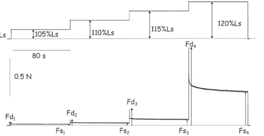
C and oxygenated with a gas mixture of 95% O2and 5% CO2 that resulted in a pH of 7.3. At the end of the experiment all animals was killed in accordance with the animal committee at Compie`gne University of Technology. The proximal part of the muscle was fixed to a force transducer and the distal extremity was linked to theextension, the muscle was suddenly released to Ls. This test enabled us to measure, for each extension stepwise, the dynamic force (Fd) that corresponded to the maximal force developed by muscle at the end of the fast extension and the steady force (Fs) at the end of the plateau in length (Fig. 1). Then, Fd and Fs were divided by the physiological cross-sectional area (PCSA) of the muscle, which yields the dynamic tension (sd) and the steady tension (ss). PCSA of
muscle was calculated using the equation PCSA = MW/ (1.066Lf), where MW is muscle mass, 1.06 is the muscle density (in g/cm3), and Lfis the fiber length. Lfcorresponds to 72% and 44% of the length of the soleus and the EDL muscles, respectively.15,16
The stretch–release test consisted of stretching the muscles at amplitude up to 125% Ls with a slow velocity (0.1 mm/s) following by a release until Ls with the same velocity (Fig. 2). From these data, stress (i.e., passive force normalized in respect of PCSA) and strain (i.e., deforma-tion/Ls) were calculated to construct the stress–strain curve. From this curve, tension at 125% Ls (F125/PCSA), stiffness at 125% Ls and tangent modulus (i.e., slope in the linear portion of the stress-strain curve) were calculated.
Statistical analysis
All data are presented as mean ¡SEM. A two-way (to evaluate the effect of the age and diet on body mass) and three-way (to evaluate the effect of age, diet and muscle on the other parameters) Analysis of variance (ANOVA) for repeated measurements followed by the Holm Sidak post hoc test were performed. A level of 95% was set as the statistical difference. The statistical treatment of the data was performed with the Sigmastat software (Systat Software, Inc., Chicago, IL).
RESULTS
The body mass of pups from UN mothers was signifi-cantly lower at 25 days (92.6¡5.18 g vs 71.3¡1.3 g for the C and UN groups, respectively; p,0.05) and 90 days (449.5 ¡9.2g vs 413.2 ¡14g for C and UN group, respectively; p,0.05).
Absolute and relative mass of the soleus and EDL muscles was significantly smaller in the UN group than in C group in weaned and young adult rats (Fig. 3).
Results of the incremental stepwise extension test indicated increases in resistance to passive stretch for each extension in both soleus and EDL muscles in weaned rats (Fig. 4). At this age, soleus muscle of UN rats showed an increase in dynamic tensions by 40%, 48%, 57%, and 52% for
Table 1 -Composition of the experimental diets.
Ingredient (g/kg)
Control diet (17% protein)
Low-protein diet (7.8% protein)
Casein ($92.5 protein) 183.8 84.3
Cornstarch 645.7 745.2
Soybean oil 70.0 70.0
Fibre 50.0 50.0
Vitamin mix* 10.0 10.0
Mineral mix{ 35.0 35.0
Choline bitartrate 2.5 2.5
L-metionine 3.0 3.0
Total 1000 1000
*American Institute of Nutrition 93G;13vitamin mix provided (mg/kg diet):
nicotinic acid 30.0, calcium pantothenate 16.0, pyridoxine-HCl 7.0, thiamin-HCl 6.0, riboflavin 6.0, folic acid 2.0, biotin 2.0, cyanocobalamin, 25.0,a-tocopherol 150.0, retinyl palmitate 8.0, cholecalciferol 2.5, and phylloquinone 0.75.
{
American Institute of Nutrition 93;13mineral mix provided (mg/kg diet):
the first, second, third, and fourth increment, respectively (Fig. 4). Similar increases were obtained in EDL muscle. In addition, undernutrition induced an increase in steady tension of about 65% in the soleus and 100% in the EDL (Fig. 4). At 90 days, no difference in either dynamic tension or steady tension was observed in the soleus and the EDL between the control group and the undernourished group.
Passive force developed at 25% strain during the stretch– release test and was not modified in the soleus and EDL muscles of weaned and young adult rat (Fig. 5). When passive force was expressed in terms of normalized tension (i.e., force divided by PCSA), there was an increase in the passive tension in both the soleus and the EDL muscles in weaned rats (Fig. 5). This increase in resistance to passive stretch observed in the soleus and EDL of the UN group was also confirmed by the increase in the tangent modulus and in normalized stiffness. In young adult rats, no difference was observed in these parameters between groups in either the soleus or the EDL muscles.
DISCUSSION
The results of present study are in accordance with numerous studies showing poor maternal nutrition during gestation affects fetal growth and development.1,3,6,8,11
Thus, the decrease in the pup weight can be associated with the availability of nutrients for transfer to the fetus, possibly involving metabolic parameters such as glucose and insulin.17 Consequent to maternal nutrient restriction, the soleus and EDL muscle weight was significantly reduced in weaned and young adult rats. This muscle atrophy is consistent with the programming of skeletal muscle insulin sensitivity during fetal development8 as it has been shown that insulin-sensitive tissues undergo important changes in response to maternal protein restric-tion.18,19
A previous study had shown that maternal protein restriction during gestation induced changes in both contractile and series elastics properties.11 In addition to these mechanical changes, the present work has demon-strated that the passive elastic properties are also changed by this early nutritional manipulation. In effect, even if passive force that developed during the slow velocity stretch was not different between nutritional groups, the normalized tension showed an increase in soleus and EDL muscles in UN weaned rats. This increase in resistance to passive stretch was also perceived by the increase in the normalized stiffness and the increase in the tangent modulus. Results of the incremental stepwise test showed that passive stiffness was increased during both the
Figure 1 -Typical recording of passive tension induced by an incremental stepwise extension test. Fd: dynamic force; Fs: steady force.
Upper trace, change in length; bottom trace, change in force.
dynamic and the static phases and for short and long stretches.
Muscle passive stiffness is a function of the parallel elastic component described in Hill’s model.20 Its properties are affected by membrane structure and specifically by the concentration and type of collagen.21–25 The effects of nutritional status on the regulation of skeletal muscle collagen content are varied. Roy et al.26 reported no influence of nutritional level in muscle collagen content in pectoralis muscle of broilers but with some differences in the collagen structure of the perimysium. In the gastro-cnemius muscle of adult mice deprived of food for 2 days, Jagoe et al.27showed a decrease in gene expression for many extracellular matrix proteins like collagen. More recently, Stevenson et al.28 studied the transcriptional profile of myotube under starvation conditions. These authors reported a downregulation of genes involved in collagen synthesis and maturation. Nevertheless, the effect of nutritional supply during fetal development seems to be different in the development of connective tissue in skeletal muscle. In swine, Karunaratne et al.29 reported that the smallest littermate, reflecting a poor level of in utero nutritional supply, contained a higher concentration of type I collagen than the largest littermate. In our study, such an increase in the content of collagen could explain the increase in the passive tension observed in the soleus and EDL muscles of undernourished rats.
In addition to collagen, other connective proteins are a source of muscle passive tension. Titin, a 3-MDa elastic filamentous protein, links the Z line to the myosin filament in sarcomeres. Wang et al.30 reported that passive elastic properties of muscle fibers are related to expression of the titin isoform. Moreover, Toursel et al.31showed a decrease in passive tension in soleus fiber of unloaded rat in relation to a decrease in titin content. Passive stiffness results also from the relation between endosarcomeric and exosarco-meric protein networks constituted by different structural proteins like desmin.32 Lastly, telethonin (Titin-cap), an important component of the N-terminal titin anchor in the Z line,33seems to act on passive stiffness.34Interestingly, it has been shown that nutritional status changes the gene expression of these proteins. Byrne et al.35 reported an upregulation of cytoskeletal proteins like desmin or tele-thonin in the muscles of steers after nutritional restriction. Oumi et al.36 reported muscle ultrastructure damages induced in rats nourished with a low-protein diet for 2 weeks after weaning. More precisely, they showed disorganization in some sarcomeres, with a disruption of the Z line appearing jagged. As postulated by Oumi et al.,36 these sarcomere damages could be the result of the ‘‘disintegration’’ of structural proteins like desmin and titin. Muscle disorganization, such as those observed by these authors, could induce an increase in passive stiffness. As a matter of fact, Anderson et al.37 reported an increase in
passive stiffness in desmin knockout mice and ascribed this mechanical change to the adaptation of passives structures consequent to the lack of desmin.
Lastly, no modification in the passive stiffness properties was observed between groups in young adult rats. The total
recovery of these elastic properties reveals that the changes observed in the weaning rats can be completely reversed after nutritional recovery before the animal reaches the adult age. Nevertheless, it will be interesting to evaluate older animals in order to confirm or invalidate that the in
Figure 4 -Effects of undernutrition on dynamic tension (sd) (left) and steady tension (ss) (right) in soleus and EDL muscles at 25 and
utero low-protein diet supply has no long-term conse-quences in muscle passive mechanical properties.
CONCLUSIONS
This study has permitted understanding of the effect of a prenatal undernutrition on the passive elastic component of the postural muscle (soleus) and a nonpostural muscle (EDL). Prenatal undernutrition showed short-term altera-tions in passive stiffness that can be explained in terms of adaptations in passive structures and/or distribution of endosarcomeric and exosarcomeric proteins in the skeletal muscle. However, further biochemical investigations are necessary to establish the effects of this particular nutri-tional manipulation in a noncontractile protein profile of skeletal muscle.
REFERENCES
1. Bedi KS, Birzgalis AR, Mahon M, Smart JL, Wareham AC. Early life undernutrition in rats. 1. Quantitative histology of skeletal muscles from underfed young and refed adult animals. Br J Nutr. 1982;47:417–31, doi: 10.1079/BJN19820053.
2. Berleze KJ, Mu¨ller AP, Schweigert ID, Longoni A, Sordi F, de Assis AM, et al. Gestational and postnatal low protein diet alters insulin sensitivity in female rats. Exp Biol Med (Maywood). 2009; 234:1437–44, doi: 10. 3181/0903-RM-111.
3. Ozanne SE, Olsen GS, Hansen LL, Tingey KJ, Nave BT, Wang CL, et al. Early growth restriction leads to down regulation of protein kinase C zeta and insulin resistance in skeletal muscle. J of Endocrinol. 2003;177:235–41, doi: 10.1677/joe.0.1770235.
4. Tygesen MP, Harrison AP, Therkildsen M. The effect of maternal nutrient restriction during late gestation on muscle, bone and meat parameters in five month old lambs. Livestock Sci. 2007;110:230–41, doi: 10.1016/j.livsci.2006.11.003.
5. Hales CN, Barker DJ. Type 2 (non–insulin-dependent) diabetes mellitus: the thrifty phenotype hypothesis. Diabetologia. 1992;35:595–601, doi: 10. 1007/BF00400248.
6. Souza LS, Orozco-Solis R, Grit I, Manha˜es-de-Castro R, Bola˜nos-Jimenez F. Perinatal protein restriction reduces the inhibitory action of serotonin on food intake. European Journal of Neuroscience. 2008;27:1400–8, doi: 10.1111/j.1460-9568.2008.06105.x.
7. Lucas A, Programming in early nutrition in man. Ciba Found Symp. 1991;156:38–50.
8. Ozanne SE, Smith GD, Tikerpae J, Hales CN. Altered regulation of hepatic glucose output in the male offspring of protein-malnourished rat dams. Am J Physiol. 1996;270:E559–64.
9. Howells KF, Jordan TC, Mathews DR. Effects of pre and perinatal malnutrition on muscle fibres from fast and slow rat muscles. Res Exp Med (Berlin). 1978;173:35–40, doi: 10.1007/BF01851372.
10. Prakash YS, Fournier M, Sieck GC. Effects of prenatal undernutrition on developing rat diaphragm. J Appl Physiol. 1993; 75:1044–52.
11. Toscano AE, Manha˜es de Castro R, Canon F. Effect of a low-protein diet during pregnancy on skeletal muscle mechanical properties of offspring rats. Nutrition. 2008;24:270–8, doi: 10.1016/j.nut.2007.12.004.
12. Rosant C, Pe´rot C. An index of spindle efficacy obtained by measuring electroneurographic activity and passive tension in the rat soleus muscle. J Neurosci Methods. 2006;150:272–8, doi: 10.1016/j.jneumeth.2005.07.001. 13. Reeves PG, Nielsen FH, Fahey GC Jr. AIN-93 purified diets for laboratory rodents: final report of the American Institute of Nutrition
Figure 5 -Effects of undernutrition on tension at 125% Ls(F125), F125/PCSA, stiffness at 125% Lsand tangent modulus in soleus and EDL
muscles at 25 and 90 days of age induced by the stretch–release test. Ls: slack length; PCSA: physiological muscle cross-sectional area; C:
ad hoc writing committee on the reformulation of the AIN-76A rodent diet. J Nutr. 1993;123:1939–51.
14. Lensel-Corbeil G, Goubel F. Series elasticity in frog sartorius muscle during release and stretch. Arch Int Physiol Biochim. 1989;97:499–509, doi: 10.3109/13813458909075081.
15. Gregorevic P, Plant DR, Leeding KS, Bach LA, Lynch GS. Improved contractile function of the mdx dystrophic mouse diaphragm muscle after insulin-like growth factor-I administration. Am J Pathol. 2002; 161:2263–72.
16. Ranatunga KW. Temperature-dependence of shortening velocity and rate of isometric tension development in rat skeletal muscle. J Physiol. 1982;329:465–83.
17. Ozanne SE, Hales CN. The long-term consequences of intra-uterine protein malnutrition for glucose metabolism. Proc Nutr Soc. 1999;58: 615–9.
18. Albuquerque KT, Sardinha FL, Telles MM, Watanabe RL, Nascimento CM, Tavares Do Carmo MG, et al. Intake of trans fatty acid-rich hydrogenated fat during pregnancy and lactation inhibits the hypopha-gic effect of central insulin in the adult offspring. Nutrition. 2006;22:820– 9, doi: 10.1016/j.nut.2006.04.009.
19. Heywood WE, Mian N, Milla PJ, Lindley KJ. Programming of defective rat pancreatic beta-cell function in offspring from mothers fed a low-protein diet during gestation and the suckling periods. Clinical Sci (London). 2004;107:37–45, doi: 10.1042/CS20030350.
20. Hill AV. The heat of shortening and the dynamic constants of muscle. Proc R Soc Lond B Biol Sci. 1938;126:136–95, doi: 10.1098/rspb.1938.0050. 21. Alnaqeeb MA, Al Zaid NS, Goldspink G. Connective tissue changes and physical properties of developing and ageing skeletal muscle. J Anat. 1984;139:677–89.
22. Kovanen V, Suominen H, Heikkinen E. Mechanical properties of fast and slow skeletal muscle with special reference to collagen and endurance training. J Biomec. 1984;17:725–35, doi: 10.1016/0021-9290(84)90103-9. 23. Kovanen C. Effects of ageing and physical training on rat skeletal
muscle. An experimental study on the properties of collagen, laminin, and fibre types in muscles serving different functions. Acta Physiol Scand. 1989;577:1–56.
24. Gosselin LE, Adams C, Cotter TA, McCormick RJ, Thomas DP. Effect of exercise training on passive stiffness in locomotor skeletal muscle: role of extracellular matrix. J Appl Physiol. 1998; 85:1011–6.
25. Ducomps C, Maurie`ge P, Darche B, Combes S, Lebas F, Doutreloux JP. Effects of jump training on passive mechanical stress and stiffness in
rabbit skeletal muscle: role of collagen. Acta Physiol Scand. 2003;78:215– 24, doi: 10.1046/j.1365-201X.2003.01109.x.
26. Roy BC, Oshima I, Miyachi H, Shiba N, Nishimura S, Tabata S, et al. Effects of nutritional level on muscle development, histochemical properties of myofibre and collagen architecture in the pectoralis muscle of male broilers. Brit Poult Sci. 2006;47:433–42, doi: 10.1080/ 00071660600828334.
27. Jagoe RT, Lecker SH, Gomes M, Goldberg AL. Patterns of gene expression in atrophying skeletal muscles: response to food deprivation. FASEB J. 2002;16:1697–712, doi: 10.1096/fj.02-0312com.
28. Stevenson EJ, Koncarevic A, Giresi PG, Jackman RW, Kandarian SC. Transcriptional profile of a myotube starvation model of atrophy. J Appl Physiol. 2005;98:1396–406, doi: 10.1152/japplphysiol.01055.2004. 29. Karunaratne JF, Ashton CJ, Stickland NC. Fetal programming of fat and
collagen in porcine skeletal muscles. J Anat. 2005;207:763–8, doi: 10.1111/ j.1469-7580.2005.00494.x.
30. Wang K, McCarter R, Wright J, Beverly J, Ramirez-Mitchell R. Regulation of skeletal muscle stiffness and elasticity by titin isoforms: a test of the segmental extension model of resting tension. Proc Nat Ac Sci U S A. 1991;88:7101–5, doi: 10.1073/pnas.88.16.7101.
31. Toursel T, Stevens L, Granzier H, Mounier Y. Passive tension of rat skeletal soleus muscle fibers: effects of unloading conditions. J Appl Physiol. 2002;92:1465–72.
32. Salviati G, Betto R, Ceoldo S, Pierobon-Bormioli S. Morphological and functional characterization of the endosarcomeric elastic filament. Am J Physiol. 1990;259:C144–9.
33. Trinick J, Tskhovrebova L. Titin: a molecular control freak. Trends Cell Biol. 1999;9:377–80, doi: 10.1016/S0962-8924(99)01641-4.
34. Lee EH, Gao M, Pinotsis N, Wilmanns M, Schulten K. Mechanical strength of the titin Z1Z2-telethonin complex. Structure. 2006;14:497–509, doi: 10.1016/j.str.2005.12.005.
35. Byrne KA, Wang YH, Lehnert SA, Harper GS, McWilliam SM, Bruce HL, et al. Gene expression profiling of muscle tissue in Brahman steers during nutritional restriction. J Anim Sci. 2005; 83:1–12.