DOI: 10.1590/0004-282X20140133
ARTICLE
Lower trait frontal theta activity in
mindfulness meditators
Reduzida atividade teta na região frontal em praticantes de meditação mindfulness
Guaraci Ken Tanaka1, Caroline Peressutti1,2, Silmar Teixeira1,2, Mauricio Cagy5, Roberto Piedade1, AntonioEgídio Nardi6,7, Pedro Ribeiro1,2,3, Bruna Velasques1,2,3,4
he physiology of meditation practices may depend on whether they involve concentrative mindfulness (CM) or open monitoring mindfulness (OM) as well as on the prac
-titioner’s expertise level. Experienced meditators are able to maintain the meditative state for a long time in an efortless way compared to beginners. Electroencephalographic (EEG) studies of CM have found increased activity in low-frequen
-cy bands (alpha and theta), which relect relaxation and at
-tentional focus, in many brain regions, including the frontal
cortex1–3
. However, in long-term OM meditators, an increase in occipital4
and frontoparietal gamma activity also occurs due to their improved sensory awareness4
. Evidence from long-term practitioners has also shown increased low-fre
-quency oscillations in conjunction with gamma5
. Lutz et al.6
have found an increased ratio of gamma to slow oscillatory activity (4–13 Hz). However, the authors did not discuss if this increased ratio was related only to higher gamma or lower slow activity or both6
. Some hypotheses have been suggested,
1Institute of Psychiatry of the Federal University of Rio de Janeiro – Brain Mapping and Sensory Motor Integration – Rio de Janeiro RJ. 2Institute of Applied Neuroscience (INA) – Rio de Janeiro RJ.
3School of Physical Education of the Federal University of Rio de Janeiro (UFRJ) Bioscience Department – Rio de Janeiro RJ.
4Institute of Psychiatry of the Federal University of Rio de Janeiro – Neurophysiology and Neuropsychology of Attention – Rio de Janeiro RJ. 5Biomedical Engineering Program, of the Federal University of Rio de Janeiro (COPPE/UFRJ) - Rio de Janeiro RJ
6Institute of Psychiatry of the Federal University of Rio de Janeiro – Panic & Respiration Laboratory – Rio de Janeiro RJ. 7National Institute of Translational Medicine (INCT-TM).
Correspondence: Guaraci Tanaka – Avenida Venceslau Brás 71 – CEP: 22290-140 Botafogo – Rio de Janeiro RJ – Brazil. E-mail: acupunturatanaka@gmail.com
ABSTRACT
Acute and long-term effects of mindfulness meditation on theta-band activity are not clear. The aim of this study was to investigate frontal theta differences between long- and short-term mindfulness practitioners before, during, and after mindfulness meditation. Twenty partic-ipants were recruited, of which 10 were experienced Buddhist meditators. Despite an acute increase in the theta activity during meditation in both the groups, the meditators showed lower trait frontal theta activity. Therefore, we suggested that this inding is a neural correlate of the expert practitioners’ ability to limit the processing of unnecessary information (e.g., discursive thought) and increase the awareness of the essential content of the present experience. In conclusion, acute changes in the theta band throughout meditation did not appear to be a speciic correlate of mindfulness but were rather related to the concentration properties of the meditation. Notwithstanding, lower frontal theta activity appeared to be a trait of mindfulness practices.
Keywords: mindfulness, meditation, EEG, frontal theta power, working memory.
RESUMO
Os efeitos agudos e de longo prazo da meditação mindfulness sobre a atividade da banda teta não são claros. O objetivo deste estudo foi investigar as diferenças da banda teta na região frontal entre praticantes de mindfulness iniciantes e experientes. Desta forma, vinte participantes foram recrutados (dez meditadores budistas experientes e dez não-meditadores). Apesar do aumento agudo da atividade teta durante a meditação para ambos os grupos, os meditadores apresentaram uma menor potência em ambas as condi-ções. Sugerimos que este achado é um correlato neural da capacidade dos praticantes especialistas em limitar o processamento de informações desnecessárias e aumentar a conscientização sobre o conteúdo essencial da experiência presente. Em conclusão, as alterações agudas na banda teta durante a meditação devem estar relacionadas ao processo de concentração típico de qualquer técnica meditativa. No entanto, a atividade teta reduzida encontrada entre meditadores experientes de mindfulness parece ser uma característica desta prática especíica.
such as frontal theta (FT) activity is more engaged in CM than OM4
as well as higher frontal alpha-1 power and coher
-ence is associated with transcendental meditation, indicat
-ing efortless meditation3
.
FT activity has been described in many nonspeciic situa
-tions as states of concentration, focused attention, emotion, working memory, drowsiness, and REM sleep (lower theta, 4–6 Hz)1,5,7,8. hus, the studies of the cognitive processes un
-derlying FT have been extensive, although its functions in meditation are still not clear. he mental states that are re
-lated to the practice of meditation at the beginner and ex
-perienced levels have often been correlated with increased theta activity9
. However, such changes are quite unspeciic because they are observed in diferent techniques of medita
-tion and relaxa-tion and during the transi-tion to a sleep state5
. Speciically, an increased FT has been observed in CM10,11,
and it was deined as a sustained focus on a speciic prese
-lected object.
In contrast, OM refers to a state of sustained attention to any thought, feeling, or sensation that arises in the mind, with an attitude of acceptance and nonjudgment11
. hese mind
-fulness properties are thought to improve self-regulation and stress management by allowing individuals to refrain from trying to control the content of the mind12,13. Over the last de
-cades, related treatments, such as mindfulness-based stress reduction14
and mindfulness-based cognitive therapy15
, have been extensively researched and applied in diferent clinical and nonclinical populations with positive re
-sults. Notwithstanding, there is a lack of consistent reports on the regulatory mechanisms of theta oscillations in this spe
-ciic meditation technique. hus, the aim of this study was to investigate FT diferences between experienced and irst-time meditators before, during, and after OM. We hypothesized that long-term meditation practice would change the oscilla
-tory pattern of the theta band in expert practitioners.
METHODS
Twenty participants were recruited. Ten were experienced meditators (4 men; mean ± standard deviation age, 49.25 ± 8.66), including monks and laymen from various Buddhist traditions, and ten were healthy controls (5 men; age, 41.38 ± 15.10) who never meditated. he meditators must have practiced regularly for at least the last ive years (11.61 ± 7.40 years), and all of them must have been living in the same city under the same conditions as the control group. All of the participants were medication-free and had no sensory, mo
-tor, cognitive, or attention deicits that could afect their per
-formance. he subjects gave their written consent (in accor
-dance with the Helsinki Declaration) to participate in the study. he experiment was approved by the Ethics Committee of the Federal University of Rio de Janeiro (IPUB/UFRJ).
Task protocol
he subjects sat with straight spines in a darkened and noise-free room to minimize sensory interference. Two med
-itators and one nonmeditator chose to sit on cushions on the loor, while the other participants sat on chairs. he subjects were asked to rest for 4 min (rest instructions were particu
-larly emphasized for the meditators in order that they refrain from entering the meditative state), and then both groups started OM for 40 min. Finally, they rested for more 4 min at the end. An auditory signal marked the beginning and end of each stage.
he mindfulness (OM) instruction for the meditative and nonmeditative practitioners was to “pay attention to what
-ever comes into your awareness. What-ever it is, a stressful thought, an emotion or body sensation, just let it pass in an efortless way, without trying to maintain it or change it in any way, until something else comes into your conscious
-ness”16
. he instructions were given immediately before the recording, as we believed this would promote higher motiva
-tion as it was a irst attempt of a new activity. Due to its sim
-plicity, the technique could be implemented, and we actual
-ly found this was particular-ly important. he subjects were evaluated after the task, and they reported any problems fol
-lowing the instructions.
EEG recording
he International 10/20 EEG electrode system (Jasper, 1958) was used with a 20-channel EEG system (Braintech-3000, EMSA Medical Instruments, Brazil). he 20 electrodes were arranged on a nylon cap (ElectroCap Inc., Fairfax, VA, USA), which yielded monopolar derivation by using the earlobes as reference. he impedance of the EEG and electrooculography electrodes was kept between 5–10 kΩ. he amplitude of the recorded data was less than 70 μV. he EEG signal was am
-pliied with a gain of 22,000 Hz, analogically iltered between 0.01 Hz (high-pass) and 80 Hz (low-pass), and sampled at 200 Hz. he software Data Acquisition (Delphi 5.0) from the Brain Mapping and Sensory Motor Integration Lab was employed with the notch (60 Hz) digital ilter.
he data analysis was performed with MATLAB 5.3 (Mathworks, Inc.) and the EEGLAB toolbox (http://sccn. ucsd.edu/eeglab). We applied a visual inspection and Independent Component Analysis (ICA) to remove possible sources of artifacts that were produced by the task (i.e., blink, muscle). he data were collected with the bi-auricular refer
-ence, and they were transformed (re-referenced) with the av
-erage reference after we conducted artifact elimination with the ICA.
First, each of the trials (rest 1, meditation, rest 2) was di
-vided into segments of 6,000 data points, which correspond
-ed to 30-s blocks. Second, as the m-editation trials last-ed as long as 40 min, we chose to analyze eight blocks (4 min) cor
consistent with the moment in which meditators recognize they are entering a deeper (concentrative) state. At that mo
-ment, we had 24 trials of 30-s blocks. A fast Fourier transform method was used to obtain the mean power amplitudes in the theta (4–7.5 Hz) band. he number of samples was 6,000 (30 s × 200 Hz) with rectangular windowing. he absolute the
-ta power was individually calculated on each lead every 4 s, thus totaling 7 excerpts for each block. hus, the total data for each group was 1,680 (24 trials × 7 absolute power samples × 10 subjects). As the data was not normally distributed, a loga
-rithmic transformation of log10 was used. Because the data did not achieve a nearly Gaussian distribution, we therefore checked it individually and decided to exclude 4 observations that showed a value above 2 times the standard deviation.
he statistical analyses of the spectral densities at the frontal sites were performed on each lead individually and av
-eraged into a single measure of the left and right hemispheres. We used a two-way mixed design analysis of variance (group by condition) and posthoc tests with a Bonferroni correc
-tion for multiple comparisons. All of the univariate analysis of variance tests were assessed for violations of the spheric
-ity assumption, and, when violated, they were corrected with the Huynh-Feldt method.
RESULTS
here was a statistically signiicant interaction between group and condition on the absolute theta power for Fp1, Fp2, F8 (p<0.001), F3 (p<0.015), F4 (p=0.033), and F7 (p=0.002). For the simple main efects result, the groups difered during rest 1 for the Fp1, F3, F4, and F8 derivations, during the medita
-tion for the Fp1, Fp2, F3, F4, and F7 deriva-tions, and during rest 2 for the F3, F4, F7, and F8 derivations. he FT activi
-ty was statistically signiicantly greater in the control group (nonmeditator) in all of the conditions, except for Fp1 during rest 1 (Tables 1 and 2).
Within the meditator group (MG), FT power was statis
-tically signiicantly greater during the meditation compared to rests 1 and 2 for the F3, F4, F7, and F8 derivations and sig
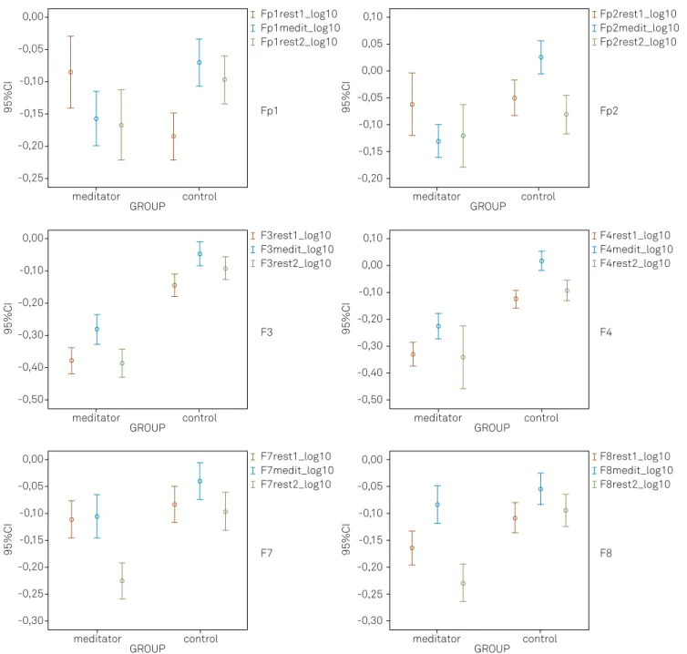
-niicantly reduced during rest 2 compared to rest 1 for the Fp1, F7, and F8 derivations (Table 3 and Figure 1). Within the control group (NMG), the FT power was statistically signii
-cantly higher during the meditation compared to rest 1 for all of the derivations studied, although it remained unchanged after the meditation (rest 2) for the Fp1 and F8 derivations (Table 3 and Figure 1).
For the averaged measure of the frontal sites, there was a statistically signiicant interaction between group and condi
-tion on the absolute theta power for the right (p<0.001) and left (p<0.001) frontal hemispheres. For the simple main efects re
-sult, the groups difered at rest 1 (p<0.001), meditation (p<0.001), and rest 2 (p<0.001) for both hemispheres (Table 4). he FT ac
-tivity was signiicantly greater in the control group (NMG) in all of the conditions (Table 5). Within the meditator group (MG), the FT power was signiicantly greater during meditation com
-pared to rest 1 and 2 for the right hemisphere and only during rest 2 for the left side. Within the control group (NMG), the FT power was signiicantly greater during meditation compared to the rest conditions for both hemispheres. However, there was an increase in the theta activity on the left side during rest 2 com
-pared to during rest 1 (Tables 5 and 6, and Figure 2).
DISCUSSION
he aim of this study was to investigate whether EEG diferences existed in the absolute FT powers between
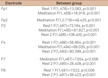
Table 1. Statistical analyses of between groups effects.
Electrode Between group
Fp1 Rest 1 F(1, 478)=13.083, p<0.001 Meditation F(1, 458)=16.916, p<0.001 Fp2 Meditation F(1,2.719)=48.425, p<0.001
F3 Rest1 F(1,497)=73.164, p<0.001
Meditation F(1,495)=61.827, p<0.001 Rest 2 F(1,498)=106.400, p<0.001 F4 Rest 1 F(1,498)=56.954, p<0.001
Meditation F(1,494)=68.035, p<0.001 Rest 2 F(1,493)=90.399, p<0.001 F7 Meditation F(1,487)=7.554, p<0.006
Rest 2 F(1,495)=28.409, p<0.001 F8 Rest 1 F(1,491)=7.522, p<0.006
Rest 2 F(1,497)=36.423, p<0.001
Table 2. Absolute theta power (μV²/Hz) means for both groups in the three conditions
Between groups
µV²/Hz Rest 1 Meditation Rest 2
Fp1
Meditator -0.085 -0.157 -0.167
Control -0.184 -0.07 -0.097
Fp2
Meditator -0.062 -0.13 -0.12
Control -0.05 0.026 -0.081
F3
Meditator -0.375 -0.279 -0.383
Control -0.142 -0.045 -0.09
F4
Meditator -0.332 -0.226 -0.363
Control -0.126 0.017 -0.094
F7
Meditator -0.11 -0.104 -0.225
Control -0.082 -0.038 -0.095
F8
Meditator -0.164 -0.082 -0.229
experienced meditators (MG) and a control group (NMG) during normal rest and OM meditation. We reported con
-sistent interactions between the group and condition for al
-most all of the derivations, indicating that the level of medi
-tation expertise ( irst-time vs. long-term meditators) had a diferential efect on the FT power depending on the condi
-tion (rest 1 vs. medita-tion vs. rest 2). Our main indings will be discussed below.
An increase in FT power during meditation has been found in many studies10,17, thus supporting our indings of an
increase in FT in both groups in the meditative state. he the
-ta band in the fron-tal area has been associated with s-tates of concentration, which is part of OM meditation practice. During OM, the attention is involuntary and directed to any stimulus that arises in the ield of perception at any given moment. Moreover, the current information is actively main
-tained only until new stimuli arise, and this new perceptual processing is not afected by the previous one.
herefore, an enhanced concentration during medita
-tion might correlate with an increase in theta oscilla-tions, independently of the level of expertise, if some level of con
-centration is reached. Perhaps the most interesting inding
of the present study was that, although an acute increase in theta activity during meditation was seen in both groups, the meditators showed a lower trait FT. Because meditation improves attention and concentration, constant training in OM limits the overuse of working memory18, and, therefore,
it may be associated with the lower levels of FT observed in the MG compared to the NMG. We suggested that this ind
-ing was a neural correlate of the expert practitioners’ ability to limit the processing of unnecessary information (e.g., dis
-cursive thought) and increase awareness of the essential con
-tent of the present experience with an attitude of acceptance. According to Chiesa et al.19
, this is a clear description of a bot
-tom-up regulatory process.
Furthermore, attention is also thought to act as a gate to working memory20
. hus, theta band increases in the fron
-tal area have been associated with the active maintenance of working memory representations8,21–23 and this relevant
information in an active state has inluence on future per
-ceptual processing, thought, and behavior20
. hereby, in this study, the NMG was unaware of OM (new processing) de
-spite the clear instructions that had been given just before the task. his processing generated an increase in mental ac
-tivity, and a high perceptual process load can impair the abil
-ity to detect stimuli in environments that overload the work
-ing memory24
. It might worsen attention and concentration, resulting in a decrease in vigilance and an increase in mental efort when associated with another low frequency, such as alpha25
, and this may partly explain our indings.
Another important inding was that we did not ind a sig
-niicant diference between the rest conditions and meditation at the prefrontal derivations (Fp1, Fp2) in the MG. he inding that the activation level in the prefrontal area remained con
-stant for the meditators, whether they were meditating or not, was an indicator that the ability of these subjects to control
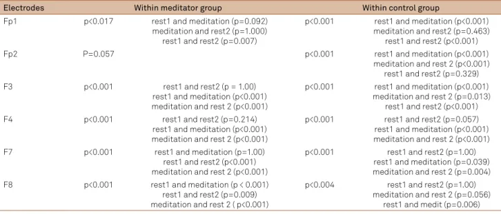
Table 3: Statistical analyses of within groups effects
Electrodes Within meditator group Within control group
Fp1 p<0.017 rest1 and meditation (p=0.092)
meditation and rest2 (p=1.000) rest1 and rest2 (p=0.007)
p<0.001 rest1 and meditation (p<0.001) meditation and rest2 (p=0.463)
rest1 and rest2 (p<0.001)
Fp2 P=0.057 p<0.001 rest1 and meditation (p<0.001)
meditation and rest 2 (p<0.001) rest1 and rest2 (p=0.329)
F3 p<0.001 rest1 and rest2 (p = 1.00)
rest1 and meditation (p<0.001) meditation and rest 2 (p<0.001)
p<0.001 rest1 and meditation (p<0.001) meditation and rest 2 (p=0.013)
rest1 and rest2 (p<0.001)
F4 p<0.001 rest1 and rest2 (p=0.214)
rest1 and meditation (p<0.001) meditation and rest 2 (p<0.001)
p<0.001 rest1 and rest2 (p=0.057) rest1 and meditation (p<0.001) meditation and rest 2 (p<0.001)
F7 p<0.001 rest1 and meditation (p=1.00)
rest1 and rest2 (p<0.001) meditation and rest 2 (p<0.001)
p<0.001 rest1 and rest2 (p=1.00) rest1 and meditation (p=0.039) meditation and rest 2 (p=0.004) F8 p<0.001 rest1 and meditation (p < 0.001)
rest1 and rest2 (p=0.009) meditation and rest 2 ( p<0.001)
p<0.004 rest1 and rest2 (p=1.00) meditation and rest 2 (p=0.056)
rest1 and medit (p=0.006)
Table 4: statistical analyses of between groups effects
Hemisphere Between group
Left
rest 1, F(1, 1472)=11.403, p<0.001 meditation F(1,1444)=73.257, p < 0.001
rest 2 F(1,1472)= 94.981, p<0.001 Right
rest 1 F(1,1485)= 28.600, p<0.001 meditation F(1,1459)=94.688, p<0.001
their narrative focus was not exclusive of formal meditation. he typical narrative focus (e.g., discursive thought) impairs at
-tentional performance and involves mental elaboration and evocation, which overload the working memory processing. he prefrontal cortex (PFC) is responsible for the coordina
-tion of all of this mental traic in working memory, and mind
-fulness training possibly enhances the ability of the PFC to
maintain high attention levels outside of formal meditation26
. his is clearly a top-down process, which involves the execu
-tive control of attention and the modulation of emotional lim
-bic structures19
. herefore, it is clear that, although bottom-up processes explain in part the role of mindfulness as an emo
-tion regula-tion strategy19
, there are also top-down processes that are important in regulating attention.
0,00 Fp1rest1_log10
Fp1medit_log10 Fp1rest2_log10
95%CI Fp1
-0,05
-0,10
-0,15
-0,20
-0,25
0,00
-0,05
-0,10
-0,15
-0,20
-0,25
meditator control GROUP
0,00 F3rest1_log10
F3medit_log10 F3rest2_log10
95%CI F3
-0,10
-0,20
-0,30
-0,40
-0,50
meditator control GROUP
Fp2rest1_log10 Fp2medit_log10 Fp2rest2_log10
95%CI
95%CI
Fp2 0,10
0,00 0,05
-0,05
-0,10
-0,15
-0,20 -0,05
-0,10
-0,15
-0,20
meditator control GROUP
F4rest1_log10 F4medit_log10 F4rest2_log10
F4 0,10
-0,10
-0,20 0,00
-0,30
-0,40
-0,50
meditator control GROUP
F7rest1_log10 F7medit_log10 F7rest2_log10
95%CI F7
meditator -0,30
control GROUP
95%CI
F8rest1_log10 F8medit_log10 F8rest2_log10
F8 0,00
-0,25
-0,30
meditator control GROUP
Figure 1. Proile plots of signiicant absolute theta power at frontal areas (μV²/Hz).
Table 5: Absolute theta power (µV²/Hz) means for both groups in the three conditions
µV²/Hz Right frontal Left frontal
Rest 1 Medit Rest 2 Rest 1 Medit Rest 2
Meditator -0.185 -0.148 -0.236 -0.185 -0.184 -0.247
0,00 -0,05 -0,10 -0,15 -0,20 -0,25
-0,05 -0,10 -0,15 -0,20 L rest1
L medit L rest2
95%CI
meditator -0,30
control Left Hemisphere
95%CI
R rest1 R medit R rest2 0,00
-0,25 -0,30
meditator 0,05
control Right Hemisphere
Figure 2. Proile plots of error bar theta power at frontal areas (μV²/Hz).
Chiesa et al.19
have suggested that mindfulness train
-ing is associated with bottom-up emotional regulation in long-term practitioners and with top-down emotional reg
-ulation in short-term practitioners according to different conceptions of mindfulness as an emotional regulation strategy. However, we suggest that top-down processing is also present in experienced practitioners, although it is reduced as bottom-up processing becomes emphasized. The top-down processing facilitates positive reapprais
-al, thus recruiting PFC regions that are associated with emotional reappraisal19,27,28. This is in accordance with our
findings because the NMG significantly increased their left prefrontal activity during meditation (with only brief meditation instructions), which was probably related to the better executive control of attention, including posi
-tive reappraisal, as this area of the cortex has been linked to positive affect29,30. Indeed, these subjects maintained
greater activity at the left PFC (Fp1) during rest 2, which can be associated with an increased mood that persists after meditation.
When we averaged the frontal sensors into a single mea
-sure of the left and right hemispheres, we found similar re
-sults in comparison to the individual prefrontal electrodes for the NMG. For the MG, we also did not ind a signiicant diference between rest 1 and meditation in the left hemi
-sphere for Fp1 (left prefrontal). hese results possibly in
-dicated an important role of the PFC in coordinating at
-tention and emotion regulation processes in the MG. his top-down processing was more emphasized in the NMG,
although it was still important for the long-term medita
-tors (MG). We suggest, based on the evidence of the emo
-tion asymmetry in the PFC30
, that the top-down regulation of positive reappraisal was related to left PFC activity, which was increased in meditators at rest and increased during meditation for the NMG. Our indings were consistent with the mindfulness-related present-moment focus, which is thought to improve well-being by allowing individuals to re
-frain from trying to control the content of the mind and to become aware of sensations, emotions, and thoughts with
-out judgment or reactivity31
.
In summary, many differences were found in this study. As we expected, long-term meditation practice altered the oscillatory pattern of the theta band, which has been as
-sociated with cognitive functions, such as executive at
-tention and working memory. Despite the acute increase in theta activity during meditation in both groups, the meditators showed a lower trait FT. This was consistent with a reduced top-down control of attention and an in
-creased present moment awareness. In conclusion, acute changes in the theta band throughout meditation did not seem to be a specific correlate of OM, but it was rather related to the concentration properties of meditation. Notwithstanding, lower FT activity appeared to be a trait of OM practice. The present study did not analyze the ra
-tio of gamma-band activity (25–42 Hz) to slow oscillatory activity (4–13 Hz), which could be enlightening. Further research is encouraged to evaluate the electrophysiologi
-cal correlates of OM meditation.
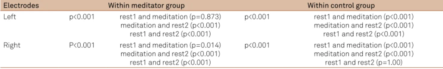
Table 6: statistical analyses of within groups effects
Electrodes Within meditator group Within control group
Left p<0.001 rest1 and meditation (p=0.873) meditation and rest2 (p<0.001)
rest1 and rest2 (p<0.001)
p<0.001 rest1 and meditation (p<0.001) meditation and rest2 (p<0.001)
rest1 and rest2 (p<0.001) Right P<0.001 rest1 and meditation (p=0.014)
meditation and rest2 (p<0.001) rest1 and rest2 (p<0.001)
p<0.001 rest1 and meditation (p<0.001) meditation and rest2 (p<0.001)
1. Lagopoulos J, Xu J, Rasmussen I, et al. Increased Theta and Alpha EEG Activity. J Altern Complement Med 2009;15:1187-1192.
2. Baijal S, Srinivasan N. Theta activity and meditative states: spectral changes during concentrative meditation. Cogn Process 2010;11:31-38.
3. Travis F, Shear J. Focused attention, open monitoring and automatic self-transcending: categories to organize meditations from Vedic, Buddhist and Chinese traditions. Conscious Cogn 2010;19:1110-1118.
4. Cahn BR, Delorme A, Polich J. Occipital gamma activation during Vipassana meditation. Cogn Process 2010;11:39-56.
5. Fell J, Axmacher N, Haupt S. From alpha to gamma: electrophysiological correlates of meditation-related states of consciousness. Med Hypotheses Res 2010;75:218-224.
6. Lutz A, Greischar LL, Rawlings NB, Ricard M, Davidson RJ. Long-term meditators self-induce high-amplitude gamma synchrony during mental practice. Proc Natl Acad Sci USA 2004;101:16369-1673.
7. Mitchell DJ, McNaughton N, Flanagan D, Kirk IJ. Frontal-midline theta from the perspective of hippocampal “theta”. Prog Neurobiol 2008;86:156-185.
8. Itthipuripat S, Wessel JR, Aron AR. Frontal theta is a signature of successful working memory manipulation. Exp Brain Res 2013;224:255-262.
9. Takahashi T, Murata T, Hamada T, et al. Changes in EEG and autonomic nervous activity during meditation and their association with personality traits. Int J Psychophysiol 2005;55:199-207.
10. Cahn BR, Polich J. Meditation states and traits: EEG, ERP, and neuroimaging studies. Psychol Bull 2006;132:180-211.
11. Lutz A, Slagter HA, Dunne JD, Davidson RJ. Attention regulation and monitoring in meditation. Trends Cogn Sci 2008;12:163-169.
12. Colzato LS, Ozturk A, Hommel B. Meditate to create: the impact of focused-attention and open-monitoring training on convergent and divergent thinking. Front Psychol 2012;3:116.
13. Perlman DM, Salomons T V, Davidson RJ, Lutz A. Differential effects on pain intensity and unpleasantness of two meditation practices. Emotion 2010;10:65-71.
14. Kabat-zinn J. Full catastrophe living: using the wisdom of your body and mind to face stress, pain and illness. New York: Delta; 1990.
15. Teasdale JD, Segal Z V, Williams JMG, Ridgeway VA, Soulsby JM, Lau MA. Prevention of relapse/recurrence in major depression by mindfulness-based cognitive therapy. J Consult Clin Psychol 2000;68:615-623.
16. Brewer JA, Worhunsky PD, Gray JR, Tang YY, Weber J, Kober H. Meditation experience is associated with differences in default mode network activity and connectivity. Proc Natl Acad Sci USA 2011;108:20254-20259.
17. Berkovich-Ohana A, Glicksohn J, Goldstein A. Mindfulness-induced changes in gamma band activity - implications for the default mode network, self-reference and attention. Clin Neurophysiol 2012;123:700-710.
18. Jha AP, Stanley EA, Kiyonaga A, Wong L, Gelfand L. Examining the protective effects of mindfulness training on working memory capacity and affective experience. Emotion 2010;10:54-64.
19. Chiesa A, Serretti A, Jakobsen JC. Mindfulness: top-down or bottom-up emotion regulation strategy? Clin Psychol Rev 2013;33:82-96.
20. Sala JB, Courtney SM. Flexible working memory representation of the relationship between an object and its location as revealed by interactions with attention. Atten Percept Psychophys 2010;71:1525-1533.
21. Jensen O, Tesche CD. Short communication frontal theta activity in humans increases with memory load in a working memory task. Eur J Neurosci 2002;15:1395-1399.
22. Benchenane K, Tiesinga PH, Battaglia FP. Oscillations in the prefrontal cortex: a gateway to memory and attention. Curr Opin Neurobiol 2011;21:475-485.
23. Roberts BM, Hsieh LT, Ranganath C. Oscillatory activity during maintenance of spatial and temporal information in working memory. Neuropsychologia 2013;51:349-357.
24. Norman G. Working memory and mental workload. Adv Health Sci Educ Theory Pract 2013;18:163-165.
25. Kamzanova T, Kustubayeva M, Matthews G. Use of EEG workload indices for diagnostic monitoring of vigilance decrement. Hum Factors J Hum Factors Ergon Soc 2012;56:203-207.
26. Slagter H, Davidson RJ, Lutz A. Mental training as a tool in the neuroscientiic study of brain and cognitive plasticity. Front Hum Neurosci 2011;5:17.
27. Ochsner KN, Gross JJ. The cognitive control of emotion. Trends Cogn Sci 2005;9:242-249.
28. Rolls ET, Grabenhorst F. The orbitofrontal cortex and beyond: from affect to decision-making. Prog Neurobiol 2008;86:216-244.
29. Aftanas LI, Golocheikine S. Human anterior and frontal midline theta and lower alpha relect emotionally positive state and internalized attention: high-resolution EEG investigation of meditation. Neurosci Lett 2001;310:57-60.
30. Davidson RJ. Anterior cerebral asymmetry and the nature of emotion. Brain Cogn 1992;20:125-151.
31. Kerr CE, Sacchet MD, Lazar SW, Moore CI, Jones SR. Mindfulness starts with the body: somatosensory attention and top-down modulation of cortical alpha rhythms in mindfulness meditation. Front Hum Neurosci 2013;7:12.