DOI: 10.1590/0004-282X20160145
ARTICLE
Assessment and nutrition education in
patients with amyotrophic lateral sclerosis
Avaliação e educação nutricional em pacientes com esclerose lateral amiotrófica
Claudinéa. S. Almeida1, Patricia Stanich2, Cristina C. S. Salvioni2, Solange Diccini3
Amyotrophic lateral sclerosis (ALS) is the most com-mon form of motor neuron disease (MND), characterized by selective loss of motor neurons in the spinal cord, brainstem and motor cortex. Symptoms include atrophy, weakness and muscle fatigue, fasciculation, dysarthria, dysphagia, sialor-rhea and emotional lability1, 2.
Muscle atrophy may mask the increased metabolic
demand characteristic of progressive diseases. he energy
being channeled to maintain pulmonary ventilation,
jus-tiies the increase in resting energy expenditure in these
patients. In recent times, the nutritional demand in ALS gained such proportions that studies sought to identify it as
predictive factor, emphasizing the importance of nutritional intervention in disease treatment3, 4.
Weight loss has multifactorial causes including oropharyn-geal dysphagia, secondary loss of appetite, loss of autonomy with emotional disorders, inappropriate use of noninvasive ventilation and hypermetabolism5. here is a direct association
between weight loss and disease severity. At the time of diag-nosis, a reduction of 5% of normal weight increased the risk of death in this population by 30%. A reduction of 1 kg/m2 in body
mass index (BMI) was associated with a 20% risk of death4.
Changes in body composition have been described since
the irst studies on nutrition in ALS. Kasarskis et al. 6 found
1Escola Paulista de Enfermagem, Departamento de Neurologia, São Paulo SP, Brasil; 2Universidade Federal de São Paulo, Departamento de Neurociências, São Paulo SP, Brasil; 3Universidade Federal de São Paulo, Departamento de Ciências da Saúde, São Paulo SP, Brasil.
Correspondence: Claudinéia S. Almeida; Universidade Federal de São Paulo, Setor de Investigação em Doenças Neuromusculares, Rua Estado de Israel, 899; 04022-020 São Paulo Sp, Brasil. E-mail: claudineianutri@gmail. com.
Conflict of interest: There is no conlict of interest to declare. Received 13 May 2016; Accepted 16 August 2016.
ABSTRACT
Neurological patients with amyotrophic lateral sclerosis (ALS)often deteriorate to a worsening nutritional status. The aim of this study was to compare the nutritional status and food intake after nutrition education in patients with ALS. Clinical, anthropometric and functional variables were analyzed. Fifty-three patients were monitored at an early stage of the disease. The average score on the functionality scale was 33 points. Initially only 3.8% were classiied as low body weight. After three months, 50% showed signiicant variation in anthropometric measures related to muscle mass and body fat reserves without association with clinical variables. After nutritional guidance, there was an increase in the intake of all food groups, especially the dairy group (p <0.05).The change of the nutritional status occurs early in patients with amyotrophic lateral sclerosis, even in those previously eutrophic or over weight. There was an increase in food intake after nutritional guidance according to the food guide adapted to the Brazilian population.
Keywords: amyotrophic lateral sclerosis; continuing education; body composition.
RESUMO
Pacientes neurológicos com esclerose lateral amiotróica frequentemente evoluem com piora do estado nutricional. O objetivo desse estudo foi comparar o estado nutricional e a ingestão alimentar depois da orientação nutricional em pacientes com ELA. Variáveis clínicas,antropométricas e funcionais foram analisadas. 53 pacientes foram avaliados na fase inicial da doença. A pontuação média da escala de funcionalidade foi de 33 pontos. Inicialmente apenas 3,8% foram classiicados como baixo peso. Após três meses, 50% apresentaram variação signiicativa nas medidas antropométricas relacionadas com reservas de massa muscular e gordura corporal, sem associação com variáveis clínicas. Após orientação nutricional, houve um aumento na ingestão de alimentos de todos os grupos com relevância para o grupo de lacticínios (p <0,05). A mudança do estado nutricional ocorre precocemente em pacientes com ELA, mesmo naqueles anteriormente eutróicos ou sobrepeso. Houve um aumento na ingestão de alimentos após orientação nutricional de acordo com o guia alimentar adaptado da população brasileira.
that men show a greater loss of skeletal muscle compared
to women, suggesting diferent nutritional requirements.
During the course of illness, decreased muscle mass is asso-ciated with lower survival, regardless of body weight7.
Based on this evidence, nutritional intervention when
applied early allows the use of this resource in the diferent stages of the disease. hus, the aim of this study was to compare
the nutritional status and food intake after nutrition education.
METHODS
his is a longitudinal study in patients with MND/ALS,
treated at the Neurology/Neurosurgery Department, of the Federal University of São Paulo (UNIFESP), during the period of
2013–2014. Fifty-three patients with conirmed diagnosis accord -ing to the El Escorial8, were included in the initial phase of
medi-cal treatment. Patients receiving any nutritional orientation were
excluded. Patients were referred by a medical staf member. he nutritional instrument was applied at both phases of the
study, and consisted of a structured interview based on clinical evaluation, nutritional assessment, analysis of food intake and functional evaluation, according to the Amyotrophic Lateral Sclerosis Functional Rating Scale (ALSFRS)9. he ALSFRS-R
is a questionnaire-based scale for activities of daily living. his scale contains 12 items grouped into three domains that
encompass appendicular function (gross motor tasks), bul-bar and respiratory function. Each item has a 5-point scale (0 for unable; 4 for normal) and scores ranging from 0 to 48. Low scores denote a serious disease status.
he assessment of nutritional status was performed using
anthropometric measurements and analyzed according to population scores10. he measures were: weight, height, arm
circumference, triceps skinfold, biceps skinfold, suprailiac skinfold, subscapular skinfold, arm muscle circumference, arm muscle area, arm fat area and calf circumference for indi-viduals over 60 years of age. For the anthropometric index, the BMI was used10. he adequacy percentile10 was calculated
using the software Decision Support System in Nutrition ver-sion 2. 5 - UNIFESP / Escola Paulista de Medicina.
For compartmented analysis of body composition, the arm muscle area measures for muscle mass and triceps skinfold for body fat were selected. For analysis of food intake, the food
consumption frequency questionnaire was applied and ana -lyzed according to the food guide for the Brazilian population11.
he study occurred in two phases: an initial evaluation at
the time of referral (T0) and after three months (T1). In the
irst evaluation, patients were divided randomly into two groups: intervention group (IG) and control group (CG). he
criterion for the division of groups was the type of nutritional
guidance provided. For the IG patients the food pyramid tool,
adapted to the Brazilian population, was used11. For the CG,
general guidelines were provided, including changes in food consistency and dividing up the components of meals.
he project was approved by the Ethics Committee in
Research of the Federal University of São Paulo under num-ber 492 239 CEP.
Statistical analysis
Categorical variables were described in absolute value and
relative frequency. Continuous variables, after being submit
-ted to the Kolmogorov-Smirnovnormality test, were described
using central tendency and dispersion measures. We considered a decline in the anthropometric measures to be the distribu-tion of values below the 25th percentile of the variadistribu-tion in both
phases of assessment (T0 and T1). he association between pre -dictor factors and the decline of selected anthropometric
mea-surements was evaluated according to Pearson’s chi-square test
or Verisimilitude ratios. Results with type I error probability of
less than 5% were considered statistically signiicant.
RESULTS
Among the characteristics of the studied population, there was a predominance of males, aged 57 years. For the type of mani-festation of the disease, an appendicular presentation was
pre-dominant (79. 2%). he factors that characterized the early stages
of the disease were the time between symptom onset and diagno-sis (360 days) and the scores found on the ALSFRS functionality scale (33 points). As for nutritional status, only 3. 8% of patients presented with low body weight, with similar proportions among those considered normal weight or overweight (Table 1).
Table 1.General characteristics of the study population.
Variable n %
Gender
Male 31 58,5
Female 22 41.5
Age (years)
Average (min – max) 57.0 (33 – 76)
< 60 years 34 64,2
≥ 60 years 19 35.8
Manifestation of thedisease
Appendicular 42 79.2
Bulbar 11 20.8
Symptom onset (days)
Average (min – max) 360 90 – 3270
ALSFRS score
Average (min – max) 33 13 – 44
Nutritional status according to BMI
Lowweight (< 18,9) 2 3,8
Eutrophy (19–24,9) 26 49.1
Overweight (≥ 25) 25 47.2
Nutritionalcounseling
Foodguide 35 66
Standard orientation 18 34
Table 2 shows the variation in anthropometric
mea-surements between the two assessments: at baseline (T0)
and after three months of follow-up (T1). Despite the biceps skinfold measures, suprailiac skinfold, subscapular skinfoldand arm muscle circumference, at least half of the subjects showed declines in measures, with a significant difference for BMI, arm circumference, biceps skinfold, triceps skinfold, and subscapular skinfold, compared with the initial phase.
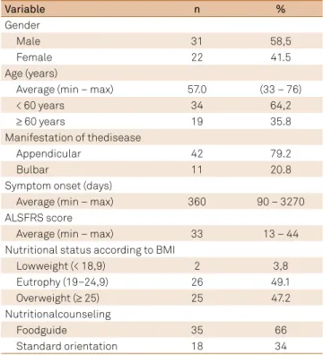
he results of variables related to the decline of body weight are shown in Table 3. We observed a decline ≥ 0. 53 kg/m2 in
BMI between the period of evaluations. he gender, age
and time of onset of symptoms showed very similar
pro-portions among categories, without signiicant diference.
We observed that individuals with bulbar involvement, and those who were overweight showed the largest decline in BMI (p < 0. 4 and p < 0. 1 respectively).
For the changes of arm muscle area and triceps skinfold, shown
in Tables 4 and 5, we found no signiicant diference between the
study variables and reducing measurement. It is worth mention-ing that the age group and functionality were the variables that were most related to the decline of the measurement.
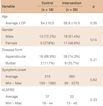
Study variables according to the type of nutritional
guid-ance are applied in Tables 6 and 7. he age group and the time of onset of symptoms were similar between the IG and CG. We found a higher frequency of female patients and with bulbar manifestation in the IG. Similar averages paired were found
between anthropometric measures highlighting averages of
BMI, arm circumference and skinfolds, but with no signiicant diference. he same was not observed for the functionality (p = 0. 03). he IG patients had lower scores on the ALSFRS
scale in both phases (T0 and T3), as shown in Figure. Table 2.Distribution of variation (T0 – T1) of
anthropometric measurements according to the percentiles of the study population.
Variable Min. P25 Average P75 Max. p**
BMI (kg/m2) -3.63 -0.53 -0.09 0.0 +1.47 0.02
AC (cm) -3.0 -1.0 -0.75 0.0 +3.0 0.04
TSF (mm) -5.0 -1.0 -0.90 0.0 +4.0 0.008
BSF (mm) -6.0 -1.0 0.0 0.0 +2.0 0.001
SISF (mm)* -8.0 -1.0 0.0 0.0 +3.0 0.003
SSF (mm) -3.0 -1.0 0.0 0.0 +3.0 0.005
AMC (cm) -3.0 -1.0 0.0 2.0 +4.0 0.69
AMA (cm2) -8.0 -3.0 -2.0 0.0 +16.0 0.94
T0: time of initial evaluation; T1: time after three months; P25: values below the 25th percentile; P75: values below the 75th percentile; BMI: body mass index;
AC: arm circumference; TSF: triceps skinfold; BSF: biceps skinfold; SISF: suprailiac skinfold; SSF: subscapular skinfold; AMC: arm muscle circumference; AMA: arm muscle area;*missing values, n = 1 (1.9%); **T-test paired.
Table 3.Body mass index decline category according to clinical, demographic and nutritional variables.
Variable BMI decline p
< 0.53 Kg/m2 ≥ 0.53 Kg/m2
Gender
Male 23 (76.7%) 7 (23.3%)
0.74
Female 16 (72.7%) 6 (27.3%)
Age group
< 60 years 25 (75.8%) 8 (24.2%) 0.86 ≥ 60 years 14 (73.7%) 5 (26.3%) Disease form
Appendicular 32 (78%) 9 (22%)
0.32
Bulbar 7 (63.6%) 4 (36.4%)
Symptom onset
≤ 360 days 24 (72.7%) 9 (27.3%) 0.61 > 360 days 15 (78.9%) 4 (21.1%) ALSFRS
≥ 33 points 23 (79.3%) 6 (20.7%) 0.42 < 33 points 16 (69.6%) 7 (30.4%) Prior nutritional status
Eutrophy/lowweight 22 (84.6%) 4 (15.4%) 0.08
Overweight 16 (64%) 9 (36%)
Nutritionalcounseling
FoodGuide 25 (71.4%) 10 (28.6%)
0.39 Standard orientation 14 (82.4%) 3 (17.6%)
BMI: body mass index; ALSFRS:amyotrophic lateral sclerosis functional rating scale.
Table 4.Arm muscle area decline categories according to clinical, demographic and nutritional variables.
Variable Arm muscle area decline p
< 3.0 cm2 ≥ 3.0 cm2
Gender
Male 20 (64.5%) 11 (35.5%)
0.31
Female 17 (77.3%) 5 (31.3%)
Age group
< 60 years 22 (64.7%) 12 (35.3%)
0.27 ≥ 60 years 15 (78.9%) 4 (21.1%)
Disease form
Appendicular 29 (69%) 13 (31.%)
0.45
Bulbar 8 (72.7%) 3 (27.3%)
Symptom onset
≤ 360 days 24 (72.7%) 9 (27.3%)
0.55 > 360 days 13 (65%) 7 (35%)
ALSFRS
≥ 33 points 19 (65.5%) 10 (34.5%)
0.45 < 33 points 18 (75%) 6 (25%)
Prior nutritional status
Eutrophy/lowweight 19 (70.4%) 8 (29.6%)
0.85
Overweight 17 (68%) 8 (32%)
Nutritionalcounseling
FoodGuide 24 (68.6%) 11 (31.4%)
0.78 Standard orientation 13 (72.2%) 5 (27.8%)
For both groups, a low frequency in food intake of legu -minous groups, cereals and tubers, vegetables and fruits was found, especially the cereal groups, tubers and vegetables. We
found a frequency greater than 50% in the consumption of dairy groups, meat and eggs, oils and fats in the IG, as shown in Figure. he qualitative nutritional guidance of the diet seems to be more efective for the consumption of dairy products and fruit groups, particularly the dairy group, which showed a signiicant increase in the recommended consumption frequency (p < 0. 05).
DISCUSSION
he population studied portayedthe epidemiological
characteristics of ALS described in the literature with a pre-dominance of males compared to females, prevalence in the sporadic form and initial appendicular involvement of the bulbarregion12. he basic characteristics between the studied
groups were shown to be compatible with the purpose of the study and randomization allowed comparison.
he median age group was 57 years old. In studies of the
Brazilian population, the average is 52 years, whereas other
countries range from 59-65 years of age. he population stud -ied was older than that described in the Brazilian literature13.
he mechanism by which the progression of the disease
is faster in older individuals is still unknown, however it seems to be associated with the natural loss of motor
neu-rons. his does not occur for the adults in balancing the
decline of motor function12.
Table 5.Triceps skinfold decline categories according to clinical, demographic and nutritional variables.
Variable TSF decline p
<1.0 mm ≥ 1.0 mm
Gender
Male 15 (50%) 15 (50%)
1.0
Female 11 (50%) 11 (50%)
Age group
< 60 years 18 (54.5%) 15 (45.5%) 0.38 ≥ 60 years 8 (42.1%) 11 (57.9%) Disease form
Appendicular 21 (51.2%) 20 (48.8%) 0.73
Bulbar 5 (45.5%) 6 (54.5%)
Symptom onset
≤ 360 days 18 (54.5%) 15 (45.5%) 0.38 > 360 days 8 (42.1%) 11 (57.9%) ALSFRS
≥ 33 points 17 (58.6%) 12 (41.4%) 0.16 < 33 points 9 (39.1%) 14 (60.9%) Prior nutritional status
Eutrophy/lowweight 14 (53.8%) 12 (46.2%) 0.48
Overweight 11 (44%) 14 (56%)
Nutritionalcounseling
FoodGuide 14 (40%) 21 (60%)
0.39 Standard orientation 12 (70.6%) 5 (29.4%)
TSF:triceps skinfold; ALSFRS:amyotrophic lateral sclerosis functional rating scale
Table 6.General and demographic data of the study population according to the nutritional guidance.
Variable Control Intervention p
(n = 18) (n = 35)
Age
Average ± DP 54 ±10.5 56.8 ±10.5 0.35
Gender
Male 13 (72.2%) 18 (51.4%)
0.14
Female 5 (27.8%) 17 (48.6%)
Disease form
Appendicular 16 (88.9%) 26 (74.3%)
0.21
Bulbar 2 (11.1%) 9 (25.7%)
Symptom onset
Average 315 360
0.62 Min – Max 150 - 1560 90 - 3270
ALSFRS
Average 37 33
0.23
Min – Max 18 - 44 13 - 45
ALSFRS: amyotrophic lateral sclerosis functional rating scale.
Table 7.Frequency of anthropometric and functional outcomes according to nutritional guidance groups
Variable Control Intervention p
(n = 35) (n = 18)
BMI* % %
Delta + orstable 9 (52.9) 17 (48.6)
0.76
Delta - 8 (47.1) 18 (51.4)
AMA
Delta < -10% 14 (77.8) 25 (71.4)
0.62
Delta ≥ -10% 4 (22.2) 10 (28.6)
AMC
Delta < -10% 17 (94.4) 34 (66.0)
0.62
Delta ≥ -10% 1 (5.6) 1 (2.9)
TSF
Delta <10% 13 (76.5) 24 (68.6)
0.55
Delta ≥10% 4 (23.5) 11 (31.4)
AC*
Delta <10% 17 (100) 34 (97.1)
Delta ≥10% 0 ( -- ) 1 (2.9)
ALSFRS
Delta + orstable 10 (55.6) 9 (25.0)
0.03
Delta - 8 (44.4) 26 (74.3)
he time between the irst symptoms and diagnosis was
360 days, similar to that described in the literature pointing to an average of 12 months, when 50% of the motor neurons have been lost; although the symptoms and severity of the
disease occur diferently between individuals14, 15.
As far as functionality goes, the patients presented with a slightly impaired function with independence for activities of daily living. So far, a prognostic score for functionality has not been described. It is known that scores greater than or
equal to 30 points may correspond with longer survival14. he
same applies to nutritional matters. he ALSFRS has been
associated with objective measures of muscle strength and lung function, however, few studies were found that assess the scale items with nutritional parameters16, 17, 18.
Unlike studies that observed low body weight as a symp-tom in the early stages of the disease, the same was not found
in this study. he change in body weight for values above the
recommended, according to the age group and stature of the Brazilian population, has been growing in recent decades.
When degenerative diseases afect individuals older than
45 years of age, being overweight and obese are commonly-observed symptoms16, 19. Another reason could be the
inclu-sion of patients in the early stages of the disease where the
respiratory functions were not afected; a fact that would help the preservation of body fat reserves. he importance
of multidisciplinary assessment in the literature is well described, including early nutritional counseling as a positive factor for maintaining the nutritional status with a preven-tive approach in the treatment of ALS14, 15, 16, 19.
Although there was a variation in BMI, it was not enough
to change the classiication of nutritional status, stressing
that as an isolated measure, the BMI is not sensitive enough to detect body alteration in the course of the disease16. Muscle
atrophy is itself the triggering factor of changes, justifying the periodic anthropometric evaluation, with a recommendation for the anthropometry of the arm20.
here was a signiicant decline of the measures from the
third month on, characterizing early changes in nutritional
status, and these indings are in agreement with other stud -ies to reduce anthropometric measurements and their main-tenance after the completion of education on the habitual diet, with calorie and protein supplements4, 20, 21.
With attention to the studied population, we did not
ind any characteristics strongly associated with the decline.
Elderly patients, with time from onset greater than 12 months and a score less than 33 points on the ALSFRS scale were the ones with the greatest loss of body fat. In the case of a diag-nostic study and nutrition education, data on a prognosis
would not it in this scope of work, but it is worth recalling
that the reduction of body fat has been described as a nega-tive prognostic factor4, 21, 22.
he initial nutritional status of individuals was proba -bly the mitigating factor for the changes in BMI. We found a
high frequency of eutrophic (within ideal weight range) indi -viduals (49%) and overweight (47%) patients. After the three months, patients with excess weight had minor variations of BMI when compared to eutrophic individuals, however
with-out statistically signiicant diference, suggesting that losses
in fat reserves can precede the decrease in body weight.
As for the frequency of anthropometric outcomes between the groups, although few signiicant diferences have
been found, both showed a decrease of anthropometric mea-surements between the study periods (T0 and T1). In 50% of patients there was a reduction of the measures, in particular the biceps skinfold, subscapular skinfoldand arm muscle
cir-cumference, with statistically signiicant diference for BMI,
arm circumference and all skin folds (biceps, triceps, and
subscapular). he same was observed by Stanich et al. 20 who
found their analysis of body composition was efective for
introducing nutritional supplementation, and also by Slowie et al. 23who analyzed body composition in patients with ALS.
Figure. Food frequency of patients in the initial phase, and after nutritional guidance.
Basaline Post Intervention Fruits
Vegetables
Cerals and tubers
Oils and fats
Legumes
Meats and eggs
Dairy products
0 20 40 60 80 100
34.3 25.7
5.7 2.9
0.0 0.0
91.4 85.7
17.1 5.6
82.9 80.0
Salvioni et al. 18also analyzed body composition using
anthropometric measures and correlated them with the time between diagnosis and nutritional assessment of patients
with ALS. he study showed that the delay in nutritional intervention negatively inluences the loss of muscle mass,
demonstrating the importance of early intervention. Initially, the dietary pattern presented characterized the
food inadequacy commonly observed in the Brazilian popu -lation, with respect to the components and composition of meals. Other studies, in particular, have demonstrated a high
frequency (70%) of subjects with ALS who had a lower food
intake, especially of calories and proteins24, 25, 26.
he importance of nutrition in ALS is so relevant that
even without a socio-demographic study analyzing factors associated with the onset of the disease, it was found that a low intake of fruit and vegetables would be a risk factor23.
In the frequency of food consumption questionnaire applied, apart from the inadequacies, the lack of knowledge about the
importance of food groups and how to incorporate them into
the daily habit was identiied.
Determining the components of the meals was not an instrument of analysis of this study, however we observed an
inadequacy related to the number of meals per day, with con -sumption of just three meals (breakfast, lunch and dinner), a fact that can combine with the non-intake of other groups recommended by the food guide. We observed that food from the dairy group, meats, eggs, oils and fats were found to have
a higher intake frequency, explaining in part the low intake
of other food groups, since the former kinds of food are often consumed during the main meals. After nutritional
coun-seling for both IG and CG patients, an increase in the fre
-quency of intake of all food groups, especially dairy products,
was observed. Anhypothesis for the increase in the intake of milk and dairy products would be their accessibility and for the taste and the eating habits of the Brazilian population. Another possibility would be the ease of dairy as a vehicle
for thickening of luids, a common solution for patients with
oropharyngeal dysphagia27.
Nutrition education, using the food guide for the Brazilian population11, has been used as a tool in an intervention strategy, as
it was noted that its use facilitated learning, thereby orienting the basic principles for a healthy diet. In general, all food groups that make up the object of intervention showed an increase in the pro-portion of consumers who met the recognized recommendations.
he intervention purpose of this study was nutritional
counseling in the early stages of the disease, in order to
mini-mize the reduction and/or inadequate diet during treatment. he guidelines were provided to the patient and caregivers during the quarterly outpatient monitoring. Periodic veriica -tion of anthropometric measurements and food intake pro-vided the patients with their own monitoring of nutritional
status. hus, there was greater involvement with the pro -posed nutritional treatment. It can be inferred that, possibly, nutritional counseling has been an opportunity for apprecia-tion of nutriapprecia-tional monitoring by patients and caregivers.
Another factor that may have positively inluenced the compli -ance of the proposed therapy, was the multidisciplinary treatment
ofered by specialists in referral centers for patients with ALS. his service difers by providing preventive guidance to patients and
caregivers, preparing them for the implications that will arise with
disease progression. he multidisciplinary team aims for the ulti
-mate goal of improving the quality of life of these individuals27, 28.
he results of this study reinforce the importance of early nutrition
counseling as part of the treatment of patients with ALS.
Under the conditions of this study, it can be concluded
that: patients present with early reduction in body weight,
in lean mass and body fat measurements; the change in the
nutritional status occurs regardless of nutritional adequacy
characterizing the hypermetabolic state of the disease; the food guide adapted for the Brazilian population, as a
nutri-tion educanutri-tion tool, has been efective in increasing the fre
-quency of consumption of food groups.
References
1. Sorarù G, Ermani M, Logroscino G, Palmieri A, D’ Ascenzo C, Orsetti V et al. Natural history of upper motor neuron-dominant ALS. Amyotroph Lateral Scler. 2010;11(5):424-9.
doi:10.3109/17482960903300867
2. Mitchell JD, Borasio GD. Amyotrophic lateral sclerosis. Lancet. 2007; 369(9578):2031-41. doi:10.1016/S0140-6736(07)60944-1
3. Genton L, Viatte V, Janssens JP, Héritier AC, Pichard C. Nutritional state, energy intakes and energy expenditure of amyotrophic lateral sclerosis (ALS) patients. Clin Nutr. 2011;30(5):553-9. doi:10.1016/j.clnu.2011.06.004
4. Marin B, Desport JC, Kajeu P, Jesus P, Nicolaud B, Nicol M et al. Alteration of nutritional status at diagnosis is a prognostic factor for survival of amyotrophic lateral sclerosis patients. J Neurol Neurosurg Psychiatry. 2011;82(6):628-34. doi:10.1136/jnnp.2010.211474 5. Braun MM, Osecheck M, Joyce NC. Nutrition assessment and
management in amyotrophic lateral sclerosis. Phys Med Rehabil Clin N Am. 2012;23(4):751- 71. doi:10.1016/j.pmr.2012.08.006
6. Kasarskis EJ, Berryman S, English T, Nyland J, Vanderleest JG, Schneider A et al. The use of upper extremity anthropometrics in the clinical assessment of patients with amyotrophic lateral sclerosis. Muscle Nerve. 1997;20(3):330-5.
doi:10.1002/(SICI)1097-4598(199703)20:3<330::AID-MUS10>3.0.CO;2-4 7. Roubeau V, Blasco H, Maillot F, Corcia P Praline J. Nutritional
assessment of amyotrophic lateral sclerosis in routine pratice: value of weighing and bioelectrical impedance analysis. Muscle Nerve. 2015;51(4):479-84. doi:10.1002/mus.24419
8. Brooks BR, Miller RG, Swash M, Munsat TL. El Escorial revisited: revised criteria for the diagnosis of amyotrophic lateral sclerosis. Amyotroph Lateral Scler Other Motor Neuron Disord. 2000;1(5):293-9. doi:10.1080/146608200300079536
10. Frisancho AR. New norms of upper limb fat and muscle areas for assessment of nutritional status. Am J Clin Nutr. 1981;34(11): 540-5. 11. Philippi ST. Pirâmide dos alimentos: fundamentos básicos de
nutrição. Barueri: Manole; 2008.
12. Oliveira AS, Pereira RD. Amyotrophic lateral sclerosis (ALS): three letters that change the people’s life. For ever. Arq Neuropsiquiatr. 2009;67(3a):750-82. doi:10.1590/S0004-282X2009000400040 13. Fga, SP, Lima JM, Alvarenga RP. Epidemiologia da esclerose lateral
amiotróica - Europa/América do Norte/América do Sul/Ásia: discrepâncias e similaridades. Rev Bras Neurol. 2009;45(2):5-10. 14. Cosmo CS, Lucena RC, Sena EP. Aspectos clínicos determinantes da
capacidade funcional na Esclerose Lateral Amiotróica. Rev Ciênc Méd Biológ. 2012;11(2):134-9.
15. Chiò A, Logroscino G, Hardiman O, Swingler R, Mitchell D, Beghi E et al. Prognostic factors in ALS: a critical review. Amyotroph Lateral Scler. 2009;10(5-6):310- 23. doi:10.3109/17482960802566824 16. Shimizu T, Nagaoka U, Nakayama Y, Kawata A, Kugimoto C, Kuroiwa Y et
al. Reduction rate of body mass index predicts prognosis for survival in amyotrophic lateral sclerosis: a multicenter study in Japan. Amyotroph Lateral Scler. 2012;13(4):363-6. doi:10.3109/17482968.2012.678366 17. Nordon DG, Esposito SB. Atualização em esclerose lateral
amiotróica. Rev Fac Med Sorocaba. 2009;11(2):1-3.
18. Salvioni CCS. Fatores Associados ao estado nutricional e a evolução da doença do neuronio motor/ esclerose lateral amiotróica [dissertação]. São Paulo: Universidade Federal de São Paulo; 2013. 19. Kaufmann P, Levy G, Thompson JLP, Delbene ML, Battista
V, Gordon PH et al. The ALSFRSr predicts survival time in an ALS clinic population. Neurology. 2005;64(1):38-43. doi:10.1212/01.WNL.0000148648.38313.64
20. Stanich P, Pereira AML, Chiappetta ALML, Nunes M, Oliveira ASB, Gabbai AA. Suplementação nutricional em pacientes com doença do neurônio motor/esclerose lateral amiotróica. Rev Bras Nutr Clin. 2004;19:70-8.
21. Silva LBDC, Mourão LF Silva AA, Lima NMFV, Almeida SR, Franca Jr MC et al. Effect of nutritional supplementation with milk whey proteins in amyotrophic lateral sclerosis patients. Arq Neuropsiquiatr. 2010;68(2):263-8. doi:10.1590/S0004-282X2010000200021
22. Limousin N, Blasco H, Corcia P, Gordon PH, De Toffol B, Andres C et al. Malnutrition at the time of diagnosis is associated with a shorter disease duration in ALS. J Neurol Sci. 2010;297(1-2):36-9. doi:10.1016/j.jns.2010.06.028
23. O’Reilly EJ, Wang H, Weisskopf MG, Fitzgerald KC, Falcone G, McCullough ML et al. Premorbid body mass index and risk of amyotrophic lateral sclerosis. Amyotroph Lateral Scler Frontotemporal Degener. 2013;14(3):205-11. doi:10.3109/21678421.2012.735240
24. Slowie LA, Paige MS, Antel JP. Nutritional considerations in the management of patients with amyotrophic lateral sclerosis (ALS). J Am Diet Assoc. 1983;83(1):44-7.
25. Okamoto K, Kihira T, Kobashi G, Washio M, Sasaki S, Yokoyama T et al. Fruit and vegetable intake and risk of amyotrophic lateral sclerosis in Japan. Neuroepidemiology. 2009;32(4):251-6. doi:10.1159/000201563
26. Silva LB, Mourão LF, Silva AA, Lima NM, Franca Junior M,
Amaya-Farfánet al. Avaliação da ingestão alimentar de indivíduos com esclerose lateral amiotróica. Rev Bras Nutr Clín. 2008;23(1):5-12. 27. Miller RG, Jackson CE, Kasarskis EJ et al. The ALS practice
parameter task force and the quality standards subcommittee of the American Academy of Neurology. Amyotroph Lateral Scler. 2010:11(Suppl 1):7-60.