Artigo Original
Original Article
Gabriela Zuin Ferreira1 Jeniffer de Cássia Rillo Dutka1,2 Melina Evangelista Whitaker3 Olivia Mesquita Vieira de Souza2 Viviane Cristina de Castro Marino4 Maria Inês Pegoraro-Krook1,2
Descritores
Fissura Palatina Insuiciência Velofaríngea Fala Avaliação Cirurgia Geral
Keywords
Cleft Palate Velopharyngeal Insuficiency Speech Evaluation General Surgery
Correspondence address:
Maria Inês Pegoraro-Krook
Departamento de Fonoaudiologia, Faculdade de Odontologia de Bauru, Universidade de São Paulo.
Alameda Octávio Pinheiro Brisolla, 9-75, Bauru (SP), Brasil, CEP: 17012-901. E-mail: [email protected]
Received: 09/01/2014
Accepted: 01/20/2015
Study carried out at the Hospital for Rehabilitation of Craniofacial Anomalies, Universidade de São Paulo – USP – Bauru (SP), Brazil.
(1) Graduate Program in Rehabilitation Sciences, Hospital for Rehabilitation of Craniofacial Anomalies, Universidade de São Paulo – USP – Bauru (SP), Brazil.
(2) Speech Language Pathology and Audiology Department, Universidade de São Paulo – USP – Bauru (SP) Brazil.
(3) Hospital for Rehabilitation of Craniofacial Anomalies, Universidade de São Paulo – USP – Bauru (SP), Brazil. (4) Speech Language Pathology and Audiology Department, School of Philosophy and Sciences, Universidade Estadual Paulista “Júlio de Mesquita Filho” – UNESP – Marília (SP), Brazil.
Financial support: Fundação de Amparo à Pesquisa do Estado de São Paulo – FAPESP.
Conlict of interests: nothing to declare.
Nasoendoscopic indings after primary palatal surgery: can
the Furlow technique result in a smaller velopharyngeal gap?
Achados nasoendoscópicos após a cirurgia
primária de palato: a técnica de Furlow pode
resultar em menor gap velofaríngeo?
ABSTRACT
Purpose: To compare the nasoendoscopic indings related to the velopharyngeal gap among patients with cleft palate who underwent the Furlow (F) technique and those who underwent the von Langenbeck (vL) technique for primary palatal surgery, who remained with velopharyngeal insuficiency (VPI). Methods: The analyzed data were retrieved from the institution’s data of recordings of nasoendoscopic exams. The sample comprised 70 recorded nasoendoscopic exams obtained from 22 patients who underwent the F technique and from 48 who underwent the vL technique during primary palatoplasty, who remained with VPI after surgery and were submitted to nasoendoscopy, between the ages of 5 and 15 years (mean age: 8 years), for deinition of the best treatment for VPI. The images were edited into a DVDin a randomized sequence to be assessed by three experienced speech language pathologists regarding displacement and excursion of the soft palate; displacement and excursion of lateral pharyngeal’s walls; displacement and excursion of the posterior pharyngeal’s wall; and presence of the Passavant ridge and size and type of velopharyngeal gap.
Results: The results of the comparison of measurements between F and vL groups were not statistically signiicant. Conclusion: The surgical technique used in primary palatoplasty was not relevant to determine the difference in the size of the velopharyngeal gap for patients who maintained VPI.
RESUMO
INTRODUCTION
Surgical corrections of cleft lip and palate, comprising the primary lip repair (surgery for lip correction) and primary pala-toplasty (reconstruction of hard and/or soft palate), are recom-mended in the irst year of life.
Primary palate surgery can be performed through vari-ous surgical techniques, of which the best for the type and the extent of the cleft is chosen, always seeking correction from the anatomic and functional point of view. Surgical failure may occur due to the surgical technique, the surgeon’s skill, and/or the extent of the cleft palate(1,2). A secondary physical interven-tion is usually needed to correct palatal istula or dysfuncinterven-tion of the velopharyngeal mechanism (VPM). Failure of the VPM is called velopharyngeal dysfunction (VPD), which occurs due to lack of tissue (velopharyngeal insuficiency – VPI) or lack of functionality (velopharyngeal incompetence)(3,4).
The ideal age for surgical correction of cleft palate remains controversial. Most recent studies argue in favor of early age, between 6 and 18 months, to promote normal speech acquisi-tion and to prevent hearing loss(5,6). Other surgeons have advo-cated two surgeries for complete cleft, in which the closure of the soft palate is performed between 3 and 8 months, whereas the hard palate is postponed until 15 months to 15 years of age(7,8).
It is through primary palatoplasty that VPD is prevented in children with cleft palate. In addition to age and the surgeon’s skill, the choice of surgical technique is one of the most impor-tant factors. It should provide not only the closing of the ana-tomical part of the cleft but also allow suficient stretching and mobility of the soft palate so that it interacts with the pharyn-geal walls when the velopharynpharyn-geal closure (VC) is required for speech(1,2).
Some studies have indicated that, even after the palate cor-rection, an incidence of VPD, which can range from 5 to 36%, may occur, even when the individual undergoes surgery under conditions considered ideal. However, this proportion may increase considerably when the patients undergo surgery late in their lives, or when the choice of surgical technique did not consider circumstances relevant to speech(3,9,10).
The repair of cleft palate was first described by von Langenbeck (vL) in 1861, whose technique, which is named after him, highlights the importance of the separation of the oral and nasal cavities, principle used in most of the techniques to date(6,11,12). Many authors pointed out that the disadvantage of this technique is that it does not lengthen the palate sufi-ciently for the reconstruction of the VPM(6,13).
The Furlow (F) technique is a palatoplasty that uses a mir-rored zetaplasty of the oral and nasal mucosa, providing greater palate elongation and restoring muscle strength(6,14). A prospec-tive clinical trial study compared the results of speech and of velopharyngeal function in patients with cleft lip and palate who underwent surgery through the F and vL techniques and found better outcomes for patients operated through the F technique(6). Other studies reported superior results on speech for patients operated through the F technique when compared with other surgical techniques(15-17).
The VPM is evaluated through a perceptual evaluation of speech(1). However, it is very common that this assessment be complemented with instrumental techniques, especially naso-endoscopy and videoluoroscopy(4,18). Nasoendoscopy, specif-ically, is a technique that allows direct visualization of velo-pharyngeal structures and their movements during speech(4,9). It complements the clinical evaluation and allows the team to deine the best approach to be used. It also allows the realiza-tion of displacement measurements of each velopharyngeal structure and to estimate, although subjectively, the size and type of velopharyngeal gap(19-21).
Because the F technique calls for greater extension of the soft palate through the repositioning of the ibers of the soft palate muscles, this study raises the following hypothesis: do patients in the study by Williams et al.(6), operated through the F technique and who remained with velopharyngeal insufi-ciency after surgery, present a smaller VP gap than those oper-ated through the vL technique, which also remained with VPI? This study aimed to compare the nasoendoscopic indings related to the VP gap between patients who underwent cleft palate surgery through the F technique and those operated through the vL technique.
METHODS
This project was approved by the Research Ethics Committee of the Hospital for Rehabilitation of Craniofacial Anomalies of Universidade de São Paulo, under protocol No. 571.365 – 25/03/2014.
The data were obtained from an analysis of the nasoendos-copy recordings databank of patients participating in the Florida Project who had suspected VPD. This project, conducted in part-nership between the Hospital for Rehabilitation of Craniofacial Anomalies of Universidade de São Paulo and the Craniofacial Center at the University of Florida (USA), between 1995 and 2006, was a prospective, randomized study inanced by the National Institute of Health - NIH/NIDCR (R01-DE10437), which aimed at comparing the speech and velopharyngeal func-tion results in patients with unilateral cleft palate, who under-went surgery through the F and the vL techniques. The speech and velopharyngeal function results generated a huge database of clinical and instrumental protocols, including nasoendoscopy. Patients in the Florida Project who were referred to nasoen-doscopy were those who had suspected VPD, who presented scores above 2/10 in the Nasal Air Emission Test and/or 2/10 in the Hypernasality Test(6).
The 187 recordings of nasoendoscopy exams in the Florida Project databank were viewed one by one and tabulated in a Microsoft® Excel table. To select which exams would com-prise the sample for this study, the exams selected were those from patients who
• presented VPI after primary palatoplasty; • did not present palatal istula; and
out between the ages of 5 and 15 years (mean age 8 years) before any secondary surgical interventions to correct VPI.
After the inclusion criteria were applied, the study sample was then composed of 70 nasoendoscopy recordings made between 2002 and 2011, of 70 patients (one per patient), 22 of whom underwent surgery through the F technique (13 boys and 9 girls) and 48 through the vL technique with intravelar velo-plasty (30 boys and 18 girls), who had palate surgery between 9 and 18 months of age.
The nasoendoscopy exams were performed by experienced professionals in the medical and speech language areas of the Florida Project, using a lexible nasoendoscope (model ENF type P4; Olympus). Samples for analysis were edited from the recordings of the selected exams. The images were edited at rest and during the emission of the syllable sequence “papapa”, replicated four times to facilitate analysis.
The nasoendoscopic indings were analyzed using an adap-tation of the protocols proposed in the literature(20,21). These protocols are models of semiquantitative measures that were developed to standardize the collection of information on the functioning of the VPM through nasoendoscopy. They are based on relative measures, with the classiication of the con-trast between the resting position and the amount of displace-ment of the pharyngeal structures.
The measures of interest were collected by three experi-enced speech language pathologists, hereinafter referred to as judges. So that the judges could have a reference to analyze the aspects of the protocol, a manual was developed (in Microsoft® PowerPoint) illustrated with guidance on every aspect analyzed, with recordings and/or images of exams that could be found in the samples. The judges could see the manual as many times as deemed necessary, until they feel secure to respond the ana-lyzed aspect in time through a consensus, and the judgment was conducted from the speech segment that represent the largest displacement of the structures under analysis. The following is a description of each of these aspects:
• soft palate movement: the judges should observe the pres-ence or abspres-ence of movement of the soft palate;
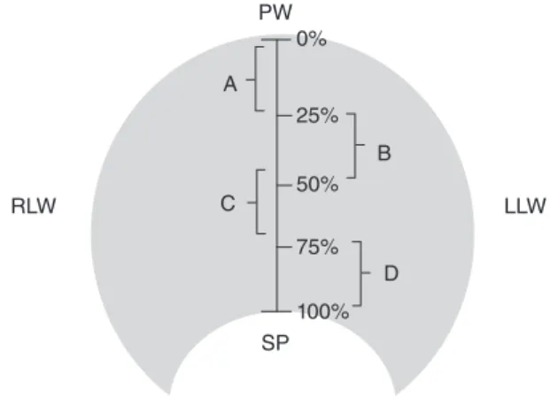
• maximum displacement of the soft palate: the judges should estimate the extent, in percentage, of the maximum displace-ment of the soft palate, considering that the latter, at rest, has its value set at 0%; the posterior pharyngeal wall, at rest, has its value set at 100%; and the center of the VPM, at rest, has its value set at 50%. According to the protocol, the maximum displacement of the soft palate could fall into four measures: 0-25%, 26-50%, 51-75%, and 76-100% (Figure 1);
• movement of the posterior pharyngeal wall: the judges were asked to identify the presence or absence of movement of the posterior pharyngeal wall;
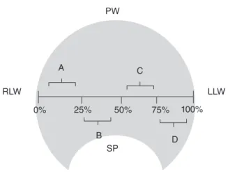
• maximum displacement of the posterior pharyngeal wall: the judges should estimate the extent, in percent-age, of the maximum displacement of the posterior wall in relation to the soft palate, considering that the wall at rest has reference value set at 0% and the soft palate at rest has reference value set at 100%. Thus, according to
the protocol, the maximum displacement of the posterior wall could fall into four measurements: 0–25%, 26–50%, 51–75%, and 76–100% (Figure 2);
• movement of the lateral pharyngeal walls: the judges should identify the presence and absence of movement of the right and left lateral walls;
• maximum displacement of the lateral pharyngeal walls: the judges should estimate the measurement, in percentage,
PW
SP 100%
75%
50%
25%
0%
RLD LLW
D
B C
A
Caption: PW = posterior pharyngeal wall; PLD = right lateral wall; PLE = left lateral wall; SP = soft palate
Figure 1. Reference scheme of displacement of the soft palate in four quadrants. A: 0–25%; B: 26–50%; C: 51–75%; D: 76–100%
PW
SP 100% 75% 50% 25% 0%
RLW LLW
D B
C A
Caption: PW = posterior pharyngeal wall; PLD = right lateral wall; PLE = left lateral wall; SP = soft palate
Figure 2. Reference scheme of the displacement of the posterior
of the maximum displacement of the right and left lateral walls, relative to one another, considering that the wall at rest has a reference value set at 0%, and the other has a ref-erence value set at 100%. Thus, according to the protocol, the maximum displacement of right and left walls could fall into four measurements: 0–25%, 26–50%, 51–75%, and 76–100% (Figure 3);
• Passavant´s pad: the judges should judge whether there was the presence of the Passavant´s pad;
• VP gap size: the judges should quantify the VP gap size according to the reference measurements, that is, quantify the space that was left of the maximum movement of velo-pharyngeal structures and not how much the structures per-chance had moved. For this reason, they should compare the image at rest with the image of the structures at maximum displacement. When there was no movement of velopha-ryngeal structures (where the image at rest is equal to the image of the maximum displacement), they should measure the gap size at 100%. The reference measures adopted for this item were subdivided on a six-point adapted scale(20,21): 0%, VP closure; 10%, VP gap with air bubble; 25%, small
PW
SP
100% 75% 50%
25% 0%
RLW LLW
D B
C A
Caption: PW = posterior pharyngeal wall; PLD = right lateral wall; PLE = left lateral wall; SP = soft palate
Figure 3. Reference scheme of the displacement of the lateral
pharyngeal wall in four quadrants. A: 0–25%; B: 26–50%; C: 51–75%; D: 76–100%. On the left wall, the measurements are reversed
Size of the velopharyngeal gap
at rest Gap at rest
closure
Gap
0%
10%
25%
50%
75%
100%
Figure 4. Reference scheme of the measurement of the velopharyngeal gap, where 0% = velopharyngeal closure; 10% = velopharyngeal gap
VP gap; 50%, medium VP gap; 75%, large VP gap; 100%, very large VP gap (Figure 4); and
• type of VP gap: the judges should classify the type of VP gap, observing the behavior of all structures, according to the following classiication(19):
• Sagittal: there is a predominance of movement of the lateral pharyngeal walls as compared to other velopha-ryngeal structures;
• Coronal or transverse: there is a predominance of move-ment of the soft palate (upward and backward) when compared to the other structures;
• Circular: there is a “homogeneous” participation of the soft palate and lateral pharyngeal walls; and
• Circular with Passavant´s pad: the circular type occurs with the formation of a Passavant´s pad in the posterior pharyngeal wall.
For statistical analysis, the results for each aspect evaluated among patients who underwent surgery through the F technique and those who underwent surgery through the vL technique were compared using Fisher’s statistical test for movement of the soft palate and of the lateral walls, maximum displacement of the soft palate and posterior wall, and type of VP gap. The χ2-test was applied to the movement of the posterior pharyngeal wall, maxi-mum displacement of the lateral pharyngeal wall, and Passavant´s pad. As for the size of the VP gap, the Mann–Whitney’s test was used. Results with p<0.05 were considered statistically signii-cant for these tests.
RESULTS
The presence of soft palate movement was observed for all (100%) patients who had surgery through the F technique and for 46 (96%) patients who had surgery through the vL tech-nique. The difference between these results was not signiicant (Fisher’s exact test, p=1.000). The maximum displacement of the soft palate between 0 and 25% occurred for 7 (32%) patients who had surgery using the F technique and 11 (23%) who underwent surgery using the vL technique; between 26 and 50% for 4 (18%) of F and 3 (6%) of vL; between 51 and 75% for 3 (14%) of F and 7 (15%) of VL; and between 76 and 100% for 8 (36%) of F and 27 (56%) of vL. The com-parison of the results was not statistically signiicant (Fisher’s exact test, p=0.282).
The movement of the posterior pharyngeal wall was observed for 10 (45%) patients of the F technique and 19 (39%) of the vL technique. The difference between the results was not sig-niicant (χ2-test, p=0.840). The maximum displacement of the posterior pharyngeal wall between 0 and 25% occurred for 19 (86%) patients of the F technique and 42 (87%) of the vL technique; between 26 and 50% occurred for 3 (14%) of F and 6 (13%) of vL. There was no displacement over 50% in both primary palatoplasty techniques for any patient. The results between the comparisons were not statistically signii-cant (Fisher’s exact test, p=1.000).
The movement of the right and left pharyngeal walls occurred for all patients of the F technique. However, in the vL
technique, the movement of the right pharyngeal wall occurred for 45 (94%) and the movement of the left pharyngeal wall occurred for 43 (93%) patients. The results of the comparisons were not statistically signiicant (Fisher’s exact test, right wall, p=0.547; left wall, p=0.546). The maximum displacement of the right and left pharyngeal wall was not statistically signii-cant (χ2-test, p=0.925; p=0.468 – Table 1).
The presence of the Passavant´s pad was observed in 7 (32%) patients of the F technique and in 14 (30%) of the vL technique. The comparison between the results was not statis-tically signiicant (χ2-test, p=0.955).
The size of the VP gap at 0% did not occur to any of the patients who underwent surgery through the F technique and occurred only in 2 (4%) who underwent the vL technique; at 10% for 5 (23%) patients of F and 16 (33%) of vL; at 25% for 7 (32%) of F and 7 (15%) of vL; at 50% for 4 (18%) of F and 12 (25%) of vL; at 75% for 6 (27%) of F and 10 (21%) of vL; and at 100% for none (0%) of F and 1 (2%) of vL. Differences between the comparisons of results were not statistically sig-niicant (Mann-Whitney’s test, p=0.531 – Table 2).
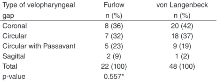
The coronal VP gap occurred for 8 (36%) patients of F and 20 (42%) of vL; sagittal occurred for 2 (9%) of F and (2%) with vL; circular occurred for 7 (32%) of F and 18 (37%) of vL; and circulate of Passavant´s pad occurred for 5 (23%) of F and 9 (19%) of vL. A comparison of the results was not statistically signiicant (Fisher’s exact test, p=0.557 – Table 3).
Table 1. Distribution of numbers and percentages of patients who
underwent surgery through the Furlow and von Langenbeck techniques, according to the values of the maximum displacement of the lateral pharyngeal walls
Maximum displacement of the lateral
pharyngeal walls
Furlow n (%)
von Langenbeck n (%)
RS LS RS LS
0–25% 16 (73) 17 (77) 37 (77) 30 (65)
26–50% 6 (27) 5 (23) 11 (23) 16 (35)
51–75% 0 (0) 0 (0) 0 (0) 0 (0)
76–100% 0 (0) 0 (0) 0 (0) 0 (0)
Total 22 (100) 22 (100) 48 (100) 46 (100)
p-value 0.925* 0.468*
Caption: RS = right side; LS = left side *Significant values (p<0.05) – χ2-test
Table 2. Distribution of the number of patients who underwent surgery through the Furlow and von Langenbeck techniques, according to the values obtained regarding the size of the velopharyngeal gap
Size of the velopharyngeal gap
Furlow n (%)
von Langenbeck n (%)
0% 0 (0) 2 (4)
10% 5 (23) 16 (33)
25% 7 (32) 7 (15)
50% 4 (18) 12 (25)
75% 6(27) 10 (21)
100% 0 (0) 1 (2)
Total 22 (100) 48 (100)
p-value 0.531*
DISCUSSION
The results of this study did not conirm the hypothesis that patients who presented VPI after the F technique and primary palatoplasty presented with a smaller VP gap than those who underwent surgery with the vL technique. All comparisons of velopharyngeal structures observed no signiicant difference.
When interpreting these indings, it is important to consider that the surgical reorganization of the palatal muscles, as con-ducted in both techniques, may have had a similar impact on the gap size. The F technique, for example, stretches the soft pal-ate through two mirrored zetaplasties involving oral and nasal laps (mirrored between planes) with transposition of the laps, which involves muscular retroposing(2,6,12). However, the vL technique with intravelar veloplasty requires the release and thorough dissection of the muscles, also allowing posterioriza-tion of the muscle ibers(6,11,22). That is, both techniques involve release and posteriorization of muscle ibers and, although the procedures for manipulation of the muscle ibers are differ-ent between the techniques, the results in terms of VP gap are similar, as observed in this study.
The palatopharyngeal ibers in patients with cleft palate run along the levator muscle of the soft palate(23). The horizontal ibers of this muscle can produce a sphincteric action, medial-izing the lateral pharyngeal walls narrowing the nasopharynx and help with VP closure(24). Some authors suggested that the palatopharyngeal muscle works as a “hydrostatic” muscle, which helps contract the posterior portion of the soft palate, molding the shape of the posterior pharyngeal wall and resulting in a better VP sealing(25,26). Therefore, the techniques used in this study can inluence the movement of the pharyngeal walls, as both involve repositions the palatopharyngeal muscle leading to functional and anatomical competence.
The ability of the surgeon to conduct the surgical technique has been cited as an important factor in the outcome of the treatment of cleft lip and palate(6-11). The surgeons involved in this study were using the vL procedure as the routine surgical technique for primary repair of cleft lip and palate while they were trained to perform the F technique for the study. Despite the greater skill with the routine technique (vL) when com-pared with the innovative procedure (F), the expected longer soft palate and, therefore, smaller gap with the F procedure, was not observed.
Table 3. Distribution of the number of patients who underwent surgery through the Furlow and von Langenbeck techniques, according to the type of velopharyngeal gap
Type of velopharyngeal gap
Furlow n (%)
von Langenbeck n (%)
Coronal 8 (36) 20 (42)
Circular 7 (32) 18 (37)
Circular with Passavant 5 (23) 9 (19)
Sagittal 2 (9) 1 (2)
Total 22 (100) 48 (100)
p-value 0.557*
*Significant values (p<0.05) – Mann-Whitney’s test
The presence of the expected pharyngeal tonsil in speak-ers aged between 9 and 13 years(27) may have been a variable that affected the indings, since nasoendoscopic examinations were conducted in patients between 5 and 15 years (mean age 8 years). The literature refers to veloadenoidal closure, at this age(28,29). It is assumed, however, that the presence of the tonsil at different stages of involution was distributed equally between the two populations. That is, if there was participation of the tonsil in VC, it occurred equally for the F and vL procedures.
The velopharyngeal function can be affected by vocal intensity and by the different levels of intraoral and subglottic pressure used by speakers during speech production(1,4,12). This study did not involve the conduction of perceptual evaluation of speech samples obtained during the nasoendoscopic exam. It is suggested that this limitation should be better controlled in future studies because the use of compensatory articulation and the presence of hypernasality and nasal air escape may interfere with the movement of velopharyngeal structures(1,12).
For this study, the selected stimulus for speech sampling production was the sequence “papapa”, since it was the most frequent speech stimulus in the database, providing a larger number of recordings for the study. Although the phonetic content of the speech sample was controlled, the ideal would be to include more representative samples of connected speech (phonetically balanced)(20,30).
Although the group studied had more nasoendoscopy tests with VPI in patients who underwent surgery with the vL tech-nique than those who underwent surgery with the F techtech-nique, the characteristics of the gaps between the groups were simi-lar. The sample size was not controlled since the purpose of this study was to evaluate all the nasoendoscopic exams in the databank.
CONCLUSION
The surgical technique used in primary palatoplasty was not relevant to determine difference in velopharyngeal gap size for patients who maintained VPI.
*GZF participated in the development and design of the study, data collection, analysis and interpretation of data, and drafting and revision of the article; JCRD carried out the study design, analysis, and interpretation of data, as well as the drafting and revision of the article; MEW, OMVS, and VCCM carried out the analysis and interpretation of data; MIPK conducted the study design, data collection, analysis, and interpretation of data and also the drafting and revision of this article.
REFERENCES
1. Ma L, Shi B, Li Y, Zheng Q. Velopharyngeal function assessment in patients with cleft palate: perceptual speech assessment versus nasopharyngoscopy. J Craniofac Surg. 2013;24(4):1229-31.
2. Smith DM, Losee JE. Cleft palate repair. ClinPlast Surg. 2014;41(2):189-210. 3. Pegoraro-Krook MI, Dutka-Souza JC, Magalhães LCT, Feniman MR.
4. Kummer AW. Speech evaluation for patients with cleft palate. Clin Plast Surg. 2014;41:241-51.
5. Bertier CE, Trindade IEK, Silva Filho OG. Cirurgias primárias de lábio e palato. In: Trindade IEK, Silva Filho OG, coordenadores. Fissuras labiopalatinas: uma abordagem interdisciplinar. São Paulo: Santos; 2007. p. 73-86.
6. Williams WN, Seagle MB, Pegoraro-Krook MI, Souza TV, Garla L, Silva ML, et al. Prospective clinical trial comparing outcome measures between Furlow and von Langenbeck palatoplasties for UCLP. Ann Plast Surg. 2011;66(2):154-63.
7. Liao Y, Mars M. Hard palate repair timing and facial morphology in unilateral cleft lip and palate: before versus after pubertal peak velocity age. Cleft Palate Craniofac J. 2006;43(3):259-65.
8. Arosarena OA. Cleft lip palate. Otolaryngol Clin North Am. 2007;40(1):27-60. 9. Kummer AW, Clark SL, Redle EE, Thomsen LC, Billmire DA. Current
practice in assessing and reporting speech outcomes of cleft palate and velopharyngeal surgery: a survey of cleft palate/craniofacial professionals. Cleft Palate Craniofac J. 2012;49(2):146-52.
10. Mahoney MH, Swan MC, Fisher DM. Prospective analysis of presurgical risk factors for outcomes in primary palatoplasty. Plast Reconstr Surg. 2013;132(1):165-71.
11. Smith KS, Ugalde CM. Primary palatoplasty using bipedicle flaps (modiied von Langenbeck technique). Atlas Oral Maxillofac Surg Clin North Am. 2009;17(2):147-56.
12. Hopper RA, Tse R, Smartt J, Swanson J, Kinter S. Cleft palate repair and velopharyngeal dysfunction. Plastic Reconstr Surg. 2014;133(6):852e-64. 13. Harris C, Dankat-Thomas L, Hill C, Leonard A. A comparison of Furlow
and von Langenbeck techniques for isolated clefts of the soft palate: a single-center review. Ann Plast Surg. 2011;66(6):679.
14. Furlow LT Jr. Cleft palate repair by double opposing Z-plasty. Plast Reconstr Surg. 1986;78(6):724-38.
15. Di Ninno CQMS, Macedo CCF, Ferreira NF, Gomes SCM, Jesus MSV, Lopes MS, et al. Resultados de fala após palatoplastia: estudo comparativo prospectivo entre as técnicas de Veau modiicada e Furlow. Rev Bras Cir Craniomaxilofac. 2012;15(2):74-8.
16. Chan EKW, Lee KH, Tsui BSY, Wong SYS, Pang KKY, Mou JWC, et al. From von Langenbeck to Furlow palatoplasty: a 16-year review of cleft palate repair. Surgical Practice. 2014;18(2):67-71.
17. Ng ZY, Young S, Por YC, Yeow V. Results of primary repair of submucous cleft palate with Furlow palatoplasty in both syndromic and non-syndromic children. Plast Reconstr Surg. 2014;133(3s):110.
18. Rajion ZA, Al-Khatib AR, Netherway DJ, Townsend GC, Anderson PJ, McLean NR, et al. The nasopharynx in infants with cleft lip and palate. J Pediatr Otorhinolaryngol. 2012;76(2):227-34.
19. Skolnick ML, McCall GN, Barnes M. The sphincteric mechanism of velopharyngeal closure. Cleft Palate J. 1973;10(3):286-305.
20. Golding-Kushner KJ, Argamaso RV, Cotton RT, Grames LM, Henningsson G, Jones DL, et al. Standardization for the reporting of nasopharyngoscopy and multiview videoluoroscopy: a report from an international working group. Cleft Palate J. 1990;27(4):337-47. 21. Souza OMV. Movimentação da velofaringe após o uso do obturador
faríngeo [dissertação]. Bauru (SP): Faculdade de Odontologia de Bauru, Universidade de São Paulo; 2009.
22. Andrades P, Espinosa-de-los-Monteros A, Shell DH 4th, Thurston TE, Fowler JS, Xavier ST, et al. The importance of radical intravelar veloplasty during two-flap palatoplasty. Plast Reconstr Surg. 2008;122(4):1121-30.
23. Fára M, Dvorák J. Abnormal anatomy of the muscles of palatopharyngeal closure in cleft palates: anatomical and surgical considerations based on the autopsies of 18 unoperated cleft palates. Plast Reconstr Surg. 1970;46(5):488-97.
24. Kummer AW. Resonance disorders and velopharyngeal dysfunction (VPD). In: Kummer AW. Cleft palate and craniofacial anomalies: the effects on speech and resonance. 2nd edition. San Diego: Thomson Delmar Learning; 2008. p. 176-212.
25. Ettema SL, Kuehn DP. A quantitative histologic study of the normal human adult soft palate. J Speech Hear Res. 1994;37(2):303-13. 26. Moon JB, Kuehn DP. Anatomy and physiology of normal and
disordered velopharyngeal function for speech. In: Bzoch KR, editor. Communicative disorders related to cleft lip and palate. 4th edition. Austin: Pro-Ed.; 1997. p. 45-7.
27. Siegel-Sadewitz VL, Sphprintzen RJ. Changes in velopharyngeal valving with age. Int J Pediatr Otorhinolaryngol. 1986;11(2):171-82.
28. Côrtes RMO, Piazentin-Penna SHA. Ocorrência de hipernasalidade após adenoidectomia. Salusvita. 2008;27(2):183-97.
29. Askar SM, Quriba AS. Powered instrumentation for transnasal endoscopic partial adenoidectomy in children with submucosal cleft palate. Int J Pediatr Otorhinolaryngol. 2014;78(2):317-22.