www.rbo.org.br/
ISSN/$–see front matter © 2013 Sociedade Brasileira de Ortopedia e Traumatologia. Published by Elsevier Editora Ltda. All rights reserved.
*Corresponding author at: Hospital Universitário Antonio Pedro, Serviço de Ortopedia, Av. Marquês de Paraná, 303, 4º andar, Niterói, RJ, Brazil. CEP: 24033-900.
E-mail: [email protected]; [email protected] ARTICLE INFO
Article history:
Received on April 22, 2012 Accepted on July 3, 2012
Keywords:
Dupuytren contracture Surgical procedures Operative hand deformities
Original Article
Open Palm Technique in Dupuytren’s Disease Treatment
Marcio Carpi Malta,
1,*Marcelo de Pinho Teixeira Alves,
2Luis Marcelo de Azevedo Malta
31PhD; Associate Professor, Head of the Orthopedics Service, Hospital Universitário Antonio Pedro, Universidade Federal Fluminense, Niterói,
RJ, Brazil.
2Master’s in Medical Sciences from the Universidade Federal Fluminense, Niterói, RJ, Brazil. 3Physician of the Orthopedics Service, Hospital Universitário Antonio Pedro, Niterói, RJ, Brazil.
Research carried out in the Hospital Universitário Antonio Pedro, Universidade Federal Fluminense, Niterói, RJ, Brazil.
doi: 10.1016/j.rbo.2012.07.005
a b s t r a c t
Objective: To evaluate the results of the open palm technique for the treatment of Dupuytren’s disease. Method: The authors used the technique described by McCash. Twelve patients (13 hands) were surgically treated, between october 2002 and september 2011. Results: The wounds healed in a medium of 25 days (variation of 17 to 30 days). There were no complications, such as infection, haematoma formation, skin necrosis, residual edema. Conclusion: The open palm technique remains a safe alternative for the treatment of Dupuytren’s disease, with satisfactory results and low risk of complications.
Introduction
Palm aponeurosis, also known as Dupuytren’s disease, was initially described by Felix Platter in 1614.1 However, according
to Rayan,2 Dupuytren was responsible for describing the
anatomical characteristics of the disease that remains named after him in 1831.
The disease is most common in Caucasian men. It is more prevalent in North America than in South America, and it is rare in China and Africa.3 Reis and Mota Júnior4 demonstrated
a relationship between Dupuytren’s disease and diabetes. Myofibroblasts, a cell type described by Gabbiani and Majno5, have the morphological characteristics of fibroblasts
and smooth muscle cells, and they are currently considered the determinant of Dupuytren’s disease.
Although recent research has suggested the local administration of substances such as collagenase6 and
corticosteroids7 for the non-surgical treatment of Dupuytren’s
disease, surgery is still the most used method.
Among the several surgical techniques employed to treat Dupuytren’s disease, the one described by McCash is characterized by its simplicity and low rate of complications.8
This report evaluated the results obtained in patients suffering from Dupuytren’s disease who were treated with the open palm technique.
Method
From October 2002 to September 2011, 12 patients (13 hands) suffering from Dupuytren’s disease underwent surgery. The patients’ ages ranged from 33 to 81 years old (average
64 years old). Two patients were female (17%), and one was of African descent (8%; the patient considered him/herself a Caucasian). Of the 13 hands that underwent surgery, 6 were (46%) left hands, and 7 were right hands (54%).
A single finger was affected in 69% of the hands (nine cases). Of these cases, 56% were on finger IV (on five hands), and 44% were on finger V (four hands). Two fingers were affected in four cases (31%), including fingers IV and V (two hands), III + IV (25%) and I + V (25%). Patient 5 had finger V involvement on both hands (the only bilateral case in our sample). In general, fingers I and III were affected in 8% of the hands, finger IV in 54%, and finger V in 54%.
Flexion deformity of the metacarpophalangeal joint greater than 30 degrees and/or deformity of the proximal interphalangeal joint greater than 15 degrees were present in all hands that underwent surgery. Chart 1 shows the general data of the patients.
All patients underwent surgery under block anesthesia with member ischemia.
The surgical technique used was previously described by McCash. The affected tissue was removed through a transverse incision in the region of the distal palm crease, which allowed the correction of the flexion deformity of the metacarpophalangeal joint. When necessary, another transverse incision was performed proximally to the first to facilitate the resection of the affected tissue. Figs. 1 to 5 illustrate the clinical case of patient number 2. As shown in Figs. 1 and 2, a flexion contracture was present in finger IV with a clearly observable longitudinal cord. Fig. 3 shows the access pathway used in the McCash technique, and the pathological tissue is evident. Figs. 4 and 5 demonstrate the postoperative evolution of the wound, with complete healing 25 days after
Patient Age (years) Side Finger(s) Affected Surgery Date
1 71 Left IV 10/05/2002
2 71 Left IV 12/09/2003
3 63 Right IV 12/02/2004
4 67 Right I, V 01/20/2005
5 69 Right V 02/16/2005
6 69 Left V 04/08/2005
7 72 Right V 08/26/2005
8 33 Right IV 09/06/2005
9 54 Left V 05/10/2006
10 72 Right IV 10/28/2008
11 59 Left IV, V 08/31/2010
12 81 Left III, IV 05/19/2011
13 59 Right IV, V 09/08/2011
Mean 65
Percentage Left 46% I 8%
Right 54% III 8%
IV 54%
V 54%
Fig. 1 - Patient number 2, preoperative. Finger IV affected, anteroposterior view.
Fig. 2 - Patient number 2, preoperative. Finger IV affected, profile view.
Fig. 3 - Patient number 2, perioperative, with identification of the longitudinal cord.
Fig. 4 - Patient number 2, 25 days postoperative, with complete extension of the finger.
Fig. 5 - Patient number 2, 25 days postoperative, with finger in flexion, demonstrating pulp-palm with minimum functional deficit.
the surgery and the absence of functional restriction of the patient’s hand.
Results
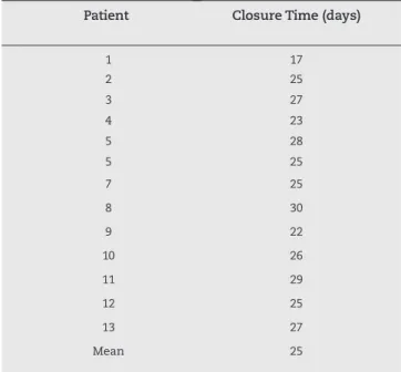
The wounds closed in an average period of 25 days, with a range of 17 to 30 days (Table 1). Infection, hematoma, skin necrosis, residual edema, and other local complications were not reported. In 12 hands (92% of the patients), the deformities were completely corrected, with no limitation of the arc of movement of the joints involved at the end of treatment.
In one of the patients (8%) with unilateral disease, the flexion deformity of the proximal interphalangeal joint of finger V exceeded 90 degrees and was treated by resection of the fibrous tissue through a skin incision and multiple z-plasty. The palmar wound was left open and healed in 22 days, but the deformity of the proximal interphalangeal joint was not corrected.
Considering the severity of the deformity and the functional aspect of the hand, finger V was amputated after receiving the patient’s approval.
aponeuroses and in the development of flexion deformity of the proximal interphalangeal and metacarpophalangeal joints, which together characterize Dupuytren’s disease.10,11
To treat Dupuytren’s disease, the literature suggests several methods, including the local injection of agents such as calcium-channel blockers 9 (verapamil and nifedipine)12 and
lytic enzymes (trypsin and hyaluronidase) in combination with the placement of the fingers in forced extension to liberate the adherences.13 Surgical treatment is the most used method, and
it is indicated when the flexion deformities of the proximal interphalangeal and metacarpophalangeal joints exceed 30 and 15 degrees, respectively.
Some authors recommend percutaneous needle fasciotomy as a minimally invasive procedure for treatment of the disease.14-17 However, these studies have demonstrated a
higher rate of recurrence after this type of procedure, which makes it more appropriate for elderly patients or patients who accept a possible early recurrence of the disease and, eventually, the deformity.15
All patients included in this study were surgically treated with the open palm technique. In 1984, one of the authors18 (MCM) published a study of 10 patients (total of 16 hands) with Dupuytren’s disease who underwent surgery using the open palm technique; that study demonstrated results similar to those presented herein. As reported by Schneider et al.19 in
a review of 49 patients, the open hand technique results in a less painful postoperative period, better mobility of the fingers, and a lower rate of complications. Lubahnn recommended this technique and reported a complication rate of 8% in the initial postoperative period, without infections. In the long term, Lubahnn reported that 20% of patients experienced residual contracture compared to 42% for patients treated by suturing the operative wounds.20,21
Galbiatti et al.22 reported satisfactory results in the
treatment of nine patients with the use of a straight incision in the palm of the hand, which was transformed during z-plasty when the wound was closed. All patients were male with a mean age of 54.2 years old, and 77% were Caucasian. In our study, we obtained similar results, without any concern for skin suturing.
Freitas et al.23 observed ten patients treated with the open
palm technique and verified that the average period for wound closure was 25 days, with a range of 15 to 45 days. In a sample of 30 patients, Silva et al.24 found the same average duration
for closure, but they reported a range of 20 to 40 days. Among our patients, the average time to closure was 25 days, with a range of 17 to 30 days, which in is in general accordance with the literature. A similar finding was observed by Skoff, who reported an average of 40 days to wound closure in patients who underwent open palm surgery. In addition, he observed the complete correction of flexion deformities of the proximal interphalangeal and metacarpophalangeal joints.25
In an analysis of 100 patients with a mean age of 52 years, including 88 men, Barros et al.26 found that the digitus
annularis (finger IV) and the little finger (finger V) were affected in 60% of cases and that the thumb (finger I) was involved in 25%. We observed a similar distribution, with fingers IV and V being the most commonly involved.
Patient Closure Time (days)
1 17 2 25 3 27 4 23 5 28 5 25 7 25 8 30 9 22 10 26 11 29 12 25 13 27 Mean 25
Table 1 – The time required for wound closure for each patient.
Discussion
Dupuytren’s disease is associated with the presence and activity of myofibroblasts, which have the features of both fibroblasts and smooth muscle cells following differentiation. They were originally described by Gabbiani and Majno5 and
later studied by Tomasek et al.9 Myofibroblasts produce the
In one of our patients, flexion deformity of the proximal interphalangeal joints exceeded 90 degrees and could not be corrected with z-plasty. After palmar wound closure, the little finger was amputated for functional reasons.
Conclusion
We conclude that the open palm technique is a safe option to treat Dupuytren’s disease and that it offers satisfactory results with low complication rates.
Conflict of Interest
The authors have no conflict of interest to declare associated with this paper.
R E F E R E N C E S
1. Elliot D. The early history of Dupuytren’s disease. Hand Clin. 1999;15(1):1-19.
2. Rayan GM. Instructional course lecture. Dupuytren disease: anatomy, pathology, presentation, and treatment. J Bone Joint Surg Am. 2007;89(1):190-8.
3. Hueston JT. Dupuytren’s diatheseis. In: Hueston JT (Ed.). Dupuytren’s contracture. Edinburgh: Livingstone, 1963, p. 51-63, E45.
4. Reis AS, Mota Júnior SC. Diabetes melito na etiologia da doença de Dupuytren. Rev Bras Ortop. 1993;28(5):329-31. 5. Gabbiani G, Majno G. Dupuytren’s contracture: fibroblast
contraction? An ultrastrutural study. Am J Patol. 1972;66(1):131-46.
6. Badalamente MA, Hurst LC. Enzyme injection as nonsurgical treatment of Dupuytren’s disease. J Hand Surg Am.
2000;25(4):629-36.
7. Ketchum LD, Donahue TK. The injection of nodules of Dupuyten’s disease with triamcinolone acetonide. J Hand Surg Am. 2000;25(6):1557-62.
8. McCash CR. The open palm technique in Dupuytren’s contracture. Br J Plast Surg. 1964;17:271-80.
9. Tomasek JJ, Gabbiani G, Hinz B, Chaponnier C, Brown RA. Myofibroblasts and mechano-regulation of connective tissue remodelling. Nat Rev Mol Cell Biol. 2002;3(5):349-63.
10. Tomasek JJ, Rayan GM. Correlation of alpha-smooth muscle actin expression and contraction in Dupuytren’s disease fibroblasts. J Hand Surg Am. 1995;20(3):450-5.
11. Tomasek JJ, Haaksama CJ. Fibronectin filaments and actin microfilaments are organized into a fibronexus in Dupuytren’s diseased tissue. Anat Rec. 1991;230(2):175-82. 12. Rayan GM, Parizi M, Tomasek JJ. Pharmacologic regulation of
Dupuytren’s fibroblast contraction in vitro. J Hand Surg Am. 1996;21(6):1065-70.
13. McCarthy DM. The long-term results of enzymic fasciotomy. J Hand Surg Br. 1992;17(3):356.
14. Becker GW, Davis TR. The outcome of surgical treatments for primary Dupuytren’s disease – A systematic review. J Hand Surg Eur. 2010;35(8):623-6.
15. Van Rijssen AL, der Linden H, Werker PM. Five-year results of a randomized clinical trial on treatment in Dupuytren’s disease: percutaneous needle fasciotomy versus limited fasciectomy. Plast Reconstr Surg. 2012;129(2):469-77.
16. Van Demark RE Jr, Van Demark RE 3rd. Needle aponeurotomy: a treatment alternative for Dupuytren’s disease. S D Med. 2011;64(11):411-5.
17. Pess GM, Pess RM, Pess RA. Results of needle aponeurotomy for dupuytren contracture in over 1000 fingers. J Hand Surg Am. 2012;37(4):651-6.
18. Malta MC, Vianna SE, Schott PC. A técnica de palma aberta na contratura de Dupuytren. Rev Bras Ortop. 1984;19(2):46-8. 19. Schneider LH, Hankin FM, Eisenberg T. Surgery of Dupuytren’s
disease: a review of the open palm method. J Hand Surg Am. 1986;11(1):23-7.
20. Lubahn JD. Open-palm technique and soft-tissue coverage in Dupuytren’s disease. Hand Clin. 1999;15(1):127-36.
21. Lubahn JD, Lister GD, Wolfe T. Fasciectomy and Dupuytren’s disease: a comparison between the openpalm technique and wound closure. J Hand Surg Am. 1984;9A(1):53-8.
22. Galbiatti JA, Fiori JM, Mansano RT, Durigan Júnior A. Tratamento da doença de Dupuytren pela técnica de incisão longitudinal reta, complementada com Z-plastia. Rev Bras Ortop. 1995;30(4):207-12.
23. Freitas AD, Pardini Júnior AG, Filho ATN. Contratura de Dupuytren: tratamento pela técnica da palma aberta. Rev Bras Ortop. 1997;32(4):301-4.
24. Silva JB, Fernandes H, Fridman M. A técnica da palma aberta na contratura grave de Dupuytren. Rev Bras Ortop. 1999;34(1):51-4.
25. Skoff HD. The surgical treatment of Dupuytren’s
contracture: a synthesis of techniques. Plast Reconstr Surg. 2004;113(2):540-4.