Comparison of airway dimensions in skeletal Class I
malocclusion subjects with different vertical facial patterns
Ana Paula Flores-Blancas1, Marcos J. Carruitero2, Carlos Flores-Mir3
Objective: The aim of this study was to compare upper airway widths among skeletal Class I malocclusion subjects with
differ-ent vertical facial patterns. Methods: The sample included a total of 99 lateral cephalograms of post pubertal individuals (18.19
± 1.76 years old). The vertical facial pattern was determined by the Vert index. The McNamara method was used to quantify upper airway widths. ANOVA test and Student’s t test for independent groups were used, when normal distribution was not
supported Kruskal-Wallis test and U-Mann-Whitney test were used. A multiple linear regression analysis was also performed.
Results: Statistically significant differences in several nasopharyngeal widths were found among the distinct vertical facial patterns.
Subjects with brachyfacial pattern presented larger nasopharyngeal widths than subjects with mesofacial (p = 0.030) or dolichofacial
(p = 0.034) patterns. The larger the Vert value, the larger the nasopharyngeal widths (R2 = 26.2%, p < 0.001). At the level of
oro-pharynx no statistically significant differences were found. Conclusion: It was concluded that nasopharyngeal linear
anteropos-terior widths in Class I malocclusion brachyfacial are larger than in mesofacial and dolichofacial individuals. The Vert index only explained 25% of the total variability. No correlation was found for the oropharyngeal widths.
Keywords: Upper airway. Vertical facial pattern. McNamara analysis. Vert index.
1 Private practice (Trujillo, Peru).
2 Universidad Privada Antenor Orrego, Facultad de Medicina Humana, Escuela
Estomatología (Trujillo, Peru).
3 University of Alberta, Division of Orthodontics (Edmonton, Canada).
» The authors report no commercial, proprietary or financial interest in the products or companies described in this article.
DOI: https://doi.org/10.1590/2177-6709.22.6.035-042.oar
How to cite: Flores-Blancas AP, Carruitero MJ, Flores-Mir C. Compari-son of airway dimensions in skeletal Class I malocclusion subjects with differ-ent vertical facial patterns. Ddiffer-ental Press J Orthod. 2017 Nov-Dec;22(6):35-42. DOI: https://doi.org/10.1590/2177-6709.22.6.035-042.oar
Submitted: September 14, 2016 - Revised and accepted: January 13, 2017
Contact address: Marcos J. Carruitero Honores Antenor Orrego Private University, 3145 América South Av. Monserrate, Trujillo/Peru – Email: [email protected]
Objetivo: o objetivo desse estudo foi comparar as dimensões das vias aéreas superiores em indivíduos portadores de má oclusão de Classe I esquelética com diferentes padrões faciais verticais. Métodos: a amostra consistiu de 99 cefalogramas laterais de in-divíduos na pós-puberdade (18,19 ± 1,76 anos). O padrão facial vertical foi determinado por meio do índice VERT. O método de McNamara foi utilizado para quantificar as dimensões das vias aéreas superiores. O teste ANOVA e o teste t de Student para grupos independentes foram utilizados e, quando a distribuição normal não era possível, o teste de Kruskal-Wallis e o teste U de Mann-Whitney foram aplicados. Foi também realizada uma análise de regressão linear múltipla. Resultados: diferenças es-tatisticamente significativas nas dimensões da nasofaringe foram encontradas entre os diferentes padrões faciais verticais. Os in-divíduos com padrão braquifacial apresentaram dimensões nasofaríngeas maiores do que os inOs in-divíduos com padrão mesofacial (p = 0,030) ou dolicofacial (p = 0,034). Quanto maior o valor do VERT, maior a dimensão nasofaríngea (R2 = 26,2%, p < 0,001).
Não foram encontradas, entretanto, diferenças estatisticamente significativas ao nível da orofaringe. Conclusão: pode-se con-cluir que as dimensões anteroposteriores lineares da nasofaringe nos indivíduos braquifaciais com má oclusão de Classe I são maiores do que nos indivíduos mesofaciais e dolicofaciais. O índice VERT foi capaz de explicar apenas 25% da variabilidade total. Não foi encontrada correlação para as dimensões da orofaringe.
INTRODUCTION
Orthodontics has been long interested in the as-sociation between mode of breathing and craniofacial growth.1-3 The pharyngeal structures play an import-ant role during breathing and swallowing functions. The pharynx can be anatomically separated in naso-pharynx and oronaso-pharynx. It has been proposed that they may vary in dimensions based on orthopedic therapy4 or craniofacial growth.5,6
Morphological upper airway obstructive process-es are factors that can lead into a partial or total upper airway obstruction. When that happens the result-ing functional imbalance could lead into a signiicant mouth-breathing pattern, which may alter the craniofa-cial morphology and dental arch shape, ultimately pro-ducing a malocclusion.7-11
The relative growth and size of the sot tissues sur-rounding craniofacial skeletal structures determine the size of the pharyngeal space. The depth of the nasophar-ynx increases as its posterior wall becomes narrower.9 There is also a natural and anatomical predisposition of airway to become thinner as it has been suggested that subjects with Class I and Class II malocclusions and ver-tical growth patterns have signiicantly narrower upper pharyngeal airways than those with normal growth pat-terns.12 If the upper airway becomes narrower, in some cases the air low resistance may increase, which may also increase the risk of snoring and, in severe cases, lead to obstructive sleep apnea.11,13,14
Altered upper airway function during the active pe-riod of craniofacial growth can also have a profound inluence on the diferent facial growth patterns.15,16 In contrast, craniofacial anomalies, including maxillary or mandibular retrognathism, short mandibular body, clockwise rotation of the jaw, high palatal vault, nar-row maxilla, and increased anterior face height have been associated with reduced pharyngeal airway.15,17 The usual unanswered question is which one may have occurred irst.
For the assessment of the dimension of the airway, some linear distances have been described.18,19 Although the association between craniofacial morphology, breathing function and pharyngeal structures has been previously explored,20-22 its association with the ver-tical facial patterns has been assessed by using various methods to determine the facial pattern like, Y-axis,13 SN-Me angle15 or SN-GoGn angle;23,24 but not using
the Vert index method, which relates both quantitative and qualitative facial vertical growth,25 being necessary a more insight in the ield.
The Vert index method is a method proposed by Ricketts which identify growth patterns taking into account ive cephalometric measurements: facial axis, facial depth, mandibular plane, anteroinferior facial height, and mandibular arch. These values let classify the face into six types: severe brachyfacial, brachyfacial, mesofacial, light dolichofacial, dolichofacial, and severe dolichofacial25 (which can also be grouped into just three types: mesofacial, brachyfacial, and dolichofacial).26,27
The aim of this study was to compare the widths of the airways in Class I malocclusion subjects with difer-ent vertical facial patterns, by using the Vert index.
MATERIAL AND METHODS
The present research was approved by the Stomatol-ogy Permanent Research Committee of the Antenor Orrego Private University (Trujillo, Peru).
Study sample
This study was conducted on 99 lateral cephalo-grams of post pubertal individuals between 16 and 22 years old (18.19 ± 1.76): 52 women (16 - 22 years
of age, 18.10 ± 1.91); and 47 men (16 - 21 years old
age, 18.30 ± 1.59). The radiographs were randomly selected from all the available radiographs during the period 2010 to 2013 in a diagnostic imaging center in Trujillo, Peru.
To determine the sample size, data from a pilot study with 24 radiographs was used. A statistical pow-er of 80% and a confidence level of 95% was finally considered. The variance for brachyfacial individuals was 8.01 mm and for the mesofacial individuals was 6.98 mm. Therefore a minimum of 15 lateral cepha-lograms per facial vertical pattern was needed. Three groups of 31, 33, and 35 radiographs for each vertical facial pattern (mesofacial, dolichofacial and brachyfa-cial, respectively) were formed.
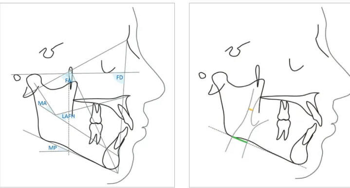
Figure 1 - Angles for Vert index: FA, Facial Axis (angle between the lines Basion-Nasion and Gnation-Pterygoid); FD, Facial Depth (angle formed by lines Nasion-Pogonion and Porion-Orbitale); LAFH, Lower Anterior Facial Height, angle formed by lines ANS-Xi and Xi-MP; MP, Mandibular Plane (angle formed by lines Porion-Orbitale and Gonial-Menton); MA, Mandibular Arch (angle formed by lines Dc-Xi and Xi-MP).
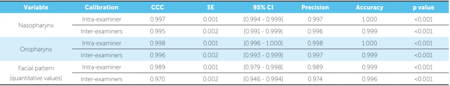
Figure 2 - Airway widths according to McNamara analysis, nasopharynx (yel-low line) and oropharynx (green line).
Tracing
The cephalometric tracings, landmark identiica-tions, and measurements were performed on acetate pa-per by one researcher.
The vertical facial pattern was determined by the Vert index.25 To determine the inal vertical facial pattern, ive cephalometric measurements were considered: [1] Fa-cial Axis (FA), angle between the lines Basion-Nasion and Gnation-Pterygoid; [2] Facial Depth (FD), angle formed by lines Nasion-Pogonion and Porion-Orbit-ale; [3] Lower Anterior Facial Height (LAFH), angle formed by lines ANS-Xi and Xi-MP; [4] Mandibular Plane (MP), angle formed by lines Porion-Orbitale and Gonial-Menton, and [5] Mandibular Arch (MA), angle formed by lines Dc-Xi and Xi-MP (Fig 1). The facial type determined by the Vert index in adults is given
by the following equation: {[(FA-90) / 3] + [(FD-90) / 3] + [(24,5-MP) / 4] + [(47-LAFH) / 4] + [(MA-28,5) / 4]} / 5. If the result was greater than +0.5, the patient was classi-ied as brachyfacial; between -0.49 and +0.49, as mesofa-cial; and smaller than -0.5, as dolichofacial.
Upper airway tracing
Method error
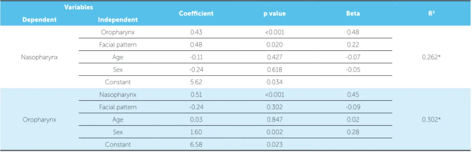
To evaluate the method error, measurements in 24 radiographic images not considered in the final study were considered. These measurements were carried out by the same researcher twice (the second time, after a week) in order to assess intra-examiner reliability for the cephalometric tracings. To assess the inter-examiners reliability, the same cases were evaluated by another researcher. The agreement between the observations of the nasopharynx, oro-pharynx, and vertical facial pattern (numerical val-ues) were evaluated by the Concordance Correla-tion Coefficient test.
Statistical analysis
Data was processed in the statistical program Sta-ta v. 12 (SSta-taSta-ta Corp. Texas, USA). Means, sSta-tandard deviations, minimum and maximum values were calculated. Before making any group comparisons, compliance with the assumptions of normality and homogeneity of variances with Shapiro-Wilk and Levene’s test were evaluated. Some groups were not normally distributed; therefore, Kruskal-Wallis test and U-Mann-Whitney test for pairwise comparisons were applied in them. To compare the means in the groups that met the assumptions, an ANOVA test was used. Comparisons between men and women were performed using Student’s t test for independent groups. An analysis of multiple linear regression, in which age and sex were included as factors, was used. Statistical significance was set at 5% in all tests.
RESULTS
Reliability was considered adequate. High con-cordance was found, with values greater than 0.93 (Table 1).
From lateral cephalograms of 99 Class I patients (considering the ANB angle), according to the fa-cial pattern: 31 were mesofafa-cial (16 to 21 years, 17.67 ±1.78), 33 dolichofacial (16 to 22 years, 18.68 ± 1.87) and 35 brachyfacial (16 to 21 years,
18.26 ± 1.54).
When comparing upper airway widths in subjects with diferent facial patterns, statistically signiicant dif-ferences in the nasopharynx where found. Subjects with brachyfacial pattern presented larger airway widths than subjects with mesofacial pattern (p = 0.030) and dolicho-facial (p = 0.034). In the oropharynx no statistically sig-niicant diferences were found (Table 2).
Table 3 shows upper airway widths according to the diferent facial patterns compared by sex. There were no statistically signiicant diferences in the nasopharyngeal widths (p > 0.05), but statistically signiicant
diferenc-es in the oropharynx of brachyfacial and dolichofacial patterns were found (p < 0.05). In both, brachyfacial and dolichofacial patterns, oropharynx average width for fe-males was minor than for fe-males (p < 0.05).
There was no diference between the nasopharyn-geal widths of males and females; therefore, the data was combined to compare the widths of the nasopharynx by facial pattern. Data were not combined in the case of the oropharyngeal widths, as sex diferences were noted. Ta-ble 4 shows the results from the multiple linear regression analysis, separated for the nasopharynx and oropharynx (both considered as continuous variables in the analy-sis), in which age and sex were included as independent variables. It was observed that facial pattern was associat-ed with nasopharyngeal widths (R2 = 26.2%, p < 0.001). The larger the nasopharyngeal widths, the higher the correlation with the facial pattern. The associated coef-icient of variance was relatively low as it explained only around 25% of the total variance.
Variable Calibration CCC SE 95% CI Precision Accuracy p value
Nasopharynx Intra-examiner 0.997 0.001 [0.994 - 0.999] 0.997 1.000 <0.001
Inter-examiners 0.995 0.002 [0.991 - 0.999] 0.996 0.999 <0.001
Oropharynx Intra-examiner 0.998 0.001 [0.996 - 1.000] 0.998 1.000 <0.001
Inter-examiners 0.996 0.002 [0.993 - 0.999] 0.997 0.999 <0.001
Facial pattern (quantitative values)
Intra-examiner 0.989 0.001 [0.979 - 0.998] 0.989 0.999 <0.001
Inter-examiners 0.970 0.002 [0.946 - 0.994] 0.974 0.996 <0.001
Table 1 - Intra and inter-examiners reliability of the cephalometric tracings for nasopharynx, oropharynx, and facial pattern (quantitative values) [n=24].
Table 2 - Comparison of airway dimensions in subjects with different vertical facial patterns.
*Kruskal-Wallis test. a,b: same letters indicate differences, U-Mann-Whitney (a: p=0.034; b: p=0.030); SD: standard deviation.
Airway Vertical facial
pattern n Mean (mm) SD Minimum Maximum p value*
Nasopharynx Brachyfacial 35 9.84
ab 2.71 5.60 18.30
0.043
Mesofacial 31 8.41a 2.14 4.30 12.70
Dolichofacial 33 8.42b 2.58 3.90 15.00
Brachyfacial 35 12.40 3.04 6.70 20.60
Oropharynx Mesofacial 31 12.22 2.24 8.80 16.40 0.971
Dolichofacial 33 12.50 3.30 5.90 18.60
Table 3 - Comparison of airway dimensions between females and males with different vertical facial patterns.
*ANOVA: for nasopharynx; Female, F=2.07, p=0.14; Male, F=1.98, p=0.15; for Oropharynx; Female, F=0.58, p=0.57; Male, F=0.37, p=0.69. Vertical facial
pattern* Airway n
Female Male
t p value
Mean SD n Mean SD
Brachyfacial Nasopharynx 19 9.53 2.23 16 10.20 3.24 -0.72 0.238
Oropharynx 19 11.44 2.78 16 13.54 3.01 -2.14 0.020
Mesofacial Nasopharynx 19 8.45 2.22 12 8.33 2.11 0.15 0.441
Oropharynx 19 11.91 2.09 12 12.71 2.47 -0.97 0.171
Dolichofacial Nasopharynx 14 7.86 2.86 19 8.84 2.35 -1.08 0.144
Oropharynx 14 10.97 2.54 19 13.63 3.40 -2.46 0.010
Total Nasopharynx 52 8.69 2.46 47 9.17 2.69 -0.93 0.178
Oropharynx 52 11.48 2.46 47 13.36 3.02 -3.41 0.001
Table 4 - Values of multiple linear regression applied to the airways with the vertical facial pattern (as a continuous variable), age and sex.
*p < 0.001; Beta, beta function probability distribution; R2, coefficient of determination. Variables
Coeicient p value Beta R2
Dependent Independent
Nasopharynx
Oropharynx 0.43 <0.001 0.48
0.262*
Facial pattern 0.48 0.020 0.22
Age -0.11 0.427 -0.07
Sex -0.24 0.618 -0.05
Constant 5.62 0.034
Oropharynx
Nasopharynx 0.51 <0.001 0.45
0.302*
Facial pattern -0.24 0.302 -0.09
Age 0.03 0.847 0.02
Sex 1.60 0.002 0.28
DISCUSSION
An increased interest in upper airway dimensions has been noted lately due to its important role during breath-ing. Upper airway has been associated to craniofacial complex growth.1,2 Changes in the normal function of the upper airway during the active period of facial growth could potentially inluence craniofacial development.15,16 However, it is not clear if an altered craniofacial growth pattern might in itself afect upper airway size and there-fore facilitate an altered breathing function.
Cephalometric analysis is of great importance to eval-uate craniofacial growth pattern both for diagnosis and planning of orthodontic treatment, it is also crucial for communication among professionals; but cephalometric studies oten present diferent interpretations on the de-scription of vertical facial types, which may lead to dis-tinct therapeutic approaches and thus diferent results.29 The Vert index, cephalometrically, distinguishes balanced facial growth (mesofacial), predominance of horizontal facial growth (brachyfacial) and predominance of vertical facial growth (dolichofacial) by using measures related to the growth direction of the mandible. This method has showed to be reliable when compared to the photometric method,30 helping to avoid diferences between facial bone and sot tissues characteristics, since it was found that hard tissues inluence the positioning of sot tissues.31
In the present study it was found that brachyfacial patterns had larger anteroposterior linear nasopharyn-geal widths in comparison to other vertical facial pat-terns. These results agree with those reported by Freit-as et al,12 who found that with a larger vertical pattern, an increased narrowing of the upper airway is expect-ed. Similarly, Ucar et al15 found statistically signiicant diferences between low angle and normal angle facial growth for nasopharyngeal airway space, palatal tongue space, upper posterior airway space, but no signiicant diferences in the oropharyngeal airway widths, simi-lar to the present study. These results may suggest that upper airway linear widths could be inluenced by the craniofacial growth pattern, especially in brachyfacial individuals. This has been reported before.32 The cur-rent sample is the largest analyzed so far.
In addition, Ceylan and Oktay33 reported that changes in the ANB angle afected nasopharyngeal air-way size, and that the oropharyngeal space was reduced in subjects with an enlarged ANB angle. Also a retru-sive chin, steep mandibular plane, vertical direction of
growth and a tendency toward Class II malocclusion could afect the airway dimensions.34 In the present study, only skeletal Class I individuals were included.
Increased nasopharyngeal linear widths in brachyfacial pattern, in comparison to other vertical facial patterns, might be the result of a deicient anteroposterior devel-opment of the craniomaxillary complex in brachyfacial pattern.35 Facial growth changes may also be related to diferences in the direction of condylar growth, and may result from diferences in development of anterior facial height and posterior facial height.36 These diferences in vertical development may lead to rotational growth or positional changes of the mandible, which could afect the airway dimensions. The problem with this hypoth-esis is that the mandibular positional changes are more likely to afect the oropharynx than the nasopharynx.
Although the indings of the present study do not sug-gest that patients with brachyfacial pattern have narrower nasopharyngeal widths, this should not be directly linked to a lower frequency of nasal obstruction, even though mouth breathing has been previously related to nasopha-ryngeal width.37 Hypothetically, the narrower the naso-pharynx, the less adenoid enlargement would be needed to partially obstruct the nasopharyngeal airway. To the best of our knowledge there has not been any previous study that has associated adenoidal hypertrophy airway obstruction with speciic craniofacial patterns.
In the present study the width of the nasopharynx was measured linearly from a point of the posterior wall of the palate to the posterior pharyngeal wall, where there was an apparent reduction of the airway. This measurement was below the anatomical location of adenoidal tissues. Adenoid hypertrophy is the most common cause of na-sopharyngeal obstruction in children and the most com-mon cause of pediatric sleep disordered breathing, which mounting emerging evidence continues to suggest the need for its multidisciplinary management.38
been suggested to have inluence on airway dimensions; consequently, they were assumed to have a healthy pha-ryngeal function, but this proxy strategy is questionable as it may not likely detect mild to moderate pharyngeal obstructions,39 however the same criteria of head and mandibular position were applied to all groups, so any misclassiication problem should have been distributed evenly in all the analyzed groups. The current sample is twice the minimal required size as a means to reduce the potential impact of this limitation. Therefore, because only relatively healthy pharyngeal patients with maloc-clusions were selected, we expected that the pharyngeal widths should only relect natural anatomical variations when no major pharyngeal pathology was present.
To eliminate the potential inluence of growth and aging, only post-pubertal subjects were selected for the current study. Lymphoid tissues are known to vary sig-niicantly during craniofacial growth. Ater puberty their size should be approximately normal.
This study was performed with two-dimensional cephalometric ilms that evaluate only pharyngeal airway linear width. A more comprehensive three-dimensional evaluation would have required an ENT assessment and more complex otorhinolaryngology equipment.40
Lateral cephalometry ofers only a 2-dimensional illustration of a 3-dimensional structure, and there are studies that have claimed that inaccurate determination of the airway size may lead to unreliable results16 and sagittal linear measurements used are weakly correlat-ed with cross sectional area measurements in CBCT, which would be more important to describe airway patency;41,42 but a recently publish systematic review concluded that no ideal diagnostic tool exists current-ly for dentists to reliabcurrent-ly screen, particularcurrent-ly, adenoid hypertrophy.43 More research to identify a low-risk, easily acceptable, highly valid diagnostic screening tool is suggested. Nevertheless, despite the use of cephalo-metric ilms, with its known limitations, the indings of the present study suggest that not only dolichofacial, but also mesofacial individuals may need to be carefully screened for potential limited pharyngeal dimensions.
Finally, the association found between pharyngeal width and facial pattern suggests that in clinical practice orthodontic, orthopedic, and orthognathic treatments should be oriented to prevent the reduction of nasopha-ryngeal anteroposterior widths, or even help to increase them, mainly in dolichofacial and mesofacial individuals.
CONCLUSIONS
Nasopharyngeal anteroposterior linear widths in skele-tal Class I malocclusion brachyfacial individuals are larger than in mesofacial and dolichofacial individuals. No difer-ences were noted for the oropharyngeal widths.
Although a positive correlation between nasopha-ryngeal widths and vertical facial pattern was found, the Vert index only explained 25% of the total variability. No signiicant correlation was found for the oropharyn-geal widths.
1. Claudino LV, Mattos CT, Ruellas AC, Sant’ Anna EF. Pharyngeal airway characterization in adolescents related to facial skeletal pattern: a preliminary study. Am J Orthod Dentofacial Orthop. 2013 June;143(6):799-809. 2. Pirilä-Parkkinen K, Löppönen H, Nieminen P, Tolonen U, Pääkkö E, Pirttiniemi
P. Validity of upper airway assessment in children A clinical, cephalometric, and MRI study. Angle Orthod. 2011 May;81(3):433-9.
3. Park J, Kim N, Kim J, Kim M, Chang Y. Volumetric, planar, and linear analyses of pharyngeal airway change on computed tomography and cephalometry after mandibular setback surgery. Am J Orthod Dentofacial Orthop. 2010 Sept;138(3):292-9.
4. Lee JW, Park KH, Kim SH, Park YG, Kim SJ. Correlation between skeletal changes by maxillary protraction and upper airway dimensions. Angle Orthod. 2011 May;81(3):426-32.
5. Sheng Ch, Lin L, Su Y, Tsai H. Developmental changes in pharyngeal airway depth and hyoid bone position from childhood to young adulthood. Angle Orthod. 2009 May;79(3):484-90.
6. Oh KM, Hong JS, Kim YJ, Cevidanes LS, Park YH. Three-dimensional analysis of pharyngeal airway form in children with anteroposterior facial patterns. Angle Orthod. 2011 Nov;81(6):1075-82.
7. Major MP, Flores-Mir C, Major PW. Assessment of lateral cephalometric diagnosis of adenoid hypertrophy and posterior upper airway obstruction: a systematic review. Am J Orthod Dentofacial Orthop. 2006 Dec;130(6):700-8.
8. Takemoto Y, Saitoh I, Iwasaki T, Inada E, Yamada C, Iwase Y, et al. Pharyngeal airway in children with prognathism and normal occlusion. Angle Orthod. 2011 Jan;81(1):75-80.
9. Martin O, Muelas L, Viñas MJ. Nasopharyngeal cephalometric studyof ideal occlusions. Am J Orthod Dentofacial Orthop. 2006 Oct;130(4):436.e1-9. 10. Bollhalder J, Hänggi MP, Schätzle M, Markic G, Roos M, Peltomäki TA.
Dentofacial and upper airway characteristics of mild and severe Class II division 1 subjects. Eur J Orthod. 2013 Aug;35(4):447-53.
11. Park SB, Kim YI, Son WS, Hwang DS, Cho BH. Cone-beam computed tomography evaluation of short- and long-term airway change and stability after orthognathic surgery in patients with Class III skeletal deformities: bimaxillary surgery and mandibular setback surgery. Int J Oral Maxillofac Surg. 2012 Jan;41(1):87-93.
12. de Freitas MR, Alcazar NM, Janson G, de Freitas KM, Henriques JF. Upper and lower pharyngeal airways in subjects with Class I and Class II malocclusions and diferent growth patterns. Am J Orthod Dentofacial Orthop. 2006 Dec;130(6):742-5.
13. Godt A, Koos B, Hagen H, Göz G. Changes in upper airway width associated with Class II treatments (headgear vs activator) and diferent growth patterns. Angle Orthod. 2011 May;81(3):440-6.
14. Zhao Y, Nguyen M, Gohl E, Mah JK, Sameshima G, Enciso R. Oropharyngeal airway changes after rapid palatal expansion evaluated with cone-beam computed tomography. Am J Orthod Dentofacial Orthop. 2010 Apr;137(4 Suppl):S71-8.
15. Ucar F, Uysal T. Orofacial airway dimensions in subjects with Class I malocclusion and diferent growth patterns. Angle Orthod. 2011 May;81(3):460-8.
16. Aboudara C, Nielsen I, Huang JC, Maki K, Miller A, Hatcher D. Comparison of airway space with conventional lateral headilms and 3-dimensional reconstruction from cone-beam computer tomography. Am J Orthod Dentofacial Orthop. 2009 Apr;135(4):468-79.
17. Katyal V, Pamula Y, Daynes CN, Martin J, Dreyer CW, Kennedy D, et al. Craniofacial and upper airway morphology in pediatric sleep-disordered breathing and changes in quality of life with rapid maxillary expansion. Am J Orthod Dentofacial Orthop. 2013 Dec;144(6):860-71.
18. McNamara JA Jr. A method of cephalometric evaluation. Am J Orthod Dentofacial Orthop. 1984;86(6):269-300.
19. Mislik B, Hänggi MP, Signorelli L, Peltomäki TA, Patcas R. Pharyngeal airway dimensions: a cephalometric, growth-study-based analysis of physiological variations in children aged 6–17. Eur J Orthod. 2014 June;36(3):331-9.
REFERENCES
20. El H, Palomo JM. An airway study of diferent maxillary and mandibular sagitalpositions. E Eur J Orthod. 2013 Apr;35(2):262-70.
21. El H, Palomo JM. Airway volume for diferent dentofacial skeletalpatterns. Am J Orthod Dentofacial Orthop. 2011 June;139(6):e511-21.
22. Grauer D, Cevidanes LS, Styner MA, Ackerman JL, Proit WR. Pharyngeal airway volume and shape from cone-beam computed tomography: relationship to facial morphology. Am J Orthod Dentofacial Orthop. 2009 Dec;136(6):805-14. 23. Memon S, Fida M, Shaikh A. Comparison of diferent craniofacial patterns with
pharyngeal widths. J Coll Physicians Surg Pak. 2012 May;22(5):302-6. 24. Zhang M, Li Y, Chen J, Yang F, Wang T. 3-D analysis of upper airway in
adult skeletal Class I patients with diferent vertical pattern. J Pract Stomatol. 2013;29(2):209-13.
25. Ricketts RM, Roth RH, Chaconas SJ, Schulhof RJ, Engel GA. Bioprogressive technique of Ricketts. Buenos Aires: Panamericana; 1983.
26. Claro CAA, Abrão J, Reis SAB. Association between overbite and craniofacial growth pattern. Braz Oral Res. 2010;24(4):425-32.
27. Araújo MC, Nahás ACR, Cotrim-Ferreira FA, Carvalho PEG. Estudo cefalométrico da correlação da anatomia da base craniana com o padrão facial e as bases apicais. Rev Dental Press Ortod Ortop Facial. 2008;13(4):67-76.
28. Baccetti T, Franchi L, McNamara JA. The Cervical Vertebral Maturation (CVM) method for the assessment of optimal treatment timing in dentofacial orthopedics. Semin Orthod. 2005;11(3):119-29.
29. Benedicto ED, Kairalla SA, Oliveira GM, Menezes Junior LR, Rosario HD, Paranhos LR. Determination of vertical characteristics with diferent cephalometric measurements. Eur J Dent. 2016;10(1):116-20.
30. Martins LF, Vigorito JW. Photometric analysis applied in determining facial type. Dental Press J Orthod. 2012;17(5):71-5.
31. Jacobson A. Planning for orthognathic surgery - art or science? Int J Adult Orthodon Orthognath Surg. 1990;5(4):217-24.
32. Tourné LP. Growth of the pharynx and its physiologic implications. Am J Orthod Dentofacial Orthop. 1991 Feb;99(2):129-39.
33. Ceylan I, Oktay H. A study of the pharyngeal size in diferent skeletal patterns. Am J Orthod Dentofacial Orthop. 1995 July;108(1):69-75.
34. Flores-Mir C, Korayem M, Heo G, Witmans M, Major MP, Major PW. Craniofacial morphological characteristics in children with obstructive sleep apnea syndrome: a systematic review and meta-analysis. J Am Dent Assoc. 2013 Mar;144(3):269-77.
35. Zhong Z, Tang Z, Gao X, Zeng XL. A comparison study of upper airway among diferent skeletal craniofacial patterns in nonsnoring Chinese children. Angle Orthod. 2010 Mar;80(2):267-74.
36. Isaacson JR, Isaacson RJ, Speidel TM, Worms FW. Extreme variation in vertical facial growth and associated variation in skeletal and dental relations. Angle Orthod. 1971 July;41(3):219-29.
37. Linder-Aronson S. Efects of adenoidectomy on dentition and nasopharynx. Am J Orthod Dentofacial Orthop. 1974;65(1):1-15.
38. Major MP, Witmans M, El-Hakim H, Major PW, Flores-Mir C. Agreement between cone-beam computed tomography and nasoendoscopy evaluations of adenoid hypertrophy. Am J Orthod Dentofacial Orthop. 2014 Oct;146(4):451-9. 39. Ung N, Koenig J, Shapiro PA, Shapiro G, Trask G. A quantitative assessment of
respiratory patterns and their efects on dentofacial development. Am J Orthod Dentofacial Orthop. 1990 Dec;98(6):523-32.
40. Major MP, El-Hakim H, Witmans M, Major PW, Flores-Mir C. Adenoid
hypertrophy in pediatric sleep disordered breathing and craniofacial growth: the emerging role of dentistry. J Dental Sleep Med. 2014;4(4):83-7.
41. Abramson ZR, Susarla S, Tagoni JR, Kaban L. Three-dimensional computed tomographic analysis of airway anatomy. J Oral Maxillofac Surg. 2010;68(2):363-71.
42. Lenza MG, Lenza MM, Dalstra M, Melsen B, Cattaneo PM. An analysis of diferent approaches to the assessment of upper airway morphology: a CBCT study. Orthod Craniofac Res. 2010;13(2):96-105.