Serum cystatin C is a sensitive early marker for
changes in the glomerular filtration rate in patients
undergoing laparoscopic surgery
Rodrigo Moreira e Lima,I*Lais Helena Camacho Navarro,I Giane Nakamura,II Daneshvari R. Solanki,III Yara Marcondes Machado Castiglia,IPedro Tadeu Galva˜o Vianna,I Eliana Marisa GanemI
IUniversidade Estadual Paulista (UNESP), Botucatu Medical School, Department of Anesthesiology, Botucatu/SP, Brazil.IIAC Camargo Cancer Hospital, Anesthesiology, Sa˜o Paulo/SP, Brazil.IIIUniversity of Texas Medical Branch, Anesthesiology Department, Galveston, TX, USA.
OBJECTIVE: Pneumoperitoneum during laparoscopy results in transient oliguria and decreased glomerular filtration and renal blood flow. The presence of oliguria and elevated serum creatinine is suggestive of acute renal injury. Serum cystatin C has been described as a new marker for the detection of this type of injury. In this study, our aim was to compare the glomerular filtration rate estimated using cystatin C levels with the rate estimated using serum creatinine in patients with normal renal function who were undergoing laparoscopic surgery.
METHODS:In total, 41 patients undergoing laparoscopic cholecystectomy or hiatoplasty were recruited for the study. Blood samples were collected at three time intervals: first, before intubation (T1); second, 30 minutes after the establishment of pneumoperitoneum (T2); and third, 30 minutes after deflation of the pneumoperitoneum (T3). These blood samples were then analyzed for serum cystatin C, creatinine, and vasopressin. The Larsson formula was used to calculate the glomerular filtration rate based on the serum cystatin C levels, and the Cockcroft-Gault formula was used to calculate the glomerular filtration rate according to the serum creatinine levels.
RESULTS: Serum cystatin C levels increased during the study (T1 = T2,T3;p,0.05), whereas serum creatinine levels decreased (T1 = T2.T3;p,0.05). The calculated eGlomerular filtration rate-Larsson decreased, whereas the eGlomerular filtration rate-Cockcroft-Gault increased. There was no correlation between cystatin C and serum creatinine. Additionally, Pearson’s analysis showed a better correlation between serum cystatin C and the eGlomerular filtration rate than between serum creatinine and the eGlomerular filtration rate.
CONCLUSION: This study demonstrates that serum cystatin C is a more sensitive indicator of changes in the glomerular filtration rate than serum creatinine is in patients with normal renal function who are undergoing laparoscopic procedures.
KEYWORDS: Cystatin C; Creatinine; Glomerular Filtration Rate; Laparoscopy.
Lima RM, Navarro LH, Nakamura G, Solanki DR, Castiglia YM, Vianna PT et al. Serum cystatin C is a sensitive early marker for changes in the glomerular filtration rate in patients undergoing laparoscopic surgery. Clinics. 2014;69(6):378-383.
Received for publication onAugust 23, 2013;First review completed onOctober 8, 2013;Accepted for publication onNovember 21, 2013 *corresponding author: [email protected]
Tel.: 55 14 3811-6222
& INTRODUCTION
The role of laparoscopic surgery has expanded in recent years. This technique’s minimally invasive approach for complex surgeries and shorter recovery time have popular-ized its use. One of the primary requirements for laparoscopic surgery is the establishment of pneumoperitoneum by the
insufflation of carbon dioxide into the abdomen to allow visualization of the abdominal structures. Pneumoperitoneum is a non-physiologic condition that causes a higher intra-abdominal pressure compared with what is present normally. This higher pressure is transmitted to the abdominal organs, including the kidneys. An intra-abdominal pressure higher than 10 mm Hg has been shown to produce transient oliguria and to decrease the glomerular filtration rate (GFR) and renal blood flow (RBF) (1-9).
Renal dysfunction can be detected early using renal function tests. The diagnosis of acute renal injury is based on the elevation of serum creatinine, the presence of oliguria (10), or both. However, a substantial increase in serum creatinine does not occur until 48-72 hours after an initial insult. The serum creatinine concentration can also be
Copyrightß2014CLINICS– This is an Open Access article distributed under
the terms of the Creative Commons Attribution Non-Commercial License (http:// creativecommons.org/licenses/by-nc/3.0/) which permits unrestricted non-commercial use, distribution, and reproduction in any medium, provided the original work is properly cited.
No potential conflict of interest was reported.
affected by other factors, such as gender, diet, exercise, hydration, and muscle mass (11).
Recently, a new marker has been described for detecting renal dysfunction (12-15): serum cystatin C (Cys C). Cys C is a protein produced at a constant rate by all nucleated cells. This protein is cleared by the glomeruli, reabsorbed by the renal tubules and metabolized; thus, it is not returned to the circulation (16-18). To our knowledge, no reports in the literature have compared Cys C and creatinine levels as markers of renal dysfunction in patients undergoing laparoscopic surgery. The goal of the present study was to compare the GFRs calculated using the serum levels of Cys C and creatinine as markers for possible early detection of renal dysfunction in patients undergoing laparoscopic surgery.
& METHODS
This study was approved by the Research and Ethics Committee of Botucatu Medical School. In total, 41 patients between the ages of 18 and 55 years and undergoing elective laparoscopic cholecystectomy or hiatoplasty were recruited for the study. All patients were ASA (risk index classifica-tion of the American Society of Anesthesiology) Class I or II and had a body mass index below 30 kg/m2and provided informed consent. All subjects had normal renal function, as determined by normal preoperative creatinine levels. Patients with thyroid abnormalities, impaired renal func-tion, uncontrolled hypertension, or diabetes mellitus were excluded. Patients with a history of using anti-inflammatory drugs, alcohol, or illicit substances were also excluded. None of the patients underwent bowel preparation prior to surgery, and all subjects were NPO for 8 hours before surgery.
The patients were monitored with standard ASA moni-tors. An intravenous catheter was placed, and a bolus of Ringer’s lactate 10 mL.kg-1 was administered prior to the
induction of anesthesia. Additionally, all subjects were preoxygenated. Anesthesia was induced with an intrave-nous injection of propofol 2-3 mg.kg-1 and remifentanil 0.5mg.kg-1 over 5 minutes. Tracheal intubation with direct
laryngoscopy was accomplished after adequate muscle relaxation following the administration of rocuronium 0.6 mg.kg-1. Anesthesia was then maintained using a continuous infusion of propofol 150-250mg.kg-1min-1, which
was titrated to maintain a bispectral index (BIS) of 50-60. Remifentanil was also infused at a rate of 0.1-0.3mg.kg-1min-1. Additional doses of rocuronium were given to maintain muscle relaxation. Moreover, a semi-closed anesthesia system with controlled ventilation was used to maintain the Pet CO2at 35-40 mm Hg.
Lactated Ringer’s solution was infused at a rate of 10 mL.kg-1hr-1for the duration of the surgery. An abdom-inal cannula was placed and carbon dioxide was insufflated into the abdomen. The intra-abdominal pressure was maintained at 15 mm Hg. At the conclusion of surgery, propofol and remifentanil infusion was discontinued. A Foley catheter was not placed because of the brevity of the procedure and the potential for urinary tract infection postoperatively. The neuromuscular blockade was reversed with atropine and neostigmine based on each subject’s weight. Subjects were extubated when the train-of-four ratio was 1 and they resumed spontaneous ventilation, were able to follow commands, and could sustain a 5-second head lift.
Each patient’s electrocardiogram, heart rate, blood pres-sure, temperature, oxygen saturation, and end-tidal carbon dioxide were monitored throughout the surgery. In addi-tion, the intra-abdominal pressure was monitored and maintained at 15 mm Hg.
Each patient’s heart rate, blood pressure, and BIS were recorded at 9 different times: (T0) before the induction of anesthesia; (T1) after the induction of anesthesia but before tracheal intubation; (T2) 5 minutes after tracheal intubation; (T3) immediately after abdominal insufflation; (T4-T6) 15, 30, and 45 minutes after insufflation; and (T7-T8) immedi-ately and 30 minutes after deflation.
Blood samples were collected at three different times:
(T1) The first sample was collected before intubation. (T2) The second sample was collected 30 minutes after the insufflation of carbon dioxide and the creation of the pneumoperitoneum.
(T3) The third sample was collected 30 minutes after deflation of the pneumoperitoneum.
These blood samples were analyzed for the serum levels of Cys C, creatinine and vasopressin at the three time periods listed.
Serum Cys C was measured by nephelometric immu-noassay using the Behring system (BN-II, Dade Behring, Marburg, Germany) (19). The modified Jaffe reaction was performed on a Beckman Coulter LX20 Pro Clinical system (Beckman Coulter Inc., Brea, CA, USA) with the recom-mended reagents to measure serum creatinine. Vasopressin was measured by a specific radioimmunoassay after extraction from the plasma using acetone and ether (20).
Renal function was determined by calculation of the GFR. The Larsson formula (eGFR-Larsson = 77.24 X Cys C mL.L-1)-1.2623(21) was used to calculate the GFR using the
serum Cys C levels. The Cockcroft-Gault (CG) formula (eGFR-CG = ([(140-age) x weight (kg)]/(plasma creatinine x 72)) (0.85 in the case of a female patient)) was also used (22).
Statistical Analysis
An analysis was performed prior to the study to determine the number of subjects required to detect a mean difference between the time points of 0.2, in association with a standard deviation of 0.2 with 80% power and an error less than 0.05. It was determined that 30 subjects needed to be enrolled in the study based on the biological viability of serum Cys C. Serum vasopressin levels were later analyzed using the Friedman test. Additionally, the Pearson correla-tion coefficient test was used to evaluate the correlacorrela-tion between the biomarkers and the GFR. Throughout the study, p values ,0.05 were considered significant. Prism software (version 4.0, GraphPad software, La Jolla, CA, USA) was used to perform the statistical analysis.
& RESULTS
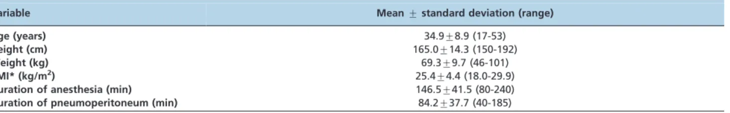
In total, 41 patients completed the study, of whom 29% were male and 71% were female. Additionally, 78% under-went cholecystectomy, and 22% underunder-went hiatoplasty. The biometric data and durations of anesthesia and pneumoper-itoneum are summarized in Table 1.
abdominal deflation. There was then an increase in heart rate and blood pressure fifteen minutes after the insufflation of carbon dioxide, and this increase was statistically significant. There was also a statistically significant increase in end-tidal CO2after the insufflation of carbon dioxide. In
contrast, vasopressin levels did not change at any time point, as shown in Figure 2.
Baseline renal function was normal in all subjects. There was a significant increase in Cys C levels 30 minutes after deflation of the abdomen (Figure 3) (p,0.001). As a result, the eGFR calculated by the Larsson formula was also lower during that time period (Figure 5) (p,0.02). Serum creatinine was lower 30 minutes after deflation of the abdomen, so the eGFR calculated using the CG formula showed a significant increase compared with baseline (Figures 4-5) (p,0.002). A comparison of these two methods of GFR calculation suggested that Cys C was more sensitive for the detection of mild to moderate changes in renal function in patients undergoing laparoscopic procedures.
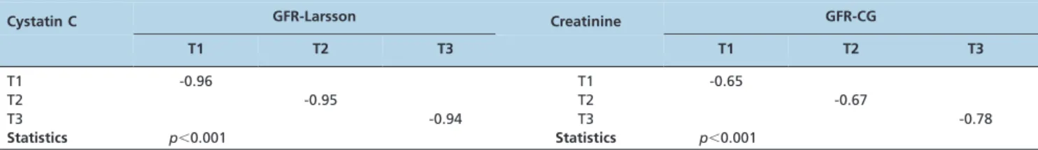
Pearson’s analysis showed a better correlation between Cys C and eGFR-Larsson than between Creatinine and eGFR-CG (Table 2). Additionally, there was no significant correlation between Cys C and serum creatinine values (p.0.05).
Our results indicate that serum Cys C and the eGFR derived from it perform better as an early marker of changes in renal function compared with serum creatinine and its
derived eGFR in patients with normal renal function who are undergoing laparoscopic surgery.
& DISCUSSION
Oliguria that is dependent on the intra-abdominal pressure is observed during laparoscopy following pneumoperitoneum Table 1 -Biometric data and the duration of anesthesia and pneumoperitoneum. The data are presented as the mean¡
standard deviation. The range of the variables is also included in the table.
Variable Mean¡standard deviation (range) Age (years) 34.9¡8.9 (17-53)
Height (cm) 165.0¡14.3 (150-192)
Weight (kg) 69.3¡9.7 (46-101)
BMI* (kg/m2) 25.4¡4.4 (18.0-29.9)
Duration of anesthesia (min) 146.5¡41.5 (80-240)
Duration of pneumoperitoneum (min) 84.2¡37.7 (40-185) *Body mass index.
Figure 1 - Heart rate and systolic blood pressure (mean ¡ standard deviation) at baseline and at the different time points in the study. (T0) before anesthesia induction; (T1) after induction and before tracheal intubation; (T2) 5 minutes after tracheal intubation; (T3) immediately after abdominal insuffla-tion; (T4-T6) 15, 30, and 45 minutes after abdominal insufflainsuffla-tion; (T7-T8) immediately after and 30 minutes after abdominal deflation. *p,0.05versusbaseline values.
Figure 2 -Vasopressin levels (mean ¡ standard deviation) at baseline (T1), 30 minutes after abdominal insufflation (T2), and 30 minutes after the end of the pneumoperitoneum (T3).
(1,23). Several mechanisms, including renal vein compression, renal parenchyma compression, and hormonal effects, are implicated in the development of oliguria (1-9). For example, Hartman et al. have shown that cardiac output decreases during laparoscopy, although normalizing cardiac output does not improve the GFR or RBF (8). Moreover, a recent study has shown that if an intra-abdominal pressure greater than 20 mm Hg is maintained for longer than 2 hours, it produces tubular damage and histologic changes in the subcapsular renal cortex (4).
Special attention must be paid to urine output and the GFR during the preoperative management of patients with decreased renal function who will undergo laparoscopic procedures. The early detection of renal dysfunction in these patients may allow better protection of the kidneys against further damage or further deterioration of function. The present prospective clinical trial was performed to determine the effectiveness of serum Cys C as a marker of renal dysfunction compared with serum creatinine. Subjects undergoing laparoscopic procedures were recruited for the study because these patients experience a transitory increase in intra-abdominal pressure due to the insufflation of CO2,
which can decrease renal function.
Several biomarkers and laboratory tests can be used to assess the GFR and tubular function (24,25). Although widely used, changes in serum creatinine are not specific; they do not indicate the nature of the insult and are relatively insensitive to mild changes in the GFR (10,11,26). The current study confirms these findings. The GFR estimated based on serum creatinine did not detect mild changes in renal function, whereas serum Cys C levels were sensitive and reflected renal changes. In particular, there was a 4% increase in serum Cys C levels and eGFR-Larsson was 4% lower than at baseline 30 minutes after pneumoper-itoneum creation (T2), with a pressure of 15 mm Hg. Thirty minutes after deflation of the abdomen (T3), serum Cys C was 9% higher, and the eGFR-Larsson was 10% lower, indicating that the kidneys required time to recover from the insult. In an experimental model, McDougall et al. demonstrated that when an intra-abdominal pressure of 15 mm Hg or more was sustained for more than 2 hours, there was a significant decrease in blood flow in the renal vein and a decrease in creatinine clearance (3). A clinical study of 40 patients undergoing colorectal surgery with laparoscopy confirmed the previous result and also showed compromised creatinine clearance 2 hours after pneumo-peritoneum deflation (27).
In our study, there were unexpected decreases in serum creatinine levels of 4% and 9% at T2 and T3, respectively. In contrast, the eGFR-CG showed increases of 6% and 17% at T2 and T3, respectively. The most likely explanation for the lower creatinine levels at this time is hemodilution secondary to aggressive volume replacement, overlapping with a decrease in renal function. Chang et al., reported similar results regarding the decrease in serum creatinine levels after laparoscopic surgery, despite the presence of oliguria (23). These authors also concluded that a high rate of intravenous hydration was the cause of this decrease in serum creatinine.
The ideal biomarker for detecting renal injury should have several qualities (28-29). It should be endogenous, nontoxic, freely filtered by the glomeruli, and excreted unchanged by the kidneys. It must also be readily available for use in clinical practice, and the measurement of such a biomarker should be easy, rapid, and inexpensive. In addition, the biomarker’s measurement should be an accurate and reliable surrogate for the GFR. Such a test should detect renal injury in a population with different renal pathologies. Moreover, the test should be sensitive
Figure 5 -The glomerular filtration rate calculated using Larsson’s formula (mean¡standard deviation) and the glomerular filtration rate calculated using the Cockcroft-Gault formula (mean ¡ standard deviation) at baseline (T1), 30 minutes after abdominal insufflation (T2), and 30 minutes after the end of the pneumoperitoneum (T3). *p,0.05versusbaseline values.
and specific for identifying small changes in renal function and detecting early injury.
A biomarker that can reflect real-time dynamic changes in the GFR is not available at present for clinical use. The serum creatinine concentration is influenced by extrarenal factors such as muscle mass, diet, gender and exercise (11). Creatinine takes time to accumulate before abnormal levels are detected, which can potentially delay the diagnosis of acute changes in the GFR. In fact, several studies have shown that serum Cys C levels are more sensitive for detecting early and mild changes in renal function com-pared with the sensitivity of serum creatinine levels (30-32). Additionally, the studies showed that Cys C levels are not affected by extrarenal factors. Recent studies, however, have shown that gender, C-reactive protein levels, height, weight, diabetes, the use of corticosteroids, abnormal thyroid function, and the presence of systemic inflammation can affect serum Cys C levels (33-35). We minimized the influence of these factors in the present study, as subjects with thyroid dysfunction, diabetes mellitus, or high body mass index were excluded. We also excluded patients who were taking steroids or anti-inflammatory drugs.
We are aware that our study clearly has certain limita-tions. First, no biomarker will ever be able to satisfy all of the criteria for an ideal marker. More importantly, no single biomarker will be able to accurately estimate the GFR and indicate the nature of a renal injury at the same time. However, this study shows that, although far from being ideal, Cys C may be more sensitive than creatinine in the detection of the early stages of renal dysfunction. Second, the equations used to estimate the GFR were developed for GFR estimation in healthy patients. Hence, the results cannot be extrapolated to patients with renal impairment. Further studies need to be performed to evaluate the clinical utility of Cys C and these formulae in estimating the GFR in patients with renal impairment who are undergoing laparoscopic procedures.
In conclusion, Cys C is a more sensitive biomarker for the detection of early impairment of the GFR during laparo-scopic surgery in patients with normal renal function. Further studies are necessary to determine whether Cys C is also an appropriate biomarker for the identification of renal dysfunction in patients with renal disease who are likely to undergo laparoscopic procedures.
Source of support: Brazilian Federal Agency for the Support and Evaluation of Graduate Education (CAPES).
& IMPLICATIONS STATEMENT
The present study demonstrated that cystatin C was more efficient than serum Cr in the detection of early changes in
the estimated GFR during laparoscopic surgery in patients with previously normal renal function.
& AUTHOR CONTRIBUTIONS
Lima RM was responsible for the study design, data collection, performance of the study, data analysis and manuscript preparation. Navarro LH, Nakamura G were responsible for study design, data collection, performance of the study, data analysis and manuscript preparation. Solanki DR was responsible for data analysis and manuscript preparation. Castiglia YM was responsible for data analysis and manu-script preparation. Vianna PT was responsible for the performance of the study. Ganem EM was responsible for study design, data analysis and manuscript preparation.
& REFERENCES
1. Dunn MD, McDougall EM. Renal physiology - Laparoscopic considera-tions. Urol Clin N Am. 2000;27(4):609-14.
2. Hamilton BD, Chow GK, Inman SR, Stowe NT, Winfield HN. Increased intra-abdominal pressure during pneumoperitoneum stimulates endothe-lin release in a canine model. J Endourol. 1998;12(2):193-8, http://dx.doi. org/10.1089/end.1998.12.193.
3. McDougall EM, Monk TG, Wolf Jr JS, Hicks M, Clayman RV, Gardner S, et al. The effect of prolonged pneumoperitoneum on renal function in an animal model. J Am Coll Surg. 1996;182(4):317-28.
4. Sassa N, Hattori R, YamamotoT, Kato M, Komatsu T, Matsukawa Y, et al. Direct visualization of renal hemodynamics affected by carbon dioxide -induced pneumoperitoneum. Urology. 2009;73(2):311-5, http://dx.doi. org/10.1016/j.urology.2008.09.047.
5. Demyttenaere S, Feldman LS, Fried GM. Effect of pneumoperitoneum on renal perfusion and function: a systematic review. Surg Endosc. 2007;21(2):152-60, http://dx.doi.org/10.1007/s00464-006-0250-x. 6. Abassi Z, Bishara B, Karram T, Khatib S, Winaver J, Hoffman A. Adverse
effects of pneumoperitoneum on renal function: involvement of the endothelin and nitric oxide systems. Am J Physiol Regul Integr Comp Physiol. 2008;294(3):R842-50, http://dx.doi.org/10.1152/ajpregu.00691. 2007.
7. Richards WO, Scovill W, Shin B, Reed W. Acute renal failure associated with increased intra-abdominal pressure. Ann Surg. 1983;197(2):183-7, http://dx.doi.org/10.1097/00000658-198302000-00010.
8. Harman PK, Kron IL, McLachlan HD, Freedlender AE, Nolan SP. Elevated intra-abdominal pressure and renal function. Ann Surg. 1982;196(5):594-7, http://dx.doi.org/10.1097/00000658-198211000-00015. 9. Dolgor B, Kitano S, Yoshida T, Bandoh T, Ninomiya K, Matsumoto T. Vasopressin Antagonist improves renal function in a rat model of pneumoperitoneum. J Surg Research. 1998;79(2):109-14, http://dx.doi. org/10.1006/jsre.1998.5409.
10. Mehta RL, Chertow GM. Acute renal failure definitions and classifica-tion: time for change?. J Am Soc Nephrol. 2003;14(8):2178-87, http://dx. doi.org/10.1097/01.ASN.0000079042.13465.1A.
11. Perrone RD, Madias NE, Levey AS. Serum creatinine as an index of renal function: new insights into old concepts. Clin Chem. 1992;38(10):1933-53. 12. Hojs R, Bevc S, Antolinc B, Gorenjak M, Puklavec L. Serum cystatin C as an endogenous marker of renal function in the elderly. Int J Clin Pharmacol Res. 2004;24(2-3): 49-54.
13. Coll E, Botey A, Alvarez L, Poch E, Quinto´ L, Saurina A, et al. Serum cystatin C as a new marker for non invasive estimation of glomerular filtration rate and as a marker for early renal impairment. Am J Kidney Dis. 2000;36(1):29-34, http://dx.doi.org/10.1053/ajkd.2000.8237. 14. Xia JH, Bing XG, An XT. Serum cystatin C assay for the detection of early
renal impairment in diabetic patient. J Clin Lab Anal. 2004;18(1):31-5, http://dx.doi.org/10.1002/jcla.20005.
Table 2 -Pearson’s correlation between Cys C levels (mg.L-1), creatinine values (mg.dL-1), and the GFR (mL.min-1) at three different studied time points (average¡standard deviation).
Cystatin C GFR-Larsson Creatinine GFR-CG
T1 T2 T3 T1 T2 T3
T1 -0.96 T1 -0.65
T2 -0.95 T2 -0.67
T3 -0.94 T3 -0.78
Statistics p,0.001 Statistics p,0.001
15. Oyabu C, Hayashi N, Sugiyama D, Umezu M, Mukai M, Kawano S, et al. Usefulness os serum cystatin C for the diagnosis of impaires renal function. Rinsho Byori. 2006;54(12):1204-8.
16. Grubb AO. Cystatin C - properties and use as diagnostic marker. Adv Clin Chem. 2002;35:63-99, http://dx.doi.org/10.1016/S0065-2423(01) 35015-1.
17. Randers E, Kristensen JH, Erlandsen E, Danielsen H. Serum cystatin C as a marker of the renal function. Scand J Clin Lab Invest. 1998;58(7):585-92. 18. Filler G, Bo¨kenkamp A, Hofmann W, Le Bricon T, Martinez-Bru C, Grubb A. Cystatin C as a marker of GRF - history, indications, and future research. Clin Biochem. 2005;38(1):1-8, http://dx.doi.org/10. 1016/j.clinbiochem.2004.09.025.
19. Finney H, Newman DJ, Gruber W, Merle P. Price CP. Initial evaluation of cystatin C measurement by particle-enhanced immunonephelometry on the Behring nephelometer system (BNA, BN II). Clin Chem. 1997;43 (6 Pt 1):1016-22.
20. Elias LL, Antunes-Rodrigues J, Elias PC, Moreira AC. Effect of plasma osmolality on pituitary-adrenal responses to corticotropin-releasing hormone and atrial natriuretic peptide changes in central diabetes insipidus. J Clin Endocrinol Metab. 1997;82(4):1243-7.
21. Larsson A, Malm J, Grubb A, Hansson LO. Calculation of glomerular filtration rate expressed in ml/min from plasma cystatin C values in mg/L. Scan J Clin Lab Invest. 2004;64(1):25-30, http://dx.doi.org/10. 1080/00365510410003723.
22. Cockcroft DW, Gault MH. Prediction of creatinine clearance from serum creatinine. Nephron. 1976;16:31-41, http://dx.doi.org/10.1159/000180580. 23. Chang DT, Kirsch AJ, Sawczuk IS. Oliguria during laparoscopic surgery. J Endourol. 1994;8(4):349-52, http://dx.doi.org/10.1089/end.1994.8.349. 24. Hoek FJ, Kemperman FAW, Krediet RT. A comparison between cystatin
C, plasma creatinine ande the Cockcroft and Gault formula for the estimation of glomerular filtration rate. Nephrol Dial Transplant. 2003;18(10):2024-31, http://dx.doi.org/10.1093/ndt/gfg349.
25. Tidman M, Sjostrom P, Jones I. A comparison of GFR estimating formulae based upon s-cystatin C and s-creatinine and a combination of the two. Nephrol Dial Transplant. 2008;23(1):154-60.
26. Christensson AG, Grugg AO, Nilsson JA˚ , Norrgren K, Sterner G, Sundkvist G. Serum cystatin C advantageous compared with serum creatinine in the detection of mild but not severe diabetic nephropathy. J Intern Med. 2004;256(6):510-8, http://dx.doi.org/10.1111/j.1365-2796. 2004.01414.x.
27. Pe´rez J, Taura´ P, Rueda J, Balust J, Anglada T, Beltran J, et al. Role of dopamine in renal dysfunction during laparoscopic surgery. Surg Endosc. 2002;16(9):1297-301.
28. Devarajan P. Emerging biomarkers of acute kidney injury. Contrib Nephrol. 2007;156:203-12, http://dx.doi.org/10.1159/000102085. 29. Bagshaw SM, Gibney N. Conventional markers of kidney function. Crit
Care Med. 2008;36(4 Suppl):S152-8, http://dx.doi.org/10.1097/CCM. 0b013e318168c613.
30. Coll E, Botey A, Alvarez L, Poch E, Quinto L, Saurina A, et al. Serum cystatin C as a new marker for noninvasive estimation of glomerular filtration rate and as a marker for early renal impairment. Am J Kidney Dis. 2000;36(1):29-34, http://dx.doi.org/10.1053/ajkd.2000.8237. 31. Hoek FJ, Kemperman FA., Krediet RT. A comparison between cystatin C,
plasma creatinine and the Cockcroft and Gault formula for the estimation of glomerular filtration rate. Nephrol Dial Transplant. 2003;18(10):2024-31, http://dx.doi.org/10.1093/ndt/gfg349.
32. Dharnidharka VR, Kwon C, Stevens G. Serum cystatin C is superior to serum creatinine as a marker of kidney function: a meta-analysis. Am J Kidney Dis. 2002;40(2):221-6, http://dx.doi.org/10.1053/ajkd.2002. 34487.
33. Knight EL, Verhave JC, Spiegelman D, Hillege HL, de Zeeuw D, Curhan GC, et al. Factors influencing serum cystatin C levels other than renal function and the impact on renal function measurement. Kidney Int. 2004;65(4):1416-21, http://dx.doi.org/10.1111/j.1523-1755.2004.00517.x. 34. Manetti L, Pardini E, Genovesi M, Campomori A, Grasso L, Morselli LL,
et al. Thyroid function differently affects serum cystatin C and creatinine concentrations. J Endocrinol Invest. 2005;28(4):346-9.