RBCCV 44205-1392 DOI: 10.5935/1678-9741.20120061
1 – Specialist in heart surgery at Brazilian Society of Cardiovascular Surgery (Assistant Physician at HC UNICAMP), Campinas, SP, Brazil.
2 – PhD in Surgery at State University of Campinas (UNICAMP), Campinas, SP, Brazil.
3 - PhD in Surgery at State University of Campinas (UNICAMP), Campinas, SP, Brazil.
4 - PhD in Surgery at State University of Campinas (UNICAMP), Campinas, SP, Brazil; Assistant Professor at Medical Sciences Medical School, State University of Campinas, SP, Brazil. 5 – Postdoctorate, Cincinnati University, Assistant Professor at Medical
Sciences Medica School, State University of Campinas, SP, Brazil. 6 – Full Professor at Medical Sciences Medica School, State University
of Campinas, SP, Brazil.
Carlos Fernando Ramos Lavagnoli
1,
Elaine Soraya Barbosa de Oliveira Severino
2,
Karlos Alexandre
de Souza Vilarinho
3, Lindemberg da Mota Silveira Filho
3,
Pedro Paulo Martins de Oliveira
4,
Orlando
Petrucci
5,
Reinaldo Wilson Vieira
6,
Domingo Marcolino Braile
7Fatores associados à sobrevida em pacientes submetidos a transplante cardíaco utilizando
microcardioplegia sanguínea retrógrada
Associated factors with survivals in patients
undergoing orthotopic heart transplant using
retrograde blood microcardioplegia
7 – Full Professor at São José do Rio Preto Medical School and Medical Sciences Medical School, State University of Campinas, SP, Brazil.
This study was carried out at State University of Campinas, SP, Brazil.
Correspondence address:
Carlos Fernando Ramos Lavagnoli. Caixa Postal 6111 – Campinas, SP, Brasil – Zip code 13083-970
E-mail: [email protected]
Article received on February 14th, 2012
Article accepted on July 17th, 2012
Abstract
Background: Several techniques and cardioplegic
solutions have been used for heart preservation during transplant procedures. Unfortunately, there is a lack of ideal method for myocardial preservation in the clinical practice. The use of retrograde cardioplegia provides continuous infusion of cardioplegic solution during the graft implantation. This strategy may provide better initial recovery of the graft. The objective of this study is to describe the experience of a single center where all patients received the same solution for organ preservation and were subjected to continuous retrograde blood microcardioplegia during implantation of the graft and to evaluate factors associated to early and late mortality with this technique.
Methods: This is a retrospective, observational and
descriptive study of a single center.
Results: During the study period were performed 35 heart
transplants. Fifteen (42.9%) patients were in cardiogenic
shock. The probability of survival was 74.8±7.8%, 60.4±11.3% and 15.1±13.4% at 1 year, 5 years and 10 years of follow-up, respectively. The median survival time was 96.6 months.
Conclusion: The use of myocardial protection with
retrograde cardioplegic solution may reduce the risks associated morbidity due to cold ischemia time during the heart transplant, and we suggest that this benefit may be even greater in cases of cold ischemia time longer ensuring protection to the myocardium.
Descriptors: Heart transplantation. Transplantation.
Heart arrest, induced. Myocardium. Follow-up studies.
Resumo
Introdução: Uma grande variedade de técnicas e soluções
INTRODUCTION
A variety of techniques and solutions are used in the preservation of the heart during transplantation, demonstrating the lack of ideal method in clinical practice [1].
In the present day, where the number of donors is not enough, in addition to the increase of high-risk recipients on the waiting list, every effort should be made towards better organ preservation. A better organ preservation leads to lower graft dysfunction and promotes better late outcomes after transplantation [2,3].
The use of retrograde blood cardioplegia has been described in several studies in the literature. There is no consensus whether this strategy promotes adequate myocardial protection [4]. Its use for the administration of blood cardioplegia during implantation is frequently reported in the literature. This strategy of cardioplegia administration allows for continuous perfusion of blood and added substrates, which can provide better initial recovery of the transplanted heart. [5]
In research performed by Wheeldon et al. [6] in 1992, the use of retrograde blood cardioplegia was performed in less than 6% of the centers. The continuous retrograde blood microcardioplegia was introduced into our country by Braile et al, proving to be safe and providing adequate myocardial protection in conventional cardiac surgery [7]. The aim of this study is to describe the experience of a single center where all patients received the same organ preservation solution and underwent retrograde continuous blood microcardioplegia during implantation of the graft and assessing factors of early and late mortality with the use of this technique.
METHODS
Ethical characteristics, patient selection and demographics
This is a prospective observational and descriptive study of a single center, with approval of the local Research Ethics Committee.
The study included patients undergoing orthotopic heart transplantation from 1998 to 2011. Patients undergoing cardiac transplantation were selected by a multidisciplinary team and the criteria followed were the same as described in the II Brazilian Consensus on Cardiac Transplantation of the Brazilian Society of Cardiology [8].
Graft explant technique
After performing a median sternotomy, the pleura and pericardium were opened and the heart exposed for macroscopic assessment. When decided by capturing the organ, the vessels and veins of the heart were dissected to permit the explant securely, as already described in literature [8]. After aortic clamping, cardioplegic solution was injected into the aortic root. In all cases, the solution at 4°C was used as described below (Braile Biomédica, São José do Rio Preto, Brazil):
• in 1000 ml of sodium chloride 0.9%;
• 50 mL of solution with the following composition: o75 mEq Potassium chloride;
o 40 mEq magnesium chloride; o 30 mM monosodium glutamate; o 30 mM monosodium aspartate;
During infusion of cardioplegia, the inferior vena cava and right superior pulmonary vein were sectioned. At the end of cardioplegia infusion, the cardiectomy was completed as described in literature [8].
Abbreviations, acronyms and symbols
AV C Stroke
ARF Renal failure requiring dialysis ACT Activated clotting time
administração da cardioplegia de forma retrógrada propicia perfusão contínua, o que pode conferir melhor recuperação inicial do coração transplantado. O objetivo deste trabalho é descrever a experiência de um único centro onde todos os pacientes receberam a mesma solução de conservação de órgão e foram submetidos a microcardioplegia sanguínea retrógrada contínua durante o implante do enxerto e avaliar fatores de mortalidade precoce e tardia com a utilização desta técnica.
Métodos: Este é um estudo retrospectivo, observacional e
descritivo, realizado em um único centro.
Resultados: No período do estudo, foram realizados 35 transplantes cardíacos, sendo que 15 (42,9%) pacientes encontravam-se em choque cardiogênico. A probabilidade de sobrevida foi 74,8±7,8%, 60,4±11,3% e 15,1±13,4% ao final de 1 ano, 5 anos e 10 anos de seguimento, respectivamente. O tempo médio de sobrevida foi de 96,6 meses.
Conclusão: A utilização da solução cardioplégica para
proteção de órgãos e a estratégia de iniciar a perfusão com microcardioplegia sanguínea retrógrada contínua forneceu proteção adequada.
Descritores: Transplante. Transplante de coração.
The graft was placed in a 0.9% sodium chloride solution, with ice for shipping.
Surgical technique of implantation and intraoperative monitoring
All patients underwent the same bicaval implantation technique, as described by Lower & Shumway modified Yacoub & Banner [9-11].
Prior to the beginning of the implant, we assessed the presence of patent foramen ovale and a purse-string suture in the coronary sinus ostium was performed to allow the infusion of retrograde blood cardioplegia. In summary form, the implant was initiated by anastomosis of the left atrium, using 3.0 polypropylene suture. Further, anastomoses were performed in the inferior vena cava and superior vena cava using 4.0 polypropylene suture. The pulmonary artery was then anastomosed in an end-to-end manner using 4.0 polypropylene suture. Finally, aortic anastomosis was performed using 4-0 polypropylene suture and a cannula was inserted into the aorta for infusion of antegrade blood cardioplegia in order to withdraw the cannula inserted into the coronary sinus ostium [12,13]. The intraoperative monitoring was performed using a catheter in the radial artery for continuously monitoring of blood pressure, pulmonary artery pressure, cardiac output and central venous oxygen saturation using the Swan Ganz catheter (Edwards Lifesciences, Irvine, USA).
Cardiopulmonary bypass technique
The cannulation was performed in the ascending aorta and vena cava were cannulated individually. The flow cardiopulmonary bypass was 2.4 L.min-1.m2 with temperature maintained between 32oC and 34oC. The
hematocrit during CPB was maintained between 25% and 28%. The membrane oxygenator was used in all cases (Braile Biomédica, São José do Rio Preto, Brazil). Systemic heparinization was performed at a dose of 300 to 400 IU/kg heparin and activated clotting time (ACT) was maintained above 480 seconds. After unclamping, we routinely used dopamine, isoproterenol and sodium nitroprusside, to maintain heart rate above 100 beats per minute and the best possible cardiac output. All patients received a temporary pacemaker wires in the right atrium and ventricle.
Retrograde blood microcardioplegia technique The retrograde blood microcardioplegia system was described by Braile [7]. The system allows blood infusion continuously with cardioplegic solution in a concentration of 1%. Blood flow to the heart during implantation was maintained between 100 and 200 ml/min, not being interrupted for periods longer than 5 minutes. The interruption was made according to the judgment of the surgeon, according to the need for better visualization of the operative field. At the start of the infusion of
cardioplegia-inducing, solution was used for 3 minutes and then the maintenance solution was used throughout the procedure.
Perioperative and postoperative management
Postoperatively, the same monitoring was used intraoperatively with blood pressure monitoring, continuous monitoring of cardiac output and central venous oxygen saturation. The administration of vasoactive drugs, dopamine, isoproterenol and sodium nitroprusside aimed heart rate above 100 beats per minute and the best possible cardiac output. Dobutamine was used in situations of inotropic support to optimize the right ventricular function when necessary. The epicardial pacemaker was used when necessary and during hospitalization in the intensive care unit. The withdrawal of ventilatory support was performed whenever possible. The removal of drains occurred generally within 48 hours and catheters were kept until inotropic support was no longer required. Transthoracic echocardiography was performed every 72 hours in the immediate postoperative period and routine laboratory tests in the first days after transplantation.
Late and hospital mortality
All patients were followed after transplantation. Hospital mortality was defined as one that occurred within 30 days after surgery or that on which the patient was not discharged up to death after surgical implantation. Late mortality was defined as one that occurred 30 days after surgery or after discharge.
Early and late complications
Complications were defined as early and late. The early complication occurred 30 days after surgery or until discharge of the patient, when this was over 30 days. The late complication was defined as that occurred 30 days after surgery or after discharge, when this was over 30 days. Among the types of early complications, we observed: cerebrovascular accident (CVA), infection of the surgical incision, renal failure that required dialysis (ARF), duration of mechanical ventilation longer than 24 hours, low cardiac output syndrome and endocarditis. Late complications were recorded: CVA, IRA, graft failure, endocarditis, infections with severe sepsis, myocardial infarction, and neoplastic diseases.
Immunosuppression protocol
of 5 mg/kg per day, aiming the level of serum cyclosporine between 350-450 ng/mL. Prednisone was started on the fourth day at a dose of 1 mg/kg/day, and progressive reduction of 2.5 mg per day to achieve a dose of 0.2 mg/kg/ day which was then kept for 6 months after procedure.
Biopsies protocol
As recommended in the II Brazilian Consensus on Cardiac Transplantation [8], biopsies were performed weekly for the first month and monthly until one year.
Statistical Analysis
Continuous variables were described as mean and standard deviation. The discrete variables are described as frequency.
For univariate analysis of factors associated with mortality in continuous variables we used the Student’ t test or Mann-Whitney test, when appropriate, and the chi-square test for discrete variables. The probability of survival was assessed by Kaplan-Meier analysis. The P value <0.05 was considered significant. SPSS version 19.0 for Macintosh was used (IBM, New York, United States).
RESULTS
During the study period, 35 heart transplants were performed, with 60% of these performed in the last 18 months. All patients were in New York Heart Association functional class III or IV. Of the total, 15 (42.9%) patients
were in cardiogenic shock in use of vasoactive drugs and/ or mechanical circulatory support.
The general data of this series are shown in Table 1. Patients who died early had shorter time in waiting list for transplantation (P <0.01) (Table 2).
Early complications were observed in seven patients: stroke, infection of the surgical incision, IRA, duration of mechanical ventilation longer than 24 hours, low cardiac output syndrome and endocarditis. Late complications were found in 10 patients: stroke, IRA, graft failure, endocarditis, infections with severe sepsis, myocardial infarction, and neoplastic diseases.
Patients who had more complications after transplantation were more likely to death (P <0.01) and this was also observed in patients who experienced complications during late follow-up (P <0.01) (Table 3).
The functional class and echocardiographic variables of the graft are shown in Table 4. Only functional class was associated with higher odds of death in the early and late after transplantation (P <0.01).
The median time free from the first rejection after transplant was 17 days, range 7-390 days.
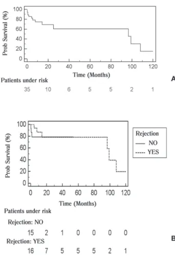
The probability of survival was 74.8 ± 7.8%, 60.4% ± 11.3 and 15.1 ± 13.4% at the end of 1 year, 5 years and 10 years of follow up, respectively (Figure 1A ). The median survival time was 96.6 months. The occurrence of rejection did not alter the probability of survival in these patients (HR 0.63 with 95% confidence interval: 0.14 to 2.87; P=0.44) (Figure 1B).
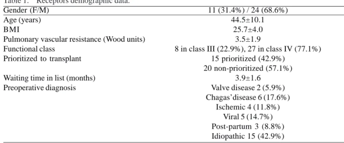
Table 1. Receptors demographic data.
Gender (F/M) Age (years) BMI
Pulmonary vascular resistance (Wood units) Functional class
Prioritized to transplant
Waiting time in list (months) Preoperative diagnosis
11 (31.4%) / 24 (68.6%) 44.5±10.1
25.7±4.0 3.5±1.9
8 in class III (22.9%), 27 in class IV (77.1%) 15 prioritized (42.9%)
20 non-prioritized (57.1%) 3.9±1.6
Valve disease 2 (5.9%) Chagas’disease 6 (17.6%)
DISCUSSION
The aim of this study was to describe the experience of a single center where all patients underwent the same type of protective solution during cold ischemia and cardiectomy. We also assessed the factors of early and late mortality with the use of these techniques.
We demonstrated that 42.9% of our patients were in cardiogenic shock and 76.5% of them in functional class IV. We had no patients with low cardiac output syndrome in the immediate postoperative period and our early mortality was 11%. We observed that patients who died in the immediate period and during follow-up were less time on the waiting list for transplantation. Patients who had more complications during the immediate postoperative period and during follow-up also were at greater risk of dying. Our survival after 5 years was 60%.
Table 2. Univariate analysis for early and late survival.
Variable
CPB time (minutes)
Aortic clamping time (minutes) Cold ischemia time (minutes) Total ischemic time (minutes) Surgery time (minutes) Operative room time (minutes) Distance from capture (Km) Donor age (years)
Donor serum sodium (mEq/L) Receptor/donor BMI relationship Intraoperative bleeding (mL) PVR (Wood)
Waiting time in list (months) Prioritized to transplant
Early death (n=4) 99.5±23.7 70.5±11.1 52.5±55.1 123.0±63.6 303.7±52.2 365.0±38.1 51.0±60.1 32.2±14.7 153.3±21.4 1.1±0.13 2450.0±2431.0 4.0±2.1 0.5±0.3 3 Early survival (n=31) 97.9±18.0 66.2±11.0 64.2±33.0 130.4±36.2 267.3±51.6 342.5±52.1 73.9±61.1 29.3±9.0 155.5±16.8 1.0±0.2 643.2±391.1 3.5±2.0 4.3±5.3 12 P 0.87 0.47 0.54 0.73 0.19 0.41 0.48 0.57 0.84 0.52 0.23 0.57 <0.01 0.14 Late death (n=10) 97.6±22.1 60.1±13.4 38.9±32.2 99.8±37.3 280.5±48.5 346.7±50.0 34.8±46.0 26.1±8.5 164.7±19.8 1.0±0.0 785.7±452.5 3.2±2.8 2.6±2.2 4 Late survival (n=21) 98.1±16.6 68.5±9.3 75.0±27.4 143.5±27.3 261.7±53.0 340.7±54.1 92.6±59.4 30.8±9.0 154.2±16.4 1.0±0.2 576.7±356 3.6±1.8 5.2±6.2 8 P 0.93 0.08 0.01 0.01 0.36 0.78 0.01 0.17 0.32 0.84 0.25 0.67 0.09 0.76
CPB = cardiopulmonar by-pass, BMI: Body Mass Index, PVR = pulmonary vascular resistance
Variable
ICU time (days)
Time of hospital stay (days) Early complications Late complications early death (n=4) 13.7±114.7 17.2±20.0 4 __ early survival (n=31) 7.2±6.6 25.0±21.8 3 __ P 0.44 0.50 <0.01 __ late death (n=9) 6.9±2.5 20.7±9.2 1 8 Late survival (n=21) 7.4±7.7 26.9±25.4 2 2 P 0.79 0.33 0.59 <0.01
ICU: intensive care unit
Table 3. Univariate analysis with hospitalization time and complications.
Variable EDLVD (mm)** ESLVD (mm)** EF (%)** MI* FC I FC II FC III FC IV Late death (n=9) 49.0±5.9 32.9±6.7 61.4±12.0 1 4 1 1 2 Late survival (n=21) 44.9±4.6 26.6±3.8 71.4±6.5 1 21 0 0 0 P 0.06 0.03 0.05 0.79 <0.01
EDVD: End diastolic left ventricular diameter . ESLVD: End systolic left ventricular diameter. EF: ejection fraction. FC: Functional Class of heart failure of New York Heart Association. MI: mitral insufficiency. (*): It was considered mitral failure that classified as moderate or severe. (**) Echocardiographic data regarding the graft. Average time of echocardiography after transplantation: 16.1 ± 31.25 months
The importance of our findings is to demonstrate the safety of the myocardial protection technique during explant and type of cardioplegia during implantation of the graft in this series and clinical outcomes in short- and long-term follow-up. Our results are generally comparable to the literature [14,15].
A variety of techniques for the preservation of heart grafts used in clinical practice reflects the lack of an ideal method, new compounds are being added to the solutions, providing better preservation and increasing cold ischemia time [16].
Several solutions have been described for organ transplants [17-19]. The solution used by us was described by Martins et al. [20]. It is routinely used in our department as cardioplegic solution in conventional cardiac surgery, and we are not aware of its use as a solution for preserving organs for transplant. Similarly, the cardioplegic solution enriched with histidine and tryptophan that was initially used in conventional cardiac surgery is now widely used as a protective solution for transplantation [19].
Fig. 1 - A: General Survival of patients undergoing transplantation after 10 years of follow-up. B: Survival of patients divided into patients without any rejection episode and patients who presented at least one episode of rejection
Although several authors described the administration of retrograde cardioplegia promoting a non-uniform distribution of the solution to the myocardium, the clinical outcomes were satisfactory in this series using this technique [21,22].
Although the data shown here, the observation should be made with caution when it states that retrograde blood reperfusion of the heart during implantation confers reduced deleterious effects of ischemic injury, given the small number of observed population, low postoperative mortality and the low number of graft dysfunction after transplantation.
The use of myocardial protection with retrograde cardioplegic solution provided adequate protection to the myocardium and, therefore, we suggest that this benefit may also be extended to cases of longest cold ischemia [23]. Furthermore, the use of this method may make possible the organ procurement further afield and can thus increase the number of transplants that currently are declined by the distance from the center to capture [24].
The cold ischemia time was not a predictor of mortality, demonstrating that the solution used provided adequate protection and not compromising the operative or late outcomes. Obviously histological cuts or contractility variables were not assessed compared with other solutions, but this is beyond the scope of this study.
The cardiopulmonary bypass time was not a factor associated with early or late mortality. The same happened to aortic clamping. We should note that the duration of cardiopulmonary bypass is very similar to that described in the literature [14,15]. The clamping time was relatively short (average 66 minutes), which may have contributed to the outcome of this series.
There were differences in times of cold ischemia and total ischemia (P=0.01 and P=0.01, respectively), demonstrating that they are greater in groups with greater survival during the late follow-up. This finding suggests that patients with greater long-term survival showed ischemia time greater than the overall group at higher risk of late death, contrary to what one might anticipate. Our explanation for this fact is that the more severe receivers, prioritized or with pulmonary hypertension were assigned to receive a graft in the best conditions of proximity to the hospital with shortest ischemic time possible. So, again, we believe that the strategy of myocardial protection did not compromise our results, but the strategy of selecting donor/recipient.
REFERENCES
1. Carrier M, Leung TK, Solymoss BC, Cartier R, Leclerc Y, Pelletier LC. Clinical trial of retrograde warm blood reperfusion versus standard cold topical irrigation of transplanted hearts. Ann Thorac Surg. 1996;61(5):1310-4.
2. Zeng Z, Jiang Z, Wang CS, Luo H, Huang YF, Jin XH. Preoperative evaluation improves the outcome in heart
conditions. We note that the three early deaths were of patients who were in the prioritization list, under use of vasoactive drugs and/or intra-aortic balloon.
A similar trend was observed with respect to time on the waiting list for transplantation and the probability of late death. The late deaths were more frequent in the group that was less time on the waiting list. We believe that the explanation is similar to death during early because when patients were more severe the selection of the donor included marginal grafts.
The early complications observed were related to rejection or infection and, less frequently, to graft dysfunction. The hospital mortality was low (11%) and comparable to data from the national literature [14,15]. Late complications were related to infection not being detected cases of organ failure or allograft vasculopathy. These findings were also similar to those observed by Jung et al. [25]. There were no patients with neoplastic disease in the late phase after transplantation.
Our overall late survival was 74% and 60% after 1 year and 5 years, respectively, rates similar to those described in the national literature [14,15].
We found no other demographic or clinical variables associated with early or late mortality in our study.
Limitations and strengths of the study
The limitations observed by us are referring to a retrospective study and the sample size, but it should be noted that 60% of the transplants were performed in the last 18 months. Therefore, follow-up time is still too small. As a strength of this study, we can mention the use of the same methodology in myocardial protection for donor cardioctomy and during graft implantation in all patients.
In conclusion, we can say that the use of cardioplegic solution used as a solution for organ protection and the strategy starting perfusion with retrograde blood microcardioplegia provided adequate protection, because we observed no increased mortality with longer ischemic time. Further studies comparing different types of organ preservation solutions are still needed.
transplant recipients with pulmonary hypertension--retrospective analysis of 106 cases. Transplant Proc. 2010;42(9):3708-10.
3. Rossi D, Pinna GD, La Rovere MT, Traversi E. Prognostic significance of tissue-Doppler imaging in chronic heart failure patients on transplant waiting list: a comparative study with right heart catheterization. Eur J Echocardiogr. 2011;12(2):112-9.
4. Lichtenstein SV, Abel JG, Panos A, Slutsky AS, Salerno TA. Warm heart surgery: experience with long cross-clamp times. Ann Thorac Surg. 1991;52(4):1009-13.
5. Juffe Stein A. New frontiers in myocardial preservation. Rev Esp Cardiol. 1995;48(Suppl 7):24-8.
6. Wheeldon D, Sharples L, Wallwork J, English T. Donor heart preservation survey. J Heart Lung Transplant. 1992;11(5):986-93.
7. Braile D. Como eu faço: cardioplegia sanguínea isotérmica retrógrada de baixo volume. Rev Bras Cir Cardiovasc. 1992;7(3):221-9.
8. Bacal F, Souza Neto JD, Fiorelli AI, Mejia J, Marcondes-Braga FG, Mangini S, et al. II Diretriz Brasileira de Transplante Cardíaco. Arq Bras Cardiol.2009;94(1 supl.1):e16-e73.
9. Yacoub MP, Mankad P, Ledingham S. Donor procurement and surgical techniques for cardiac transplantation. Semin Thorac Cardiovasc Surg. 1990;2(2):153-61.
10. Sievers HH, Weyand M, Kraatz EG, Bernhard A. An alternative technique for orthotopic cardiac transplantation, with preservation of the normal anatomy of the right atrium. Thorac Cardiovasc Surg. 1991;39(2):70-2.
11. Dreyfus G, Jebara V, Mihaileanu S, Carpentier AF. Total orthotopic heart transplantation: an alternative to the standard technique. Ann Thorac Surg. 1991;52(5):1181-4.
12. Aziz T, Burgess M, Khafagy R, Wynn Hann A, Campbell C, Rahman A, et al. Bicaval and standard techniques in orthotopic heart transplantation: medium-term experience in cardiac performance and survival. J Thorac Cardiovasc Surg. 1999;118(1):115-22.
13. Trento A, Czer LS, Blanche C. Surgical techniques for cardiac transplantation. Semin Thorac Cardiovasc Surg. 1996;8(2):126-32.
14. Branco JNR, Teles CA, Aguiar LF, Vargas GF, Hossne Junior MA, Andrade JCS, et al. Transplante cardíaco ortotópico: experiência na Universidade Federal de São Paulo. Rev Bras Cir Cardiovasc. 1998;13(4):285-94.
Instituto Dante Pazzanese de Cardiologia: análise da sobrevida. Rev Bras Cir Cardiovasc. 2001;16(4):289-304.
16. Loganathan S, Radovits T, Hirschberg K, Korkmaz S, Koch A, Karck M, et al. Effects of Custodiol-N, a novel organ preservation solution, on ischemia/reperfusion injury. J Thorac Cardiovasc Surg. 2010;139(4):1048-56.
17. Corps CL, Attia MS, Potts D, Lodge JP. PBSH: a new improved cardiac preservation solution in comparison with three clinically proven solutions. Transplant Proc. 2010;42(5):1587-90.
18. Lee S, Huang CS, Kawamura T, Shigemura N, Stolz DB, Billiar TR, et al. Superior myocardial preservation with HTK solution over Celsior in rat hearts with prolonged cold ischemia. Surgery. 2010;148(2):463-73.
19. Wu K, Türk TR, Rauen U, Su S, Feldkamp T, de Groot H, et al. Prolonged cold storage using a new histidine-tryptophan-ketoglutarate-based preservation solution in isogeneic cardiac mouse grafts. Eur Heart J. 2011;32(4):509-16.
20. Martins AS, Silva MA, Padovani CR, Matsubara BB, Braile DM, Catâneo AJ. Myocardial protection by continuous, blood,
antegrade-retrograde cardioplegia in rabbits. Acta Cir Bras. 2007;22(1):43-6.
21. Carrier M, Grégoire J, Khalil A, Thai P, Latour JG, Pelletier LC. Myocardial distribution of retrograde cardioplegic solution assessed by myocardial thallium 201 uptake. J Thorac Cardiovasc Surg, 1994;108(6):1115-8.
22. Ikonomidis JS, Yau TM, Weisel RD, Hayashida N, Fu X, Komeda M, et al. Optimal flow rates for retrograde warm cardioplegia. J Thorac Cardiovasc Surg. 1994;107(2):510-9.
23. Fiocchi R, Vernocchi A, Mammana C, Iamele L, Gamba A. Continuous retrograde warm blood reperfusion reduces cardiac troponin I release after heart transplantation: a prospective randomized study. Transpl Int. 2000;13 Suppl 1:S240-4.
24. Suzuki S, Sasaki H, Matsuo T, Tomita E, Sada M, Mizuochi I, et al. Experimental heart transplantation in dogs: preservation of isolated hearts for 36 hours by retrograde coronary sinus microperfusion. Nippon Geka Gakkai Zasshi. 1984;85(6):541-7.