The aim of this study was to compare the biological activity of lipopolysaccharides (LPS) purified from Fusobacterium nucleatum and Porphyromonas gingivalis strains, both isolated from primary endodontic infection (PEI) in the levels of IL-1β and TNF-α released by macrophage cells. Moreover, LPS was purified from F. nucleatum andP. gingivalis American Type Collection (ATCC) and its biological activity was compared to respectively clinical isolates strains. F. nucleatum and P. gingivalis strains clinically isolated from PEI had their identification confirmed by sequencing the 16S rRNA gene. LPS from F. nucleatum
and P. gingivalis and their respective ATCC strains were extracted by using Tri-reagent method. Macrophages (Raw 264.7) were stimulated with LPS at 100 ng/mL for 4, 8 and 12 h. Secretion of IL-1 and TNF-α was also determined. Paired t-test, repeated measures ANOVA and one-way ANOVA were employed.All LPS induced significant production of IL-1β and TNF-α, with the former being secreted at higher levels than the latter in all time-points. F. nucleatum induced a higher expression of both cytokines compared to P. gingivalis (p<0.05). No differences were observed between clinical and ATCC strains, as both presented the same potential to induce pro-inflammatory response.It was concluded that F. nucleatum and P. gingivalis LPS presented different patterns of activation against macrophages as seen by the IL-1β and TNF-α production, which may contribute to the immunopathogenesis of apical periodontitis. Moreover, clinical and ATCC strains grown under the same in vitro environment conditions presented similar biological activity.
C o m p a r i s o n o f
F u s o b a c t e r i u m
n u c l e a t u m
a n d
Po r p h y r o m o n a s
gingivalis
L i p o p o l y s a c c h a r i d e s
Clinically Isolated from Root Canal
Infection in the Induction of
Pro-Inflammatory Cytokines Secretion
Frederico C. Martinho1,2, Fábio R. M. Leite3, Letícia M.M. Nóbrega2, Marcos S.
Endo2, Gustavo G. Nascimento3, Richard P. Darveau4, Brenda P. F. A. Gomes2
1Department of Restorative Dentistry,
Endodontics Division, São José dos Campos Dental School, UNESP - Universidade Estadual de São Paulo, São José dos Campos, SP, Brazil 2Department of Restorative
Dentistry, Endodontics Division, Piracicaba Dental School, UNESP - Universidade Estadual Campinas, Piracicaba, SP, Brazil 3Postgraduate Program in Dentistry,
Dental School, UFPel - Universidade Federal de Pelotas, RS, Brazil
4Department of Periodontics and Oral
Biology, University of Washington, Seattle, WA, United States
Correspondence: Profa. Dra. Brenda P.F.A. Gomes, Av. Limeira 901, 13414-903 Piracicaba, SP, Brasil. Tel: +55-19-2106-5343. e-mail: [email protected]
Key Words:Fusobacterium nucleatum, Porphyromonas gingivalis, bacteria, endodontics, endotoxin, LPS, macrophages, cytokines
Introduction
Apical periodontitis is an inflammatory disorder of the periradicular tissue caused by bacterial infection of endodontic origin and characterized by periapical bone resorption (1). Primary endodontic infection is a polymicrobial infection caused predominantly by Gram-negative anaerobic bacteria (2, 3), which activate different intracellular signaling pathways culminating in soft tissue breakdown and, later, in bone resorption (4).
Fusobacterium nucleatum and Porphyromonas gingivalis, both Gram-negative anaerobic rod-shaped bacteria, are highly detected in root canal infection, playing a critical role in the endodontic disease (5, 6). Both species possess a large number of putative virulence determinants (7-9). However, accumulated evidence indicates that the pathology of endodontic infection is remarkably due to the actions of host-derived cytokines induced by lipopolysaccharide molecules (4, 10), also known as LPS, being the major components of the outer leaflet of the
outer membrane of Gram-negative bacteria (11). LPS has been shown to interact with toll-like receptors (TLRs) through its greater affinity to TLR4 (12), leading rapidly to the activation of different pathways responsible for the production of pro-inflammatory cytokines, such as interleukin (IL)-1β and tumor necrosis factor (TNF)-α, the latter controlling the tissue remodeling in pathological conditions (4).
LPS structure shows considerable heterogeneity among different bacterial species (13), thus activating host cells in different ways. Previous studies showed that bacterial LPS structure could be modulated by environmental conditions, such as hemin concentration, temperature, pH, among others (14-16).
Pro-inflammatory cytokine induction by LPS from F
. nucleatum and P
. gingivalis
to induce the release of pro-inflammatory cytokines. Macrophages are the primary defense line responsible for initiation and maintenance of the inflammatory process (18), which amplifies the immune response, recruits immune cells, activates immune and non-immune cells, and may cause significant tissue damage by inducing collagenase production in fibroblasts and activation of osteoclasts (4,19,20).
In order to better understand the inflammatory process involved in apical periodontitis, reflected by the response of macrophages to LPS, the aim of this study was to compare the biological activity of lipopolysaccharides (LPS) purified from F. nucleatum and P. gingivalis strains, both isolated from primary endodontic infection (PEI) in the levels of IL-1β and TNF-α released by macrophage cells. Moreover, LPS was purified from F. nucleatum and P. gingivalis American Type Collection (ATCC) and its biological activity was compared to respectively clinical isolates strains.
Material and Methods
Bacterial Strains and Growth Conditions
F. nucleatum and P. gingivalis strains were clinically isolated from primary endodontic infection in patients with apical periodontitis who attended the Piracicaba Dental School, Brazil, for endodontic treatment. The Human Research Ethics Committee of the Piracicaba Dental School approved the protocol describing sample collection for this investigation, and all voluntary patients signed an informed consent form. The clinical strains were grown at 37 °C in anaerobic conditions (80% N2, 10% H2, 10% CO2), examined
for purity and primarily identified by using the Rapid ID 32A test (BioMérieux SA, Marcy l’Etoile, France) for phenotypic identification, which was further confirmed by sequence analysis before LPS extraction. F. nucleatum (ATCC 25586) and P. gingivalis (ATCC 33277) strains were purchased from the American Type Culture Collection (Manassas, VA, USA) and grown under required conditions in enriched trypticase soy broth (TSB) supplemented with yeast extract, hemin and vitamin K (menadione) (TYHK). The TYHK medium consisted of trypticase soy broth (30 g/L), yeast extract (5 g/L), hemin (0.005 g/L), and vitamin K3 (menadione) (0.001 g/L) at pH 7.2, with the material being autoclaved. Bacterial growth was monitored by following the optical density at 600 nm, with cells being harvested in the stationary phase of growth and the final bacterial yield being determined by wet weight after centrifugation and washing.
DNA Sequencing
Before LPS extraction, the isolates as well as the ATCC bacterial strains were sequenced and analyzed. The DNA of all bacteria tested was extracted by using the QIAamp DNA Minikit (Qiagen, Valencia, CA, USA), according
to the manufacturer’s instructions. After extraction, 5 µL of template DNA were used in 50 µL amplification reactions consisting of 1 µL Platinum Taq polymerase (Invitrogen, São Paulo, SP, Brazil), 5 µL buffer (10X), 3.0 mmol/L MgCl2, 4.0 µL dNTP solutions (25 mmol/L each),
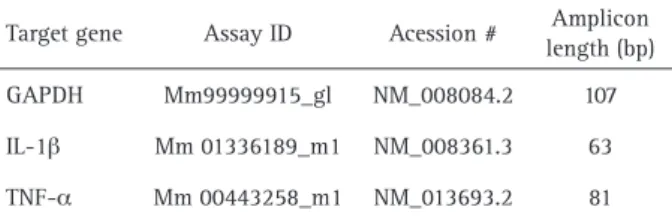
and 1.0 µL of 16S rRNA bacterial primer (0.5 mmol/L) (UnivF [5’- GAGAGTTTGATYMTGGCTCAG-3’] and UnivR [5’- GAAGGAGGTGWTCCARCCGCA-3’]). Genomic DNA of P. gingivalis (ATCC 33277) (Table 1) and water were used as positive and negative controls, respectively. Polymerase chain reaction (PCR) was performed in a DNA thermocycler (MJ Research, Waltham, MA, USA) adjusted to initial denaturation step at 94 °C for 4 min followed by 30 cycles at 94 °C for 45 s, 60 °C for 45 s, 72 °C for 90 s and a final step at 72 °C for 15 min. Two independent PCR reactions were performed for each sample. The 1500-bp fragments were revealed after electrophoresis in 1% agarose gel and purified by using QIAquick Gel Extraction (Qiagen, North Rhine-Westphalia, Germany). The purified PCR products were sequenced by using an ABI 3730 DNA Analyzer (Applied Biosystems), with Big-Dye Terminator Cycle Sequencing Kit using primer 533R (5’- TKACCGCGGCTGCTG -3’). The phylogenetic position was obtained by comparing it to sequences obtained from the GenBank database by using the Basic Local Alignment Search Tool (BLAST) algorithm at a 98% similarity level. The DNA sequences from ATCC strains were aligned respectively to the clinically isolated strains by using the WineHQ – BioEdit for Windows 7.0.9 (Carlsbad, CA, USA).
LPS Extraction and Purification
EachLPS was prepared according to the Tri-Reagent procedure, as previously described (21). Following the final ethanol precipitation, LPS was lyophilized to determine the yield and then re-suspended in distilled water to 1 mg/ mL. The LPS was further purified by the following steps: 1 mL of lyophilized LPS was suspended in 1 mL of cold (stored at 20 °C) 0.375 M MgCl2 in 95% ethanol (EtOH) and
transferred to a 1.5-mL tube. After complete mixing, the suspension was centrifuged at 2300 g for 5 min. This step was repeated twice. The second supernatant was decanted and 1 mL of 100% EtOH was added, with the suspension being thoroughly mixed and centrifuged at 2300 g for 5
Table 1. Inventoried TaqMan primers and probe (TaqMan Gene Expression Assays, Applied Biosystems)
F
.C. Martinho et al.
min. This process was repeated twice. The final pellet was re-suspended in 0.1 mL of endotoxin-free water. Limulus amebocyte lysate (LAL) was used to determine the amount of LPS extracted. The phenol-purified LPS preparation was submitted to sodium dodecyl sulfate-polyacrylamide gel electrophoresis and stained for protein by using the enhanced colloidal gold procedure, as described previously (22). As expected, the colloidal gold procedure revealed a 0.1% protein contamination in either of the LPS preparations based on both amount of LPS loaded into gel and intensity of major protein band relative to that of a known bovine serum albumin standard.
Cell
Cultureand
Cytokine ExpressionMacrophages (Raw 264.7) were cultured in 100-mm culture plates containing Dulbecco’s modified Eagle’s minimal essential medium (DMEM) and supplemented with 100 IU/mL of penicillin, 100 µg/mL of streptomycin and 10% heat-inactivated fetal bovine serum before being maintained in a humidified atmosphere at 37 °C and 5% CO2 up to 90% confluence. All tissue culture reagents were
obtained from Invitrogen (Carlsbad, CA, USA). Macrophages were released from the 100-mm plates with 0.25% trypsin-EDTA and counted in a Newbauer chamber. A total of 104
macrophages were grown for 48 h in each well of the six-well plates, de-induced by incubation for 8 h in culture medium (DMEM) containing 0.3% fetal bovine serum, and then stimulated with LPS at 100 ng/mL after 4, 8 and 12 h of incubation. Subsequently, the supernatants were stored for cytokine analysis.
Assessment of IL-1β and TNF-α Cytokines by Enzyme-Linked Immunoassays (ELISA)
The amounts ofIL-1β and TNF-alpha released into the culture media following LPS stimulation were measured by enzyme-linked immunosorbent assay with a Duoset kit (ELISA; R&D, Minneapolis, MN, USA). Briefly, standard or sample solution was added to the wells, which had been pre-coated with specific monoclonal capture antibody. After being gently shaken for 3 h, polyclonal anti-IL-1β and anti-TNF-α antibodies conjugated with horseradish peroxidase were added to the solution and incubated for 1 h. Substrate solution containing hydrogen peroxidase and chromogen were added and allowed to react for 20 min. The levels of cytokines were assessed by using a micro-ELISA reader (Ultramark, Bio-Rad, CA, USA) at 450 nm and normalized to the absorbance of standard solution. Each densitometric value expressed as mean and standard deviation (SD) was obtained from three independent experiments.
Statistical Analysis
Data were tabulated into a computer spreadsheet and
statistically analyzed by using the Stata software 12.0 (STATA Corp., College Station, TX, USA). As data presented normal distribution, they were analyzed by using the paired t-test and repeated measures ANOVA to evaluate the cytokine production in different time-points. One-way ANOVA and post-hoc Bonferroni’s test were used to compare the cytokine production between the groups (clinical versus ATTC samples) according to a specific time-point (4, 8 or 12 h). p<0.05 was considered to be statistically significant.
Results
Bacterial sequence analysis
The bacterial identification of F. nucleatum and P. gingivalis isolates, which had been obtained from clinical strains by using phenotypic assay (Rapid ID32A), was confirmed by sequencing the 16S rRNA gene. Accession number and percentage of similarity with sequences of GenBank database (BLAST) are NR 042755.1 and 99% of similarity for F. nucleatum and NR 040838.1 and 99% of similarity for P. gingivalis. The DNA sequence alignment of F. nucleatum and P. gingivalis isolates with their respective ATCC strains revealed few altered regions in the DNA sequence, as shown in Figure 1.
Measurement of IL-1β and TNF-α secretion
All LPS from F. nucleatum and P. gingivalis isolates induced significant production of IL-1β and TNF-α (Table 2). Secretion of IL-1β occurred at higher levels than that of TNF-α for all incubation times (4, 8 and 12 h). The levels of IL-1β and TNF-α secretion increased with incubation time (4, 8 and 12 h). At 4 h of incubation time for IL-1β and 8 h for TNF-α, F. nucleatum demonstrated a relatively higher antigenic activity compared to P. gingivalis. No differences in macrophage activation were observed between clinical and ATCC strains for all time-points (Table 2).
Discussion
Our findings indicated relative differences in the production of IL-1β and TNF-α in response to specific LPS, which may significantly contribute to pathophysiology of the infected site and process of repair. Both proteins are primarily related to the bone resorption process present in the chronic apical periodontitis in humans (2). Martinho et al. (2) reported a positive correlation between higher levels of IL-1 β and larger area of bone destruction. Thereby, higher levels of these cytokines are intimately involved in the pathogenesis of endodontic infection.
Pro-inflammatory cytokine induction by LPS from F
. nucleatum and P
. gingivalis
by a single gene (23). It is believed that LPS-response elements control a cluster of genes involved in the initial inflammatory reaction (24). Holtmann and Wallach (24) reported that IL-1 decreases the number of TNF receptors. F. nucleatum LPS was found to be relatively more stimulatory of IL-1β and TNF-α secretion compared to P. gingivalis LPS, as indicated by the ELISA assay. Differences may be explained by the variation in composition of the LPS molecular structure among the bacterial species (13),
which can possibly facilitate the recognition of the LPS receptor-dependent mechanism (25). This variation includes differences in the number of phosphate groups as well as in the amount and position of lipid A fatty acids (13). Particularly, the endotoxic activity of P. gingivalis LPS has been suggested to be due to the unique chemical structure of its lipid A, which is easily modulated by levels of hemin in the environment (13).
Lipid A structure is determinant for the degree of
Figure 1. DNA sequences alignment from clinically isolated F. nucleatum (A) and P. gingivalis (B) with their respective ATCC strains. (Location of non-matched pair bases between ATCC and clinical strains are indicated by different peaks).
Table 2. Mean values of interleukin1-beta (IL-1β) [pg/mL] and tumor necrosis factor-alpha (TNF-α) [pg/mL] production from macrophages (RAW 264.7) stimulated with different bacterial LPS strains for 4, 8 and 12 h
IL-1β TNF-α
4h 8h 12h 4h 8h 12h
F. nucleatum clinical 669.45 (58.13)Aa 992.15 (56.95)Ba 1459.39 (171.27)Ca 31.71 (6.91)Aa 43.16 (5.88)Ba 59.67 (7.34)Ca
F. nucleatum ATCC 662.74 (55.47)Aa 972.68 (56.49)Ba 1454.33 (176.43)Ca 33.27 (6.97)Aa 40.02 (5.55)Ba 57.16 (6.98)Ca
P. gingivalis clinical 433.75 (19.50)Ab 951.07 (45.85)Ba 1467.32 (308.49)Ca 28.78 (3.00)Aa 30.48 (2.47)Ab 56.02 (3.95)Ba
P. gingivalis ATCC 425.24 (31.70)Ab 948.15 (46.25)Ba 1409.39 (184.89)Ca 26.99 (5.68)Aa 30.35 (8.50)Ab 51.17 (9.28)Ba
F
.C. Martinho et al.
inflammation observed after TLR4 binding. A main group of phosphorylated diglucosamine with a number of fatty acid side chains composes this structure. These side chains attach to a hydrophobic pocket of the MD2 co-receptor and the complex associates with a TLR4 monomer (26). At the same time, the diglucosamine group binds with TLR4. Many studies showed that differences in both number and length of side chain groups are relevant not only for the signaling strength of TLR4, but also for the release of cytokines and chemokine (11,27).
In addition, phosphorylation of lipid A affects its ability to engage TLR4. In general, the lipid A structure presents two phosphates at the glucosamine halves. An unphosphorylated lipid A is unable to activate TLR4 (11,28). Thus, each loss of phosphate decreases the production of pro-inflammatory cytokines. Lipid A structure/phosphorylation can be influenced by different environmental conditions, such as hemin concentration, temperature, pH, among others (14-16).
Even though TLR4 has been implicated in the recognition of the LPS from Gram-negative bacteria, the other member of the TLR family, TLR2, is also a signaling receptor for bacterial cell wall component. TLR2 recognizes a wide variety of PAMPs (pathogen-associated molecular patterns), such as lipoproteins and peptidoglycans from both Gram-positive and Gram-negative bacteria, as well as lipoteichoic acid from Gram-positive bacteria (29). TLR2 has also been shown, by structural and functional studies, to heterodimerize with either TLR1 or TLR6 (30).
Until recently P. gingivalis was thought to stimulate TLR2 by its LPS and lipid A variants, fimbriae, the lipoprotein PG1828 and phosphoceramides (13). However, a recent work by the same group (30) has shown that TLR2 activation is independent of lipid A structural variants. Instead, activation of TLR2 and TLR2/TLR1 by LPS is in large part due to copurifying molecules that are sensitive to the action of the enzyme lipoprotein lipase.
Overall, F. nucleatum and P. gingivalis LPS presented different patterns of activation against macrophages as seen by the production of IL-1β and TNF-α. Apparently, there were no differences in the LPS characteristics and in the cell activation by clinical and ATCC samples. These findings are of interest for researchers in the field, since the access to ATCC samples is easier and more practical than the access to a clinical isolated. However, it is necessary to keep in mind that strains can differ in several features therefore results from a strain are not always transferable to other. For instance, whenever possible, it is advisable to work with both clinical microorganisms and those purchased from collections in order to compare their patterns. Further studies should focus on the molecular characterization of F. nucleatum and P. gingivalis LPS to
create an inert molecule which can compete for TLR, reduce the proliferation of bacteria in root canal, and slow down the progression of the disease.
It was concluded that F. nucleatum and P. gingivalis LPS presented different patterns of activation of macrophages as seen by the IL-1β and TNF-α production, which may contribute to the immunopathogenesis of apical periodontitis. Moreover, clinical and ATCC strains grown under the same in vitro environment conditions presented similar biological activity.
Resumo
O objetivo deste estudo foi comparar a atividade biológica de lipopolissacarídeos (LPS) purificados a partir de linhagens de
Fusobacterium nucleatum e Porphyromonas gingivalis, ambas isoladas
de infecções endodônticas primárias (IEP) nos níveis de IL-1β e TNF-α
produzidos por macrófagos. Adicionalmente, LPS foi purificado de F.
nucleatum e P. gingivalis “American Type Collection” (ATCC) e sua atividade comparada às respectivas linhagens clinicamente isoladas. Linhagens de
F. nucleatum e P. gingivalis isoladas clinicamente de IEP tiveram sua
identificação confirmada por sequenciamento do gene 16S rRNA. LPS de F.
nucleatum e P. gingivalis e das respectivas linhagens foram extraídos com o uso do método “Tri-reagent”. Macrófagos (Raw 264.7) foram estimulados
com LPS a 100 ng/mL por 4, 8 e 12 h. A secreção de IL-1β e de TNF-α
foi determinada. Foram usados os testes t-pareado, ANOVA de medidas
repetidas e ANOVA de um fator. Todos os LPS induziram a produção
significante de IL-1β e TNF-α, sendo o primeiro secretado em mais altas
concentrações que o último em todos os tempos avaliados. F. nucleatum
induziu uma maior expressão de ambas as citocinas comparativamente ao
P. gingivalis (p<0,05). Não foram observadas diferenças entre as linhagens clínica e ATCC, uma vez que ambas apresentaram o mesmo potencial de
indução da resposta pró-inflamatória. Conclui-se que F. nucleatum e
P. gingivalis possuem diferentes padrões de ativação dos macrófagos,
como visto pela produção de IL-1β e TNF-α, o que pode contribuir para
a imunopatogênese da periodontite apical. Ainda, linhagens clínica e
ATCC mantidas no mesmo ambiente in vitro apresentaram ativação
biológica semelhante.
Acknowlegdements
We would like to thank Ana Regina de Oliveira Polay for their support. We are also thankful to Cambrex for the Kinetic-QCL equipment. This work was supported by the Brazilian agencies FAPESP (10/19136-1;
10/17877-4; 11/50051-5, 11/50510-0; 11/09047-4) & CNPq (302575/2009-0;
150557/2011-6, 308162/2014-5).
References
1. Hong CY, Lin SK, Kok SH, Cheng SJ, Lee MS, Wang TM, et al.. The role of lipopolysaccharide in infectious bone resorption of periapical lesion. J Oral Pathol Med 2004;33:162-169.
2. Martinho FC, Chiesa WM, Leite FR, Cirelli JA, Gomes BP. Antigenic activity of bacterial endodontic contents from primary root canal infection with periapical lesions against macrophage in the release of interleukin-1beta and tumor necrosis factor alpha. J Endod 2010;36:1467-1474.
3. Fabricius L, Dahlen G, Holm SE, Moller AJ. Influence of combinations of oral bacteria on periapical tissues of monkeys. Scand J Dent Res 1982;90:200-206.
4. Aquino SG, Leite FRM, Stach-Machado DR, Silva JAF, Spolidorio LC, Rossa C, Jr. Signaling pathways associated with the expression of inflammatory mediators activated during the course of two models of experimental periodontitis. Life Sci 2009;84:745-754.
Pro-inflammatory cytokine induction by LPS from F
. nucleatum and P
. gingivalis
6. Martinho FC, Chiesa WM, Leite FR, Cirelli JA, Gomes BP. Antigenicity of primary endodontic infection against macrophages by the levels of PGE(2) production. J Endod 2011;37:602-607.
7. Bolstad AI, Jensen HB, Bakken V. Taxonomy, biology, and periodontal aspects of Fusobacterium nucleatum. Clin Microbiol Rev 1996;9:55-71. 8. Ferraz CC, Henry MA, Hargreaves KM, Diogenes A. Lipopolysaccharide from Porphyromonas gingivalis sensitizes capsaicin-sensitive nociceptors. J Endod 2011;37:45-48.
9. Sousa EL, Martinho FC, Nascimento GG, Leite FR, Gomes BP. Quantification of endotoxins in infected root canals and acute apical abscess exudates: monitoring the effectiveness of root canal procedures in the reduction of endotoxins. J Endod 2014;40:177-181. 10. Schein B, Schilder H. Endotoxin content in endodontically involved
teeth. 1975. J Endod 2006;32:293-295.
11. Reife RA, Coats SR, Al-Qutub M, Dixon DM, Braham PA, Billharz RJ, et al.. Porphyromonas gingivalis lipopolysaccharide lipid A heterogeneity: differential activities of tetra- and penta-acylated lipid A structures on E-selectin expression and TLR4 recognition. Cell Microbiol 2006;8:857-868.
12. Poltorak A, He X, Smirnova I, Liu MY, Van Huffel C, Du X, et al.. Defective LPS signaling in C3H/HeJ and C57BL/10ScCr mice: mutations in Tlr4 gene. Science 1998;282: 2085-2088.
13. Darveau RP, Pham TT, Lemley K, Reife RA, Bainbridge BW, Coats SR, et al.. Porphyromonas gingivalis lipopolysaccharide contains multiple lipid A species that functionally interact with both toll-like receptors 2 and 4. Infect Immun 2004;72:5041-5051.
14. Suomalainen M, Lobo LA, Brandenburg K, Lindner B, Virkola R, Knirel YA, et al.. Temperature-induced changes in the lipopolysaccharide of Yersinia pestis affect plasminogen activation by the pla surface protease. Infect Immun 2010;78:2644-2652.
15. Albers U, Tiaden A, Spirig T, Al Alam D, Goyert SM, Gangloff SC, et al.. Expression of Legionella pneumophila paralogous lipid A biosynthesis genes under different growth conditions. Microbiology 2007;153:3817-3829.
16. Al-Qutub MN, Braham PH, Karimi-Naser LM, Liu X, Genco CA, Darveau RP. Hemin-dependent modulation of the lipid A structure of Porphyromonas gingivalis lipopolysaccharide. Infect Immun 2006;74:4474-4485.
17. Tang Y, Sun F, Li X, Zhou Y, Yin S, Zhou X. Porphyromonas endodontalis lipopolysaccharides induce RANKL by mouse osteoblast in a way different from that of Escherichia coli lipopolysaccharide. J Endod 2011;37:1653-1658.
18. Grenier D, Mayrand D. Functional characterization of extracellular vesicles produced by Bacteroides gingivalis. Infect Immun 1987;55:111-117.
19. Guimaraes MR, Leite FR, Spolidorio LC, Kirkwood KL, Rossa C, Jr. Curcumin abrogates LPS-induced pro-inflammatory cytokines in RAW 264.7 macrophages. Evidence for novel mechanisms involving SOCS-1, -3 and p38 MAPK. Arch Oral Biol 2013;58:1309-1317.
20. Horiba N, Maekawa Y, Abe Y, Ito M, Matsumoto T, Nakamura H, et al.. Cytotoxicity against various cell lines of lipopolysaccharides purified from Bacteroides, Fusobacterium, and Veillonella isolated from infected root canals. J Endod 1989;15:530-534.
21. Yi EC, Hackett M. Rapid isolation method for lipopolysaccharide and lipid A from gram-negative bacteria. Analyst 2000;125:651-656. 22. Manthey CL, Vogel SN. Interactions of lipopolysaccharide with
macrophages. Immunol Ser 1994;60:63-81.
23. Vogel SN, Havell EA. Differential inhibition of lipopolysaccharide-induced phenomena by anti-tumor necrosis factor alpha antibody. Infect Immun 1990;58:2397-2400.
24. Holtmann H, Wallach D. Down regulation of the receptors for tumor necrosis factor by interleukin 1 and 4 beta-phorbol-12-myristate-13-acetate. J Immunol 1987;139:1161-1167.
25. Ulevitch RJ, Tobias PS. Recognition of gram-negative bacteria and endotoxin by the innate immune system. Curr Opin Immunol 1999;11:19-22.
26. Park BS, Song DH, Kim HM, Choi BS, Lee H, Lee JO. The structural basis of lipopolysaccharide recognition by the TLR4-MD-2 complex. Nature 2009;458:1191-1195.
27. Stover AG, Da Silva Correia J, Evans JT, Cluff CW, Elliott MW, Jeffery EW, et al.. Structure-activity relationship of synthetic toll-like receptor 4 agonists. J Biol Chem. 2004;279:4440-4449.
28. Coats SR, Jones JW, Do CT, Braham PH, Bainbridge BW, To TT, et al.; Human Toll-like receptor 4 responses to P. gingivalis are regulated by lipid A 1- and 4'-phosphatase activities. Cell Microbiol 2009;11:1587-1599.
29. Lin J, Bi L, Yu X, Kawai T, Taubman MA, Shen B, et al.. Porphyromonas gingivalis exacerbates ligature-induced, RANKL-dependent alveolar bone resorption via differential regulation of Toll-like receptor 2 (TLR2) and TLR4. Infect Immun 2014;82:4127-4134.
30. Jain S, Coats SR, Chang AM, Darveau RP. A novel class of lipoprotein lipase-sensitive molecules mediates Toll-like receptor 2 activation by Porphyromonas gingivalis. Infect Immun 2013;81:1277-1286.
![Table 2. Mean values of interleukin1-beta (IL-1 β ) [pg/mL] and tumor necrosis factor-alpha (TNF- α ) [pg/mL] production from macrophages (RAW 264.7) stimulated with different bacterial LPS strains for 4, 8 and 12 h](https://thumb-eu.123doks.com/thumbv2/123dok_br/15424294.591615/4.892.86.809.954.1108/interleukin-necrosis-production-macrophages-stimulated-different-bacterial-strains.webp)
