Oral Cancer
Rogério Ribeiro de Paiva(a) Paulo Tadeu de Souza Figueiredo(b)
André Ferreira Leite(b) Maria Alves Garcia Silva(c) Eliete Neves Silva Guerra(d)
(a) Department of Oral Radiology, School of Dentistry, University Center of Anapolis (UniEvangélica), Anápolis, GO, Brazil. (b) Department of Oral Radiology, Department
of Dentistry, School of Health Sciences, University of Brasília, Brasília, DF, Brazil. (c) Department of Stomatological Sciences,
Federal University of Goiás, Goiânia, GO, Brazil.
(d)Department of Oral Pathology, Department of Dentistry, School of Health Sciences, University of Brasília, Brasília, DF, Brazil.
Corresponding Author: Rogério Ribeiro de Paiva
E-mail: [email protected]
Received for publication on Jun 18, 2011 Accepted for publication on Sep 30, 2011
Oral cancer staging established by
magnetic resonance imaging
Abstract: The aim of this study was to compare clinical staging and magnetic resonance imaging (MRI) staging for oral cancer, and to assess inter-observer agreement between oral and medical radiologists. A total of 10 patients diagnosed with oral cancer were assessed before treatment. A head and neck surgeon performed clinical TNM staging. Two medical radiologists and two oral radiologists performed a new staging assess-ment by interpreting MRI scans, without prior knowledge of the clinical staging. They evaluated the extent of the primary tumor (T), metastasis to regional lymph nodes (N) and grouping by stages. The data were ana-lyzed using the Kappa Index. There was signiicant agreement (p < 0.05) between the clinical and MRI staging assessments made by one oral radi-ologist for N stage, and between those made by one medical radiradi-ologist for the T and N stages and for the grouping by stages. In the MRI assess-ment, there was signiicant agreement among all four observers for both T stage and grouping by stages. For the N stage, there was no signii-cant agreement between one oral radiologist and one medical radiologist or between both medical radiologists. There was signiicant agreement among the remaining radiologists. There was no agreement between the clinical and MRI staging. These results indicate the importance of using MRI for the diagnosis of oral cancer. Training initiatives and calibration of medical and oral radiologists should be promoted to provide an im-proved multidisciplinary approach to oral cancer.
Descriptors: Magnetic Resonance Imaging; Mouth Neoplasms; Head and Neck Neoplasms.
Introduction
The prognosis of carcinoma of the maxillofacial region is inluenced by a variety of factors, such as the degree of cellular differentiation, size, location, presence of iniltration into the bone tissue, immune response, age, gender, patient’s socio-economic status and the presence of cervical lymph node metastasis, the latter being considered the most signiicant factor when determining the prognosis.1-3 Incidence and mortality rates
vary from one country to another and even within countries, because of differences in customs, especially tobacco use and alcohol consump-tion, environmental factors and the quality of medical care.4 Oral cancer
is diagnosed after clinical examination, biopsy and anatomic pathology examination of the lesion have been carried out. After the diagnosis has been established, an assessment is then needed of the extent and spread Declaration of Interests: The authors
of the disease. Staging, which can be deined as the quantiication of the clinical parameters of the dis-ease, helps in making therapeutic decisions and in establishing a prognosis for the patient.1-3,5,6 The
TNM system classiies the anatomical extent of the disease in any part of the body, by using clinical ob-servation and histological and surgical complemen-tation, or diagnostic imaging methods.
The choice of appropriate treatment for patients with oral cancer depends largely on accurate pre-treatment staging and, above all, on the detection of cervical lymph node involvement.2,3,7 In cases of
clinically negative necks (N0), the clinical examina-tion may present up to a 40% failure rate in detect-ing lymph node metastases.2,3,8,9 A combination of
clinical and imaging examinations is essential for detecting metastatic lymph nodes and establishing the prognosis.10,11 Of the imaging modalities,
com-puted tomography (CT) and magnetic resonance im-aging (MRI) seem to be the most appropriate for the pre-therapeutic staging of head and neck tumors, because they provide information on the extent of the lesion, iniltration of large vessels and metasta-ses in lymph nodes.3,12-14
The major advantage of MRI is that it provides excellent soft tissue detail visualization and does not involve any biological risks for the patient. Research that can facilitate or provide further information on staging means that patients will be adequately treat-ed and consequently have a greater chance of being
cured. The aim of this study was to compare the staging (TNM classiication) established by clinical and MRI examinations for oral cancer, and to as-sess inter-observer agreement between medical and oral radiologists when analyzing MRI scans.
Methodology
Sample
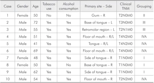
A total of 10 patients seen at the Oral Cancer Center at the Division of Dentistry in the University Hospital of Brasília (UnB), from October 2005 to December 2008, with a histologically proven diag-nosis of oral squamous cell carcinoma were exam-ined and submitted to MRI before treatment. This study was approved by the Ethics Committee at UnB (no. 025/2007) and informed consent was ob-tained from all patients. Table 1 presents the clinical characteristics of the patients.
After clinical examination and biopsies, the patients were referred to a head and neck surgeon to establish their clinical TNM stage and undergo treatment. They concomitantly underwent the CT and MRI examinations. The CT data were pub-lished in a previous study.15 Four observers
inter-preted the MRI scans. Observers 1 and 2 were den-tal specialists holding a Master’s in oral radiology, and Observers 3 and 4 were medical radiologists. These four radiologists established staging based on the MRI scans without any prior knowledge of the clinical staging already established by the head and
Case Gender Age Tobacco use consumptionAlcohol Primary site – Side Clinical TNM Grouping
1 Female 50 No No Gum – R T2N0M0 II
2 Male 72 Yes Yes Base of tongue – L T3N0M0 III
3 Male 55 Yes Yes Retromolar region – L T2N1M0 III
4 Male 51 Yes Yes Floor of mouth – R/L T4N2M0 IVA
5 Male 41 Yes Yes Tongue – R/L T4N2M0 IVA
6 Male 69 Yes Yes Floor of mouth – R/L T4N0M0 IVA
7 Female 48 Yes Yes Side of tongue – R T1N0M0 I
8 Female 50 Yes No Base of tongue – R T1N0M0 I
9 Male 62 Yes Yes Side of tongue – R T1N0M0 I
10 Male 54 Yes Yes Floor of mouth – R T2N2M0 IVA
R = right; L = left.
neck surgeon.
Three parameters were evaluated, namely, the extent of the primary tumor (T), the presence/ab-sence and extent of metastasis in regional lymph nodes (N) and the grouping by stages. The criteria established by Prehn et al.16 were used to ascertain
which cervical lymph nodes were affected when cer-vical metastasis occurred.
Magnetic resonance imaging and interpretation
MRI was performed with a Signa Excite 1.5 high-ield device (1.5 Tesla) (General Electric Healthcare Inc., Milwaukee, USA), following the protocol of the institution. Images of the head and neck region were taken to assess cervical lymph node involvement. The examinations included T1 (TR/ TE, 350/13.1 ms; FOV 24 × 24 mm; slice width/gap, 3,5/1 mm; slice number, 30), T1 with contrast (TR/ TE, 300/4.8 ms; FOV 24 × 24 mm; slice width/gap, 3,5/1 mm; slice number, 30) and T2-weighted (TR/ TE, 4600/99.4 ms; FOV 26 × 26 mm; slice width/ gap, 5/1 mm; slice number, 30) sequences on three anatomical planes (axial, coronal and sagittal). A gadolinium-based contrast agent (Gd/DTPA - Dieth-ylene Triamine Pentaacetic Acid) was used.
The MRI scans were interpreted on a Toshiba Satellite A65 laptop computer (Toshiba America Information Systems, Inc., Irvine, USA), with a 14-inch screen. Printed ilms were not used. The eFilm 2.0 program (Merge Healthcare Inc., Chicago, USA), which provides a DICOM (Digital Imaging Communication in Medicine) reading, was used to visualize and analyze the images. The MR
im-ages were considered the standard parameter in this study.
Statistical analysis
A descriptive analysis was performed using mean, median, standard deviation, and both maxi-mum and minimaxi-mum values. The SPSS for Windows program, version 13.0 (SPSS Inc., Chicago, USA) was used for all the statistical tests. Inter-observer agreement as regards MRI staging, T stage, N stage and grouping by stages was analyzed using Cohen’s kappa index. A statistical signiicance level of 95% (p value < 0.05) and a 5% error level were consid-ered for the analyses. Interpretation criteria of the kappa index recommended by Landis and Koch17
were used to analyze the results.
Results
Table 2 shows that agreement for the T stage was excellent (k = 0.85) between Observers 1 and 2 (oral radiologists) and moderate (k = 0.47) between Ob-servers 3 and 4 (medical radiologists).
All four observers presented different results for the T stage using the clinical and MRI examinations. The highest rate of agreement (k = 0.46 = moderate) was presented by Observer 4.
Table 3 shows that agreement for the N stage was substantial (k = 0.69) between Observers 1 and 2 (oral radiologists) and considerable (k = 0.38) be-tween Observers 3 and 4 (medical radiologists).
All four observers presented different results for the N Stage using the clinical and MRI examina-tions.
Table 4 shows that the agreement for grouping by
Obs. 1 Obs. 2 Obs. 3 Obs. 4
Clinical T (p = 0.116)k = 0.29 (p = 0.100)k = 0.31 (p = 0.366)k = 0.17 (p = 0.012)*k = 0.46
Obs. 1 (p < 0.000)*K= 0.85 (p = 0.024)*k = 0.42 (p = 0.001)*k = 0.58
Obs. 2 (p = 0.003)*k = 0.57 (p < 0.000)*k = 0.73
Obs. 3 (p = 0.006)*k = 0.47
k = kappa value; *p < 0.05 = statistically significant agreement; Obs. = observer; 1 and 2 = oral radiolo-gists; 3 and 4 = medical radiologists.
stages was substantial (k = 0.72) between Observers 1 and 2 (oral radiologists) and moderate (k = 0.44) between Observers 3 and 4 (medical radiologists).
All four observers presented different results for the grouping by stages using the clinical and MRI examinations. The highest rate of agreement (k = 0.44 = moderate) was presented by Observer 4.
Table 5 shows that there was signiicant agree-ment (p < 0.05) between the clinical and MRI stag-ing assessments made by one oral radiologist (Ob-server 2) for N stage, and signiicant agreement between those made by one medical radiologist (Observer 4) for T and N stages and for grouping by stages.
As to MRI staging, there was signiicant agree-ment (p < 0.05) among all four observers for T stage and for grouping by stages, and there was no sig-niicant agreement between Observers 1 and 4, or between 3 and 4, for N stage. In all other compari-sons, there was signiicant agreement (p < 0.05) for N stage (Table 6).
Discussion
The results of the present study contribute to rec-ognizing the importance of MRI examination in es-tablishing the staging of oral cancer.
Carcinoma lesions in the oral cavity are very ag-gressive and usually iniltrate the surrounding tissue and lymph vessels, producing metastasis in the cer-vical region.2,18
CT and MRI examinations are the most signii-cant methods of diagnostic imaging in the preopera-tive staging of head and neck tumors, because they provide information on the extent of the lesion, in-iltration of large vessels and lymph node metastasis, thereby facilitating treatment planning and progno-sis.12-14,19 During the preoperative phase of a patient’s
squamous cell carcinoma, CT scans are essential for evaluating the primary lesion and the possibility of bone invasion, and especially for deining involve-ment in cervical lymph node chains.12-14,19 In the
preoperative treatment of oral cancer, it has been shown that MRI is better for evaluating soft tissue, bone marrow involvement and perineural invasion,
Obs. 1 Obs. 2 Obs. 3 Obs. 4
Clinical N (p = 0.115)k = 0.35 (p = 0.003)*k = 0.69 (p = 0.056)k = 0.47 (p = 0.001)*k = 0.69
Obs. 1 (p = 0.003)*k = 0.69 (p < 0.000)*k = 0.84 (p = 0.274)k = 0.24
Obs. 2 (p < 0.000)*k = 0.84 (p = 0.014)*k = 0.55
Obs. 3 (p = 0.075)k = 0.38
k = kappa value; *p < 0.05 = statistically significant agreement; Obs. = observer; 1 and 2 = oral radiolo-gists; 3 and 4 = medical radiologists.
Table 3 - Inter-observer and clinical and MRI examination agreement for N Stage.
Obs. 1 Obs. 2 Obs. 3 Obs. 4
Clinical
Grouping (p = 0.100)k = 0.31 (p = 0.100)k = 0.31 (p = 0.509)k = 0.13 (p = 0.022)*k = 0.44
Obs. 1 (p < 0.000)*k = 0.72 (p < 0.000)*k = 0.72 (p = 0.017)*k = 0.44
Obs. 2 (p = 0.001)*K = 0.58 (p < 0.000)*K = 0.72
Obs. 3 (p = 0.014)*K = 0.44
k = kappa value; *p < 0.05 = statistically significant agreement; Obs. = observer; 1 and 2 = oral radiolo-gists; 3 and 4 = medical radiologists.
and has been particularly decisive in the diagnosis of small lesions.7,20,21
The discrepancies found in the comparisons be-tween the staging established by clinical and MRI examinations demonstrate the importance of this study. A clinical staging assessment lower than the real staging could result in ineffective treatment and/ or increase the possibility of recurrence in a partic-ular case, whereas a higher clinical staging assess-ment could lead to more radical treatassess-ment, thereby increasing treatment aftereffects. The related litera-ture states that appropriate staging of a lesion is es-sential for decision-making during surgical and/or radiotherapy planning, for predicting the prognosis and for deciding how to carry out patient follow-up to guarantee greater life expectancy and cure.1,2,7
Different results were found between the cal and MRI examinations for T staging. If a clini-cal staging assessment establishes a primary tumor as smaller than it really is, this could result in in-effective surgical margins and in an incomplete re-moval of the lesion.7 Figures 1 through 3 show case
number 8 clinically staged as T1, in which the MRI shows the primary tumor measuring 3.4 cm (T2). Surgical and anatomic pathology conirmation would be necessary to determine the actual size of the primary tumor.
Agreement among all four observers was
sig-niicant for MRI T staging. This agreement shows that MR images provide greater interpretation stan-dardization. Calibration of the observers may have been decisive in achieving the agreement levels and should be used in joint training programs that pre-pare medical and oral radiologists to diagnose oral cancer at reference centers providing multidisci-plinary care.15
There was substantial and signiicant agreement between the clinical and MRI staging performed by Observers 2 and 4 for N stage. Nevertheless, case number 3, clinically staged as N1, was staged as N0 by two observers (1 and 3) in the MRI examination. A higher clinical staging assessment, establishing a false-positive for regional metastasis, may lead to more radical treatment and increase morbidity. Ac-cording to Malard et al.10 and Scully and Bagan,11 a
combination of both clinical and imaging examina-tions is essential for the detection of metastatic cer-vical lymph nodes, and could improve staging and prognosis determination.
Agreement between Observers 1 and 2, 1 and 3, 2 and 3, and 2 and 4 in the MRI staging was sig-niicant, indicating that the pre-established criteria and image interpretation guide could also have been crucial for the level of agreement achieved for the N stage.
Different results were observed for the clinical
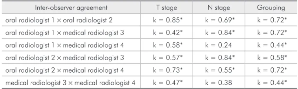
Inter-observer agreement T stage N stage Grouping oral radiologist 1 × oral radiologist 2 k = 0.85* k = 0.69* k = 0.72* oral radiologist 1 × medical radiologist 3 k = 0.42* k = 0.84* k = 0.72* oral radiologist 1 × medical radiologist 4 k = 0.58* k = 0.24 k = 0.44* oral radiologist 2 × medical radiologist 3 k = 0.57* k = 0.84* k = 0.58* oral radiologist 2 × medical radiologist 4 k = 0.73* k = 0.55* k = 0.72* medical radiologist 3 × medical radiologist 4 k = 0.47* k = 0.38 k = 0.44*
k = kappa value; *p < 0.05 = statistically significant agreement.
Table 6 - Inter-observer agreement for MRI staging.
Agreement between clinical and MRI staging T stage N stage Grouping clinical × oral radiologist (Obs. 1) k = 0.29 k = 0.35 k = 0.31 clinical × oral radiologist (Obs. 2) k = 0.31 k = 0.68* k = 0.31 clinical × medical radiologist (Obs. 3) k = 0.17 k = 0.47 k = 0.13 clinical × medical radiologist (Obs. 4) k = 0.46* k = 0.69* k = 0.44*
k = kappa value; *p < 0.05 = statistically significant agreement; Obs. = observer.
and MRI staging, and agreement was signiicant only for Observer 4. Among these results, three cases were staged as belonging to the IVA grouping, which represents lesions which are larger and at a more ad-vanced stage, thereby facilitating diagnosis.15 This
conirms the greater importance of the MRI
exami-nation for smaller lesions. The deinition of grouping by stages is critical in determining the patient’s treat-ment plan, prognosis and survival span.
Agreement among all four observers for stage grouping using MRI was signiicant, and greater be-tween the oral radiologists. Based on these results and on literature data, it could be stated that clini-cal examination, anatomic pathology testing and diagnostic imaging modalities (which could include CT and MRI) are necessary to establish the staging of patients with oral cancer. According to Weber et al.13 and Scully and Bagan,22 CT and MRI
examina-tions seem to be the most important diagnostic tools when establishing pre-therapeutic staging of head and neck tumors. CT is essential, insofar as it pro-vides a better evaluation of cervical lymph node in-volvement and invasion of bone cortices adjacent to the primary tumor area. The MRI examination pro-vides a better evaluation of the soft tissues affected by the lesion and allows a more thorough evaluation of small tumors. This examination should be part of the treatment protocol of patients with oral cancer, depending on its availability, accessibility and the possibility of carrying out the examination. Accord-ing to Warnakulasuriya,4 improvement in the
qual-ity of healthcare helps to reduce mortalqual-ity rates.
Figure 2 - MRI axial slice, T1-weighted, with contrast, show-ing a tumor of hyper-signal intensity (enhanced with contrast) at the base of the tongue (arrowhead), on the right side.
Figure 3 - MRI coronal slice, T2-weighted, showing a tumor of isosignal intensity at the base of the tongue (arrowhead), on the right side.
While the results would indicate the importance of using MRI in the diagnosis of oral cancer, there is also a very obvious need for a combination of re-search and surgical and pathological information to identify sources of error in pretreatment staging. Joint training initiatives and calibration of medical and oral radiologists should be promoted to provide an improved multidisciplinary approach to oral can-cer.
Conclusion
There was no agreement between the staging es-tablished by clinical and MRI examinations for oral cancer. MRI examination is useful to provide a bet-ter assessment of TNM staging, and the examina-tions should be analyzed by different professionals (physicians and dentists) in a multidisciplinary ap-proach.
References
1. Shah JP, Gil Z. Current concepts in management of oral cancer – Surgery. Oral Oncol. 2009 Apr-May;45(4-5):394-401. 2. Gil Z, Carlson DL, Boyle JO, Kraus DH, Shah JP, Shaha
AR, et al. Lymph node density is a significant predictor of outcome in patients with oral cancer. Cancer. 2009 Dec 15;115(24):5700-10.
3. Liao CT, Lee LY, Huang SF, Chen IH, Kang CJ, Lin CY, et al. Outcome analysis of patients with oral cavity cancer and extracapsular spread in neck lymph nodes. Int J Radiat Oncol Biol Phys. 2010 Oct 7:1-8.doi:10.1016/j.ijrobp.2010.07.1998. 4. Warnakulasuriya S. Global epidemiology of oral and
oropha-ryngeal cancer. Oral Oncol. 2009 Apr-May;45(4-5):309-16. 5. Gospodarowicz MK, Miller D, Groone PA, Greene FL, Logan
PA, Sobin LH. The process for continuous improvement of the TNM classification. Cancer. 2004 Jan;100(1):1-5. 6. Patel SG, Shah JP. TNM Staging of cancers of the head and
neck: striving for uniformity among diversity. CA Cancer J Clin. 2005 Jul-Aug;55(4):242-58.
7. Park JO, Jung SL, Joo YH, Jung CK, Cho KJ, Kim MS. Di-agnostic accuracy of magnetic resonance imaging (MRI) in the assessment of tumor invasion depth in oral/oropharyngeal cancer. Oral Oncol. 2011 May;47(5):381-6.
8. Carvalho AL, Kowalski LP, Borges JA, Aguiar S, Magrin J. Ipsilateral neck cancer recurrences after elective supraomohy-oid neck dissection. Arch Otolaryngol Head Neck Surg. 2000 Mar;126(3):410-2.
9. Freire AR, Lima EN, Almeida OP, Kowalski LP. Computed tomography and lymphoscintigraphy to identify lymph node metastases and lymphatic drainage pathways in oral and oro-pharyngeal squamous cell carcinomas. Eur Arch Otorhino-laryngol. 2003 Mar;260(3):148-52.
10. Malard O, Toquet C, Jegoux F, Bordure P, Beauvillain de Montreuil C, Gayet-Delacroix, M. Computed tomography in TN stage evaluation of oral cavity and oropharyngeal cancers. Clin Imaging. 2004 Sep-Oct;28(5):360-7.
11. Scully C, Bagan JV. Recent advances in Oral Oncology. Oral Oncol. 2007 Feb;43(2):107-15.
12. Lell M, Baum U, Greess H, Nömayr A, Nkenke E, Koester, M, et al. Head and neck tumors: imaging recurrent tumor and
post-therapeutic changes with CT and MRI. Eur J Radiol. 2000 Mar;33(3):239-47.
13. Weber A, Romo L, Hashmi S. Malignant tumors of the oral cavity and oropharynx: clinical, pathologic and radiologic evaluation. Neuroimaging Clin N Am. 2003 Aug;13(3):443-64.
14. Albuquerque MA, Kuruoshi ME, Oliveira IR, Cavalcanti MG. CT assessment of the correlation between clinical ex-amination and bone involvement in oral malignant tumors. Braz Oral Res. 2009 Apr-Jun;23(2):196-202.
15. Figueiredo PT, Leite AF, Freitas AC, Nascimento LA, Caval-canti MG, Melo NS, et al. Comparison between computed tomography and clinical evaluation in tumour/node stage and follow-up of oral cavity and oropharyngeal cancer. Dentomax-illofac Radiol. 2010 Mar;39(3):140-8.
16. Prehn R, Pasic T, Harari P, Brown W, Ford C. Influence of computed tomography on pretherapeutic tumor staging in head and neck cancer patients. Otolaryngol Head Neck Surg. 1998 Dec;19(6):628-33.
17. Landis JR, Koch GG. The measurement of observer agreement for categorical data. Biometrics. 1977 Mar;33(1):159-74. 18. Walker DM, Boey G, Mcdonald LA. The pathology of oral
cancer. Pathology. 2003 Oct;35(5):376-83.
19. Vidiri A, Guerrisi A, Pellini R, Manciocco V, Covello R, Mattioni O, et al. Multi-detector row computed tomography (MDCT) and magnetic resonance imaging (MRI) in the evalu-ation of the mandibular invasion by squamous cell carcinomas (SCC) of the oral cavity. Correlation with pathological data. J Exp Clin Cancer Res. 2010 Jun 17;29:73. doi: 10.1186/1756-9966-29-73.
20. Lenz M, Greess H, Baum U, Dobritz M, Kersting-Sommerhoff B. Oropharynx, oral cavity, floor of the mouth: CT and MRI. Eur J Radiol. 2000 Mar;33(3):203-15.
21. Rumboldt Z, Day TA, Michel M. Imaging of oral cavity can-cer. Oral Oncology. 2006 Oct;42(9):854-65.