Oral Cancer
Joana Mattos Ozi(a) Ivana Barbosa Suffredini(b) Mateus Paciencia(c) Sergio Alexandre Frana(b) Luciano Lauria Dib(a)
(a) Graduate Program in Dentistry, Paulista University, São Paulo, SP, Brazil. (b) Center for Research in Biodiversity,
Extraction Laboratory, Paulista University, São Paulo, SP, Brazil.
(c) Center for Research in Biodiversity, UNIP Herbarium, Paulista University, São Paulo, SP, Brazil.
Corresponding author: Joana de Matos Ozi E-mail: joana.ozi@gmail.com
Received for publication on Aug 13, 2011 Accepted for publication on Oct 22, 2011
In vitro
cytotoxic effects of Brazilian
plant extracts on squamous cell
carcinoma of the oral cavity
Abstract: Squamous cell carcinoma (SCC) is the most prevalent cancer of the oral cavity and the ifth most prevalent of all malignancies in males. Many researchers have attempted to develop new treatments that will improve the prognosis of SCC patients. Over 20% of the world’s biodiversity is located within the Brazilian forests, but little is known about the chemical and/or pharmacological potential of these plants. Certain extracts obtained from Amazon and Atlantic Forest plants have previously been shown to have cytotoxic activity against various can-cers. The aim of this study was to screen these extracts for cytotoxic activity against oral SCC cells. The extracts were analyzed for activ-ity against the KB-ADL#12 cell line at various concentrations up to a maximum dose of 100 µg/mL. Comparisons with a control group were performed using one-way ANOVA. Signiicant cytotoxicity was induced by the extracts obtained from the aerial parts of Picrolemma sprucei
(Simaroubaceae), from the leaves and stems of Laetia suaveolens (Sali-caceae), from the aerial parts of Abarema auriculata (Fabaceae-Mimo-soideae) and from the stem of A. auriculata.
Descriptors: Mouth Neoplasm; Carcinoma, Squamous Cell; Plant Extracts.
Introduction
Squamous cell carcinoma (SCC) is the most prevalent cancer of the oral cavity and one of the most frequent cancers in the world. Annually, approximately 350,000 new cases of oral and oropharyngeal SCCs are diagnosed worldwide.1
In Brazil, the prevalence of oral cancer is high, ranking ifth among all malignant tumors. Smoking and alcohol consumption are considered to be the most important etiological factors contributing to the develop-ment of this disease.2 The stage at diagnosis is an important prognostic indicator for oral SCC.3 Despite aggressive treatment interventions, the ive-year survival rate for patients with SCC remains low.4
Several studies have been conducted recently to develop new therapeu-tics that can improve the prognosis of SCC patients, in particular those derived from nature.5 With more than 56,000 species, Brazil has one of the richest loras in the world, harboring nearly 19% of the world lora.6 Plants are considered to be a potential source of new anti-cancer com-pounds; however, little is known about the chemical and pharmacological
potential of these plants, including those occurring in the Amazon and Atlantic Forests. Screening assays that are rapid and cost effective play an important role in the identiication of new active compounds. For more than a decade, our group has been screen-ing Brazilian plant extracts7-9 against prostate, breast, lung, colon and central nervous system cancers as well as leukemias. From those studies, 72 extracts were discovered to have activity against one or more cancer cell lines. The aim of this in vitro study was to screen these extracts obtained from Amazon and Atlantic Forest plants against SCC cells (KB-ADL # 12).
Methodology
Plant collection and extract preparation
Plants were collected in the Amazon and in the Atlantic Forests (licenses were obtained from IB-AMA/MMA and Cgen/MMA). The plants were identiied, and vouchers were deposited at the UNIP Herbarium (Paulista University). Plant organs were collected and processed as follows.
Each plant material was dried and ground be-fore being submitted to a 24-h maceration with a methanol:dichloromethane (1:1) solution; a second 24-h maceration was performed with water, result-ing in two extracts containresult-ing non-polar (organic extracts) and polar (aqueous extracts) substances, respectively. Seventy-two extracts had been previ-ously investigated for activity against cancer cells from the prostate,6 breast,7 lung and the central ner-vous system in addition to leukemia cells.8 For that reason, these extracts were selected to be analyzed
in vitro against SCC cells.
Extracts were diluted to an initial concentration of 40 mg/mL, which was further diluted to 100 µg/ mL for experimentation. Organic extracts were di-luted with 50% dimethylsulfoxide; aqueous extracts were diluted with distilled water.
Cell culture
The KB-ADL#12 tumor cell line (SCC) was ob-tained from the National Cancer Institute (Fred-erick, Maryland, United States). The cells were maintained in DMEM supplemented with 20% fe-tal bovine serum (FBS), 1% glutamine, and 0.2%
gentamicin in tissue culture lasks (Costar Corn-ing, Lowell, USA) in an incubator (Thermo For-ma, Asheville, USA) at 37 °C, 5% CO2 and 100% relative humidity. Cell counts were obtained by the trypan blue exclusion method to calculate cell den-sities. Experiments were performed in 96-well mi-croplates (Costar Corning, Lowell, USA) at cell den-sities of 27,500, 60,000 or 100,000 cells per well. The cells were incubated for 24 h before the drug or plant extract was added. After treatment, the cells were incubated for 48 h prior to analysis with the sulforhodamine B (SRB) assay.10
Sulforhodamine B assay
Viable cells were ixed in the microplates with 50 µl of cold 50% trichloroacetic acid solution. The microplates were washed four times with running water to completely remove non-viable cells and air dried for 24 h. A volume of 100 µl of SRB was add-ed to each well and incubatadd-ed for 10 min. The plates were washed ive times with 1% acetic acid using a microplate washer (Biotek Winooski, USA) for the complete removal of unbound SRB. The plates were air dried for 24 h. The stain was resuspended in 100 µl of Trisma Buffer. Viable cells were mea-sured by obtaining the optical densities (ODs) with a microplate spectrophotometer reader (Biotek 408x Winooski, USA) at a wavelength of 515 nm.10
Experimental groups and assay strategy
The experiments were designed to identify the most potent cytotoxic extracts from the 72 previ-ously selected extracts.
Cells that were not treated with extract were des-ignated as the control group.
The drug control group was the group of cells treated with Doxorubicin (Sigma-aldrich, Mis-souri, USA), which is also known by the commercial name Adriamycin.
The extracts group was the group of cells treated with the 72 selected plant extracts.
to determine the background OD. The cell growth at 24 h (T0) was measured (n = 24); T0 was deined as the time immediately prior to the addition of ex-tract. OD values were obtained as described. The KB-ADL#12 cell line was used at various densities, according to the stage of screening.
Statistical analyses
The mean OD readings were obtained to evaluate cell growth inhibition after various treatments, i.e., the extracts, reference substance and experimental controls. A primary battery of experiments was used to screen the extracts according to an “inhibitory eficacy”. Mean OD values that were signiicantly reduced compared with the controls were used to identify active extracts. The assays performed with a reduced number of extracts were analyzed using the same statistical analyses. Depending on the case, a one-way or two-way ANOVA was used followed by Tukey’s post-test.11,12 A p value < 0.05 was con-sidered statistically signiicant (StatSoft Incorpora-tion 2001, Oklahoma, USA).
Control drug and extracts
DOXO was used at the following concentrations:
• 2.5 × 10⁻⁵,
• 2.5 × 10⁻⁶,
• 2.5 × 10⁻⁷,
• 2.5 × 10⁻⁸,
• 2.5 × 10⁻⁹ and
• 2.5 × 10⁻¹⁰ M.
Plant extracts were initially analyzed at a single dose of 100 µg/mL, and the four extracts selected for further characterization were analyzed at con-centrations of 100, 10, 1, 0.1, 0.01 and 0.001 µg/ mL. Data from each concentration were compared with the control groups (ANOVA).
Results
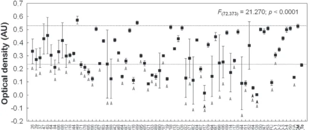
Screenings were performed on all 72 extracts and on the standard drug (DOXO) (p < 0.05). Sig-niicant differences were observed for 41 plant ex-tracts out of the 72 analyzed when compared with T0 and the control groups. DOXO showed signii-cant levels of cytotoxicity at all concentrations (Fig-ures 1, 2 and 3). Six of the 41 extracts were excluded for showing a false positive result, likely due to fun-gal contamination. Therefore, 35 extracts induced signiicant differences in OD values (Figure 1) and were selected for further assessment.
The remaining 35 extracts were investigated for
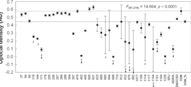
activity against KB cells at densities of 27,500 and 60,000 cells/well; of these, 18 extracts were consid-ered active (Figure 2; Table 1). The 18 extracts were again analyzed, and nine extracts (Figure 3) were considered signiicantly cytotoxic to the KB cell line.
Four out of the nine extracts exhibited a strong ability for inhibition and were subjected to more
rig-orous experimental conditions using the same pro-tocol but with a cell density of 60,000 cells/well; the trend toward cell inhibition was conirmed. These four extracts were obtained from Laetia suaveolens
(Salicaceae, extract 719; p < 0.0002), Picrolemma sprucei (Simaroubaceae, extract 1151; p < 0.0002) and Abarema auriculata (Fabaceae, extracts 633
Figure 3 - The optical density (OD) of cells (60,000 cells/well) after exposure to the 18 plant extracts (one-way ANOVA). Ex-tracts and/or standard substances that induced an OD lower than the controls are indicated by arrows (p < 0.05). Upper line represents the OD mean ± s.d. of the control group; lower line represents the OD mean ± s.d. of the T0 group. Empty arrows indicate the OD mean ± s.d. of treatments that were lower than control group, and full arrows indicate the OD mean ± s.d. of treatments that were lower than T0.
and 689, both p < 0.0002).
Extracts 633, 689, 719 and 1151 were then ana-lyzed at six different concentrations of 100, 10, 1, 0.1, 0.01 and 0.001 µg/mL against cell densities of 27,500, 60,000 and 100,000 cells/well. Signiicant differences were observed for extract 689 at a con-centration of 100 µg/mL at cell densities of 60,000 and 100,000 cells/well, relative to the control groups. DOXO inhibited cell growth in a similar manner, although at a concentration lower than the extract concentrations. Evaluations of extract 719 demonstrated differences between the treated and control cells only at a concentration of 100 µg/mL at cell densities of 27,500, 60,000 and 100,000 cells/ well. Finally, extract 1151 showed signiicant differ-ences at concentrations of 1 µg/mL, 10 µg/mL and 100 µg/mL at a cell density of 27,500 cells/well and at concentrations of 10 µg/mL and 100 µg/mL at cell densities of 60,000 and 100,000 cells/well com-pared with the control groups.
Discussion
The treatment of oral cancer relies on surgery, radiotherapy, chemotherapy or a combination of these methods.13 Poor survival rates still occur, particularly for patients in advanced stages of the disease.14 Approximately 40% of patients fail to re-spond to treatment.15 Therefore, the development of new strategies for the early diagnosis and treat-ment of this malignancy is of great importance.16-20 Natural products display a wide range of diversity in terms of their chemical structures and pharma-cological properties. Several important antitumor drugs have been isolated from plants, such as pacli-taxel, the vinca alkaloids, camptothecin, podophyl-lin and others.21 Studies concerning the activities of natural products against SCC have drawn attention in the past few years, such as Aloe-emodin, which inhibit KB cells in a dose-dependent manner.22 Cur-cumin has also been studied as a treatment for oral cancer.23
The cytotoxic analyses of 72 Amazon and At-lantic forest plant extracts against the SCC
KB-Table 1 - List of the 18 plant extracts that showed activity against the KB-ADL#12 cell line in vitro.
Collect Date Extracts# Family Species Organ
30/05/97 37 Fabaceae Mimosoideae Pithecellobium sp. Aerial Organs
18/04/97 64 Hypericaceae Vismia guianensis (Aubl.) Choisy Stem
05/12/97 84 Olacaceae Chaunochiton lorantoides Benth. Stem
25/06/98 171 Fabaceae Caesalpinioideae Macrolobium multijugum (DC.) Benth. Leaves
18/04/97 178 Meliaceae Trichilia pleeana (A. Juss.) C.DC. Aerial Organs
27/06/98 205 Fabaceae Caesalpinioideae Hymenaea courbaril L. Stem
27/06/98 206 Fabaceae Caesalpinioideae Hymenaea courbaril L. Stem
19/04/97 257 Fabaceae Caesalpinioideae Cynometra spruceana Benth. Aerial Organs
19/04/97 364 Polygonaceae Cimeria sp. Aerial Organs
19/04/97 416 Fabaceae Faboideae Taralea opositifolia Aubl. Stem
18/04/97 505 Lecythidaceae Gustavia augusta L. Stem
09/09/98 621 Salicaceae Laetia cobymbulosa Spruce ex Benth. Stem
22/01/99 633 Fabaceae Mimosoideae Abarema auriculata (Benth.) Barneby & J. W. Grime Aerial Organs 22/01/99 689 Fabaceae Mimosoideae Abarema auriculata (Benth.) Barneby & J. W. Grime Stem
02/04/99 719 Salicaceae Laetia suaveolens (Poepp.) Benth. Leaves and Stem
22/01/00 1017 Fabaceae Faboideae Aldina reticulata R. S. Cowan Aerial Organs
22/01/00 1117 Lauraceae Ocotea cymbarum Kunth Stem
25/02/00 1151 Simaroubaceae Picrolemma sprucei Ducke Aerial Organs
ADL#12 cell line produced four active extracts, namely 633, 689, 719 and 1151, which were selected based on results from the dose-response analysis; the data were supported by one-way ANOVA and Tukey’s test. Comparisons between the effectiveness of DOXO and the extracts were performed. Howev-er, the level of cell growth inhibition from a single, concentrated substance was dificult to be surpassed by a complex mixture of substances, such as an ex-tract, due to the concentration of active compounds present in the extract. Nevertheless, the four ex-tracts showed highly signiicant cytotoxicities and will be further analyzed.
Extracts 633 and 689 were obtained from A. auriculata; little is known about chemical or phar-macological studies related to this plant, although it has been used as timber for some time. Studies re-lated to Abarema species are rare. Gastroprotective and ulcer-healing activities24 and anti-inlammatory intestinal activity25 have been reported for A.
cochli-acarpusi, a plant traditionally used in Northeastern Brazil. The ethanolic extract obtained from A. ellip-tica showed anti-oxidant activity and is the subject of two Chinese patents.26,27
Extract 719, obtained from the leaves and stems of L. suaveolens, has not been reported, although studies with plants from the same genus have been conducted. One of these studies reported that meth-yl ester derivatives isolated from the leaves of L.
thamna exhibited cytotoxic activity against pros-tate cancer cells, although the derivatives were ana-lyzed in colon and breast cancer cells.28 An extract from L. procera revealed the presence of diterpenes, which inhibited breast cancer cell growth.29 Ex-tract 1151 was obtained from the aerial organs of P. sprucei, a species that has been studied previously. Quassinosides have been isolated from P. sprucei, and several synthetic derivatives were obtained and evaluated against human tumor cells in vitro, exhib-iting strong activity against the HL-60 cell line.30
Conclusions
In conclusion, we highlight the signiicant cyto-toxic activity of the aerial parts of P. sprucei and
A. auriculata, from the leaves and stems of L. sua-veolens and from the stem of A. auriculata, which introduces promising expectations for new projects in chemistry, pharmacology and toxicology. These results may aid in achieving the development of an anticancer medicine obtained from the rain forest.
Acknowledgements
The authors thank the Fundação de Amparo à Pesquisa do Estado de São Paulo - FAPESP (grant #08/58706-8), and the Coordenação de Aper-feiçoamento de Pessoal de Nível Superior (CAPES) for a scholarship to JMO.
References
1. Jemal A, Siegel R, Ward E, Hao Y, Xu J, Murray T, et al.
Cancer Statistics, 2008. CA Cancer J Clin. 2008 Mar-Apr;58(2):71-96.
2. Scully C, Field JK, Tanzawa H. Genetic aberrations in oral or head and neck squamous cell carcinoma (SCCHN):1. Car-cinogen metabolism, DNA repair and cell cycle control. Oral Oncol. 2000 May;36(3):256-63.
3. Dantas DD, Ramos CC, Costa AL, Souza LB, Pinto LP. Clin-ical-pathological parameters in squamous cell carcinoma of the tongue. Braz Dent J. 2003;14(1):22-5.
4. Liu Sy, Lu CL, Chiou CT, Yen Cy, Liaw GA, Chen YC, et al. Surgical outcomes and prognostic factors of oral cancer associated with betel quid chewing and tobacco smoking in Taiwan. Oral Oncol. 2010 Apr;46(4):276-82.
5. Manoharan S, Kavitha K, Senthil N, Renju GL. Evalua-tion of anticarcinogenic effects of Clerodendron inerme
on 7,12-dimethybenz(a) anthracene-induced hamster buccal pouch carcinogenesis. Singapore Med J. 2006 Dec;47(12):1038-43.
6. Giulietti AM, Harley RM, Queiroz LP, Wanderley MGL, Berg CVD. Biodiversity and Conservation of Plants in Brazil. Conserv Biol. 2005 Jun; 19(3):632-9.
7. Suffredini IB, Paciencia ML, Varella AD, Younes RN. In vitro prostate cancer cell growth inhibition by Brazilian plant extracts. Pharmazie. 2006 Aug;61(8):722-4.
8. Suffredini IB, Paciencia ML, Frana SA, Varella AD, Younes
RN. In vitro breast cancer cell lethality of Brazilian plant
extracts. Pharmazie. 2007 Oct;62(10):798-800.
9. Suffredini IB, Paciencia ML, Varella AD, Younes RN. In vitro
10. Monks A, Scudiero D, Skehan P, Shoemaker R, Paull K, Vistica D, et al. Feasibility of a high-flux anticancer drug screen us-ing a diverse panel of cultured human tumor cell lines. J Natl Cancer Inst. 1991 Jun;83(11):757-66.
11. Sokal RR, Rohlf F J. Biometry. 3rd ed. New York: W.H. Free-man and Company; 1995. 880 p.
12. Zar JH. Biostatiscal analysis. 4th ed. Upper Saddle River:
Prentice Hall; 1999.663 p.
13. Hsu S, Singh B, Schuster G. Induction of apoptosis in oral cancer cells: agents and mechanisms for potential therapy and prevention. Oral Oncol. 2004 May;40(5):461-73.
14. Yao M, Epstein JB, Modi BJ, Pytynia KB, Mundt AJ, Feldman LE. Current surgical treatment of squamous cell carcinoma of the head and neck. Oral Oncol. 2007 Mar;43(3):213-23. 15. Bilde A, von Buchwald C, Dabelsteen E, Therkildsen MH,
Dabelsteen S. Molecular markers in the surgical margin of oral carcinomas. J Oral Pathol Med. 2009 Jan;38(1):72-8. 16. Forastiere A, Koch W, Trotti A, Sidransky D. Head and neck
cancer. N Engl J Med.2001 Dec 27;345(26):1890-900. 17. Wang SJ, Peyrollier K, Bourguignon LY. The influence of
hyaluronan-CD44 interaction on topoisomerase ii activity and etoposide cytotoxicity in head and neck cancer. Arch Otolaryngol Head Neck Surg. 2007 Mar;133(3):281-8. 18. Sun Z, Sood S, Li N, Yang P, Newman RA, Yang CS, et
al. Chemoprevention of 7,12-dimethylbenz(a) anthracene (DMBA)-induced oral carcinogenesis in hamster cheek pouch by topical application of a dual inhibitor of epidermal growth factor receptor (EGRF) and ErbB2 tyrosine kinases. Oral On-col. 2008 Jul;44(7):652-7.
19. Scully C, Bagan JV. Recent advances in Oral Oncology 2007: imaging, treatment and treatment outcomes. Oral Oncol. 2008 Mar;44(3):211-5.
20. Torres-Pereira C. Oral cancer public policies: is there any evidence of impact? Braz Oral Res. 2010;24 Suppl 1:37-42. 21. Wall ME, Wani MC. Camptothecin and taxol: discovery to
clinic-thirteenth Bruce F. Cain Memorial Award Lecture. Cancer Res. 1995 Feb 15;55(4):753-60.
22. Xiao B, Guo J, Liu D, Zhang S. Aloe-emodin induces in vitro
G2/M arrest and alkaline phosphatase activation in human oral cancer KB cells. Oral Oncol. 2007 Oct;43(9):905-10. 23. Chen JW, Tang YL, Liu H, Zhu ZY, Lü D, Geng N, et al.
[Anti-proliferative and anti-metastic effects of curcumin on
oral cancer cells]. Hua Xi Kou Qiang Yi Xue Za Zhi. 2011 Feb;29(1):83-6. Chinese.
24. da Silva MS, de Almeida AC, de Faria FM, Luiz-Ferreira A, da Silva MA, Vilegas W, et al. Abarema cochliacarpos: gas-troprotective and ulcer-healing activities. J Ethnopharmacol. 2010 Oct 28;132(1):134-42.
25. da Silva MS, Sánchez-Fidalgo S, Talero E, Cárdeno A, da Silva MA, Villegas W, et al. Anti-inflammatory intestinal activity of Abarema cochliacarpos (Gomes) Barneby & Grimes in TNBS colitis model. J Ethnopharmacol. 2010 Mar 24;128(2):467-75. 26. Wu R, Huang J, Lei J, Tang Z. Abarema elliptica extract
with antiviral and anti-oxidation effects, and medicinal or nutraceutical composition containing th esame. (Guangzhou Lifetech Pharmaceuticals Co., Ltd., Peop. Rep. China). Faming Zhuanli Shenqing Gongkai Shuomingshu (2006a), CODEN: CNXXEVCN1850146A20061025 Patent written in Chi-nese. Application: CN2006-1003369820060221. Priority: AN2006: 1141651 CAPLUS(Copyright (C) 2009 A C Son Sci Finder (R)).
27. Wu R, Wu X, Lei J, Chen D, Huang J. inflammatory buccal tablet comprising extract of Abarema elliptica. Guangzhou Lifetech Pharmaceutical Co., Ltd., Peop. Rep. China). Fam-ing Zhuanli ShenqFam-ing Gongkai ShuomFam-ingshu (2006b), 14pp. CODEN: CNXXEV CN1718222A20060111 Patent writ-ten in Chinese. Application: CN2004-1002791320040706. Priority: CA N145: 50910A N20 06: 453479CA PLUS (Copyright(C)2009 AC Son Sci Finder (R)).
28. Henry GE, Adama LS, Rosales JC, Jacobs H, Heber D, See-ram NP. Kaurene diterpenes from Laetia thamnia inhibit the growth of human cancer cells in vitro. Cancer Lett. 2006 Dec 8;244(2):190-4.
29. Jullian V, Bonduelle C, Valentin A, Acebey L, Duigou AG, Prévost MF, et al. New clerodane diterpenoids from Laetia procera (Poepp.) Eichler (Flacourtiaceae), with antiplasmodial and antileishmanial activities. Bioorg Med Chem Lett.2005 Nov 15;15(22):5065-70.